Journal List > Prog Med Phys > v.35(2) > 1516087711

Jegal, Park, Kang, Choi, and Kim: Motion Management and Image-Guided Technique in Photon Radiation Therapy: A Review of an Advanced Technology

Abstract

Herein, we provide a concise review of the critical role of motion management in radiation therapy, with a focus on photon radiation therapy, real-time control of respiratory motion, and image-guided radiation therapy (IGRT) in lung stereotactic body radiation therapy (SBRT). The dynamic nature of human anatomy, particularly in regions prone to movement such as the thoracic and abdominal areas, poses significant challenges in accurately targeting tumors during radiation therapy. This review explores the implications of organ and tumor motion, emphasizing the necessity for precise treatment delivery. We assess the advancements in four-dimensional (4D) imaging techniques such as 4D computed tomography, which provide time-resolved images for enhanced treatment planning. The review highlights various motion management strategies, including motion-encompassing methods, respiratory-gating, breath-hold techniques, and real-time tumor tracking, discussing their implementation and impact on treatment efficacy. The role of IGRT in lung SBRT is particularly emphasized, showcasing how real-time imaging and advanced targeting techniques enhance the precision of high-dose radiation delivery while minimizing exposure to surrounding healthy tissues. This comprehensive review aims to underscore the significance of integrating motion management in radiation therapy, highlighting its pivotal role in improving treatment accuracy, reducing toxicity, and ultimately enhancing patient outcomes in cancer care.

Introduction

Radiation therapy is a cornerstone in treating various cancers, offering the promise of targeted tumor control while sparing surrounding healthy tissues. However, the dynamic nature of human anatomy, caused mainly by respiratory and organ motion, is a significant challenge in the precise delivery of therapeutic radiation. The challenge is compounded in treating thoracic, abdominal, and pelvic tumors, where organ motion can significantly affected the accuracy and efficacy of radiation therapy [1-4].
The advent of motion management techniques in radiation therapy has been a pivotal development in addressing these challenges. These techniques include strategies from advanced imaging methods, such as four-dimensional computed tomography (4DCT) [5,6], to real-time control of respiratory motion and sophisticated image-guided radiation therapy (IGRT) [7,8].
This review seeks to provide an overview of the current state of motion management in radiation therapy. We explore the effects of organ and tumor motion on treatment outcomes, delve into various imaging and motion management strategies currently employed, and consider the application of these techniques in different treatment contexts such as photon radiation therapy and lung stereotactic body radiation therapy (SBRT). This review also aims to assess the future of motion management, anticipating advancements that may further enhance the precision and effectiveness of radiation therapy.
Understanding and managing motion in radiation therapy is both a technical challenge and a clinical necessity as this directly influences treatment accuracy, toxicity profiles, and, ultimately, patient outcomes. As such, this review seeks to underscore the importance of motion management in pursuing more effective and safer radiation treatments.

Motion in Photon Radiation Therapy

In photon radiation therapy, addressing the challenge of motion is crucial for treatment efficacy, precision, and patient safety. This section covers the impact of motion, imaging techniques, and motion management strategies in photon radiation therapy.

1. Geometric uncertainties

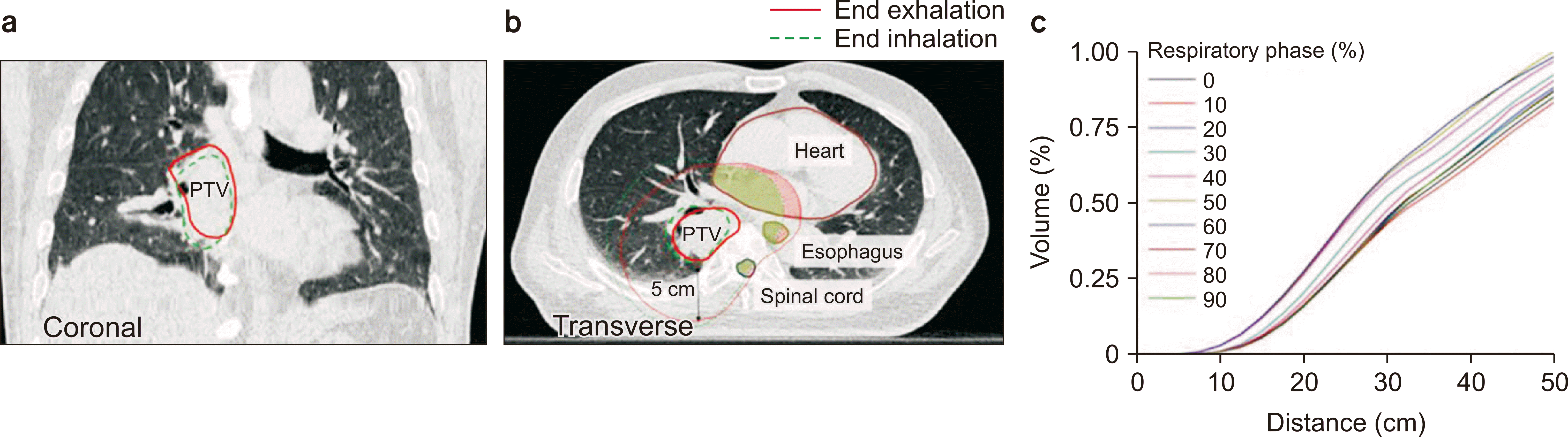
The primary challenge posed by motion is the variability produced in tumor position [9-11]. Movements caused by breathing, heartbeat, or patient shifts can cause the tumor to move out of the planned radiation field, leading to a potential geometric miss [4]. Fig. 1 shows an example where geometrical realtionships between the planning target volume (PTV) and organs at risk (OARs) changed from respiratory phase to phase (end of inhalation to end of exhalation) [9]. Langen and Jones (2001) [4] reviewed respiration-induced organ movements of the liver, diaphragm, kidneys, and pancreas and of tumors located in the lung with their reported variation of movement. To account for this variability, larger margins are often added around the tumor. Although this approach aims to ensure that the tumor remains within the radiation field, this also increases the volume of healthy tissue exposed to radiation. Other suggestions have been investigated and reported to manage the organ motion such as the use of radiopaque [12], gold marker detection [13], control of organ position in initial planning scan [14], adaptive radiation therapy [15], and respiration management. These techniques, especially respiration management, will be discussed later.

2. Dosimetric implications

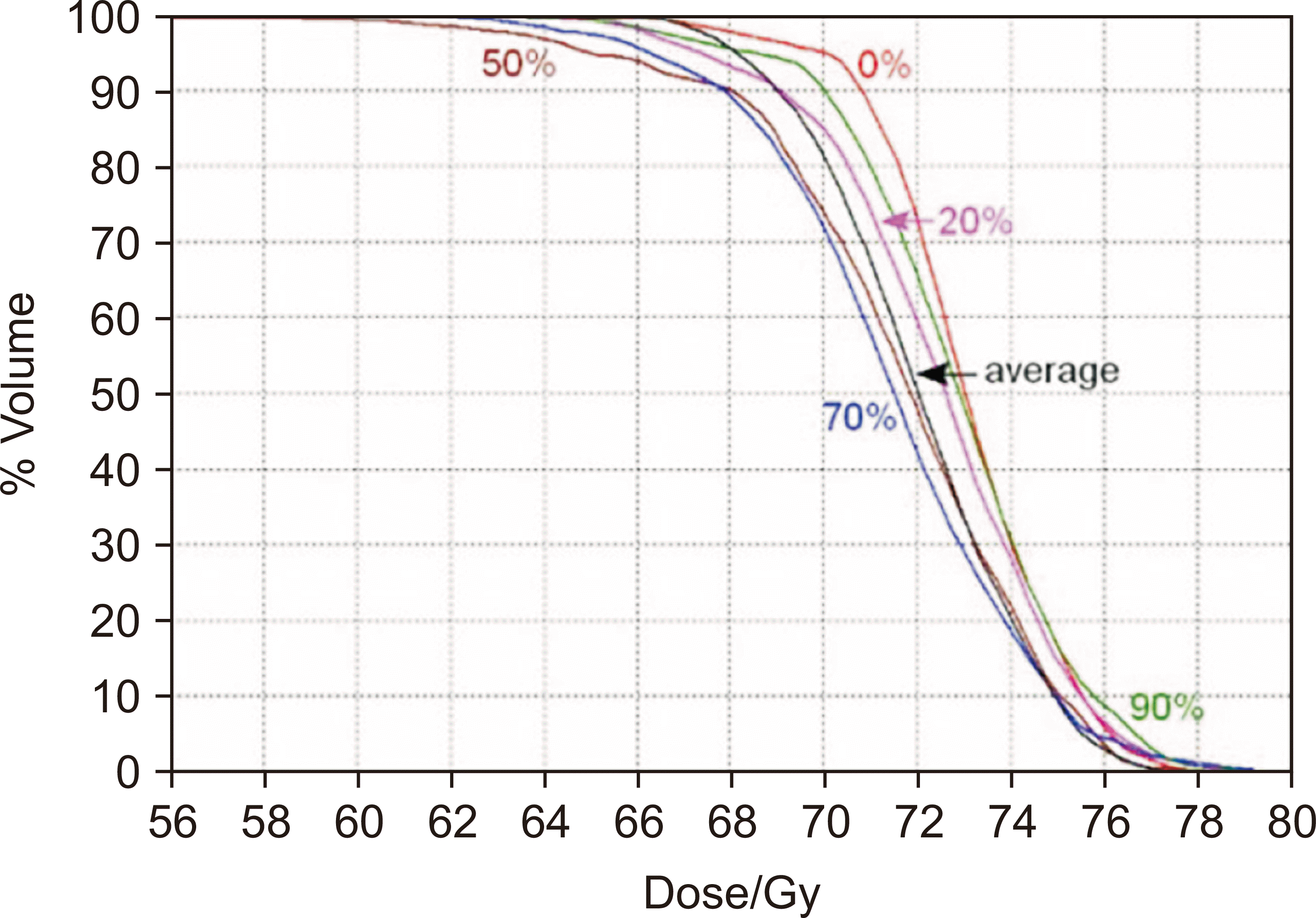
Organ movement can cause variations in distributing the radiation dose within the tumor, generating areas of both under- and over-dosing [16-18]. The dose variation due to respiration has been reported to be up to 30% for a single field in one fraction of lung intensity modulated radiation therapy (IMRT) [19]. Single arc IMRT showed a dose variation of 9% due to the respiration effect [20]. The dosimetric effect of respiration has been measured in lung IMRT and showed a 6% reduction of the minimum target dose [21]. In prostate cancer, daily anatomical variation produced a dose reduction of up to 15% to the target and a dose increase of >5% to the rectum and bladder [22]. Additionally, as shown in Fig. 2 [23], even in one respiratory cycle, each respiratory phase has a different dose-volume histogram. This heterogeneity can compromise the effectiveness of the treatment and increase the risk of radiation-induced side effects [4,24]. For tumors located near critical structures or OARs, motion can cause unintended radiation exposure to these sensitive areas, potentially leading to significant complications.

3. Image artifacts and blurring

Diagnostic and treatment planning images, such as computed tomography (CT) and positron emission tomography scans, are susceptible to motion artifacts caused representatively by the heartbeat, respiration, and bowel movement (Fig. 3) [25]. These artifacts can cause blurring and double images or even streaks on the images, increasing the difficulty of accurately delineating the tumor boundaries and defining the target volume. This can be critical since the radiation treatment plan is established based on CT images. The motion artifact of the CT image can also affect the accuracy of hounsfield unit values, which is related to the accuracy of patient dose calculation [26]. Inaccuracies in imaging due to motion can cause suboptimal treatment planning, where the radiation dose may not be optimally conformed to the tumor geometry.

4. Challenges in adaptive radiation therapy

Adaptive radiation therapy, which involves adjusting the treatment plan in response to changes in tumor size or position, is particularly challenged by motion [27,28]. Accurately tracking these changes is crucial for effectively adapting the treatment plan. The dynamic nature of tumor and organ motion necessitates real-time monitoring and rapid adaptation of the treatment plan to ensure continued precision and efficacy. The relationship between external and internal motion varies over time during the treatment. This correlation can not only be affected by respiration but also by internal anatomical changes, such as abdominal contents and tumor growth or shrinkage [27]. The tumor should be located within acceptable discrepancies from what was predicted based on the external monitor signal. Real-time or daily tracking of organ geometry with high precision is also challenging. The short-term motion of the bladder due to urinary inflow can change the position of the tumor over 0.5–1.0 cm in 10 minutes [28,29]; furthermore, the bladder and bladder tumors are not visible on portal imaging devices [29]. The delineation of the lung structure is considerably affected by its variability [30,31], which also produces target definition uncertainty.

5. Patient-specific factors

The extent and type of motion can vary significantly among patients and are influenced by factors such as the tumor location, patient-specific anatomy, and physiology (e.g., respiratory patterns [32-34]). Tumor location and size can vary according to each patient even within the same treatment site. In breast carcinoma in the U.S., the size of the tumor varied from <1 cm to >5 cm [33]. Anatomical variation is also considered an important patient-specific factor since this is related to the distance of organ motion [35-37]. Respiration can considerably differ in terms of both the thoracic movement distances and breathing patterns, according to age, gender, and other patient-specific factors [38]. The breathing pattern can vary daily and even with each breath [39]. The mean inter-fractional breath amplitude has been reported to be >3 mm and up to 4.3 mm among patients undergoing lung SBRT (13 out of 145 patients) [40]. This variability requires a tailored approach to motion management, where strategies are customized based on individual patient characteristics and tumor dynamics.

Patient Respiratory Training

Patient respiratory motion can significantly impact the accuracy and efficacy of photon radiation therapy. This can be improved through the regular breathing of the patient and realized through breathing training for the patient. Personalized breathing protocols are necessary to enable patients can breathe comfortably and independently, and these should be tailored to their abilities and specific treatment needs. Alternatively, before actual treatment, patients can practice and improve their breathing techniques under the guidance of a radiation therapist through simulation sessions. The following section explores the importance, methods, and outcomes of breathing exercises for patients receiving radiation therapy.

1. Importance of respiratory training

Respiratory movement can cause significant tumor displacement, especially in the thoracic and upper abdominal regions [41,42]. The maximum difference between the inspiration and expiration phases is noticeably evident in the 4DCT plan images [43]. This displacement produces a deviation between the intended and delivered dose distribution [44-46]. With breast and nodal irradiation, an organ motion of up to 8.8 mm has been observed during free breathing while interphase mean dose variations of 2.2%, 1.2%, and 1.4% were observed for the PTV, ipsilateral lung, and heart, respectively [46]. Training patients to control or synchronize their breathing can significantly enhance the precision of radiation delivery [47-49]. Breathing training can improve the ability to control breathing and increase the breath-hold time [50]. Consistent breathing patterns facilitate the use of advanced techniques such as respiratory-gating and breath-hold methods, making radiation delivery more predictable and accurate.

2. Methods of respiratory training

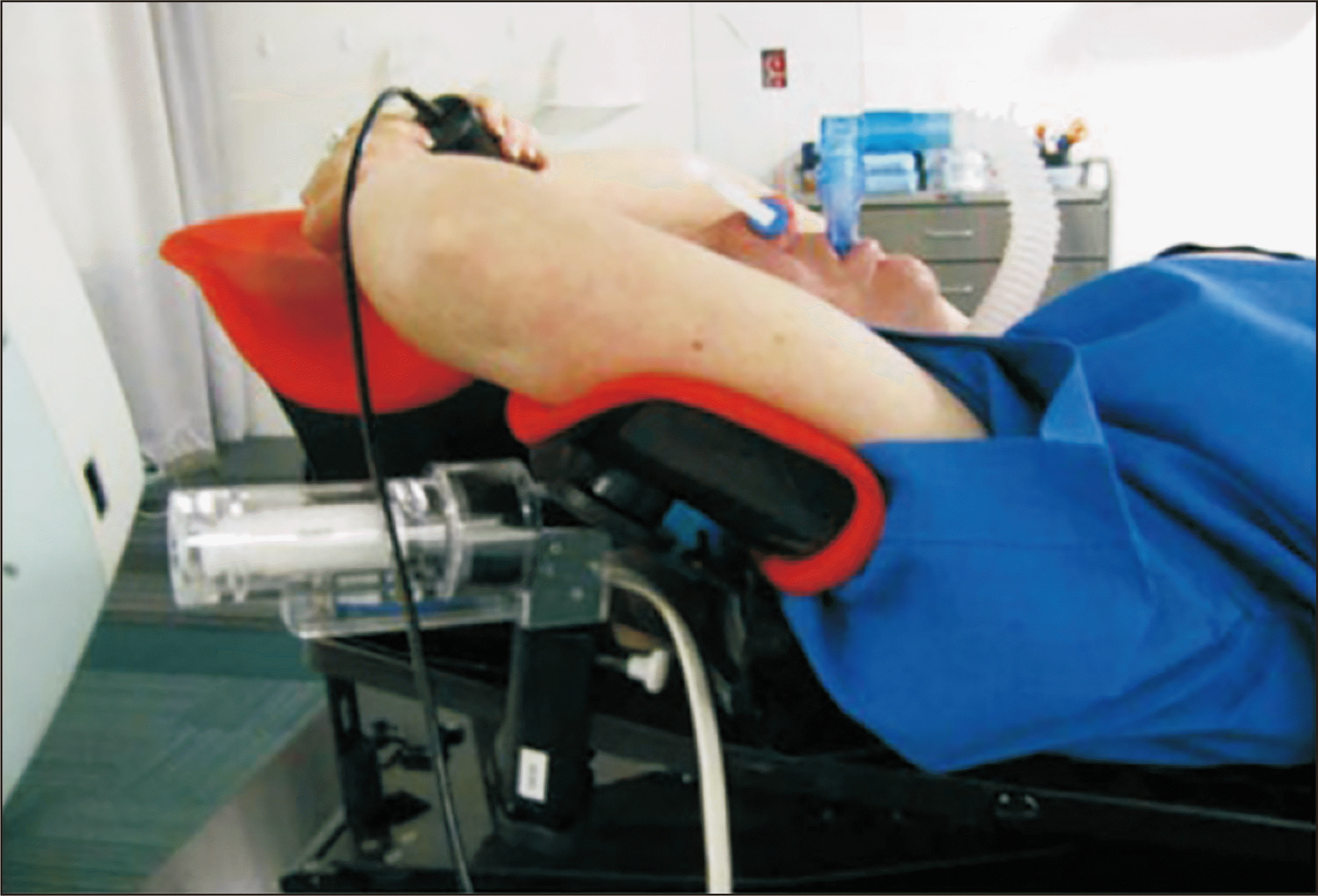
Breath-hold techniques: Patients are trained to hold their breath at a certain lung volume. This can be achieved through self-controlled breath-hold [51,52] or using devices such as active breathing coordinators (ABC) [53-55] shown in Fig. 4 [56]. A mouthpiece is attached to a spirometer while the nose of the patient is pegged to ensure they are breathing only through the device. The radiation therapist can visualize the inspiration level of the patient through the connected computer. When the patient reaches the required threshold, the pinch valve of the spirometer closes remotely, preventing the patient from exhaling or inhaling beyond the required threshold.
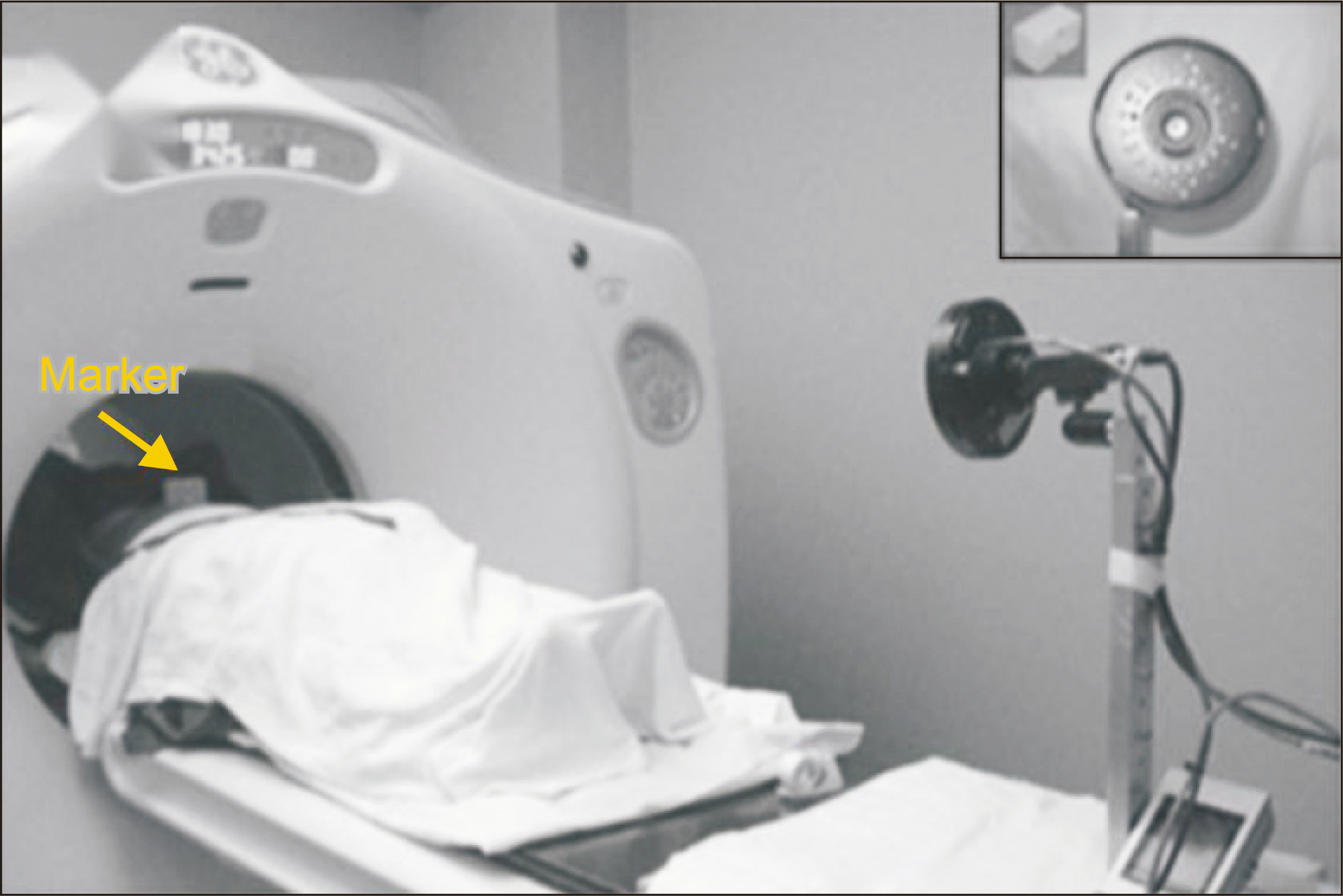
Respiratory-gating training: Patients learn to breathe consistently to align with the gating window during which radiation is delivered [49]. A marker is placed on the midpoint of the patient between the xiphoid and the umbilicus as shown in Fig. 5. Tattoos, permanent ink, or measurements may be used for precise repositioning of this marker daily. Infrared light is used as the camera records the reflection of the dots on the marker through the computer.
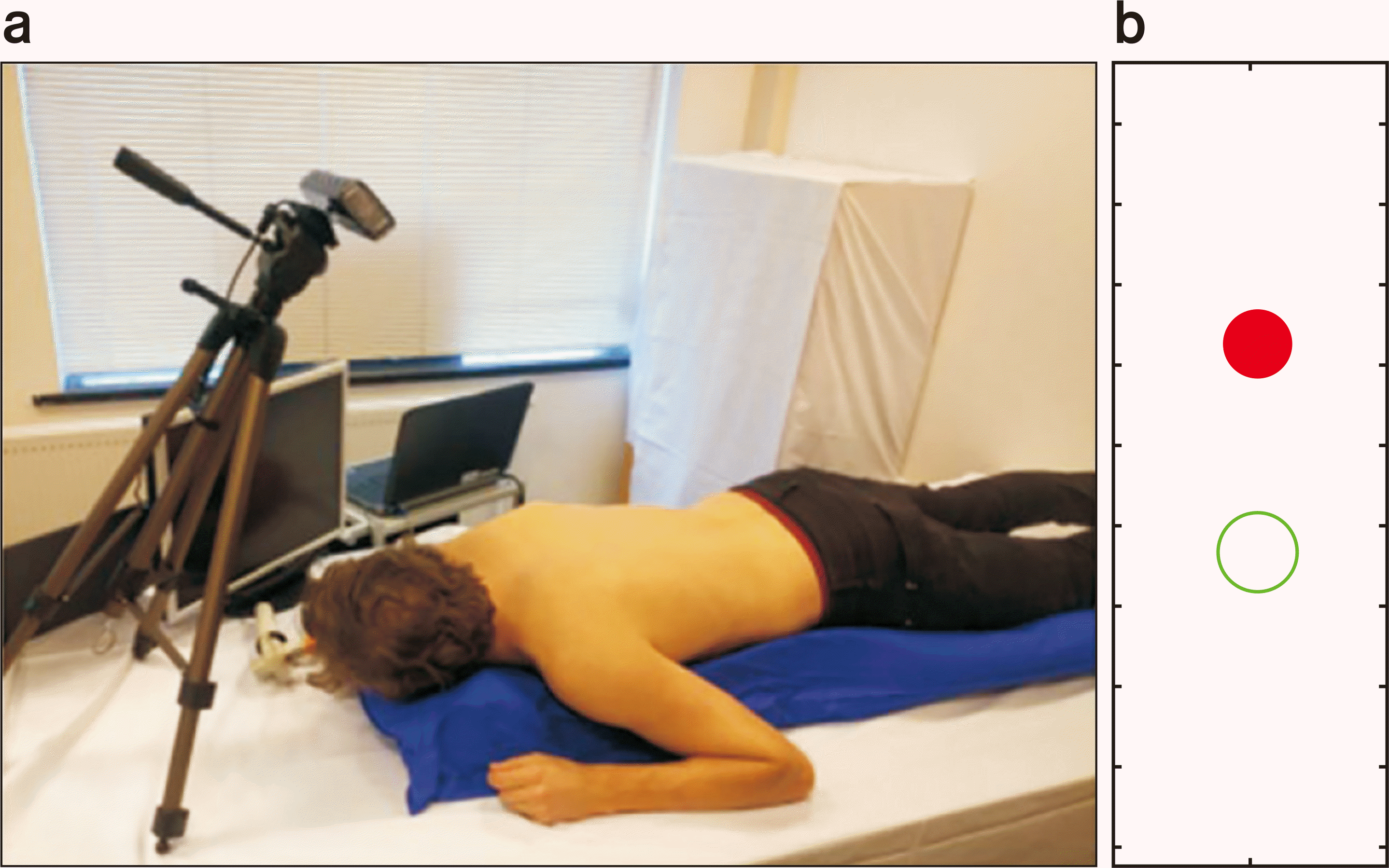
Visual biofeedback: This uses real-time visual biofeedback (BFB) systems where patients can monitor their respiratory patterns and adjust accordingly to maintain consistency [57,58]. In a study [58], using BFB significantly improved the reproducibility of the breath-hold level. Fig. 6a shows the experiment setup and a volunteer could see the visual BFB on the additional screen. On a screen next to the volunteer, a red circle moved up and down with respiration and a green circle corresponded to the respiratory level as shonw in Fig. 6b.

3. Challenges in patient training

Not all patients can adequately control their breathing or maintain breath-hold for the required duration, necessitating alternative strategies for some individuals. The anxiety of treatment and discomfort from the position or immobilization devices can affect the ability of a patient to follow breathing instructions. They observed positive correlations between the individual anxiety score and respiratory rate [59].

4. Clinical outcomes and benefits

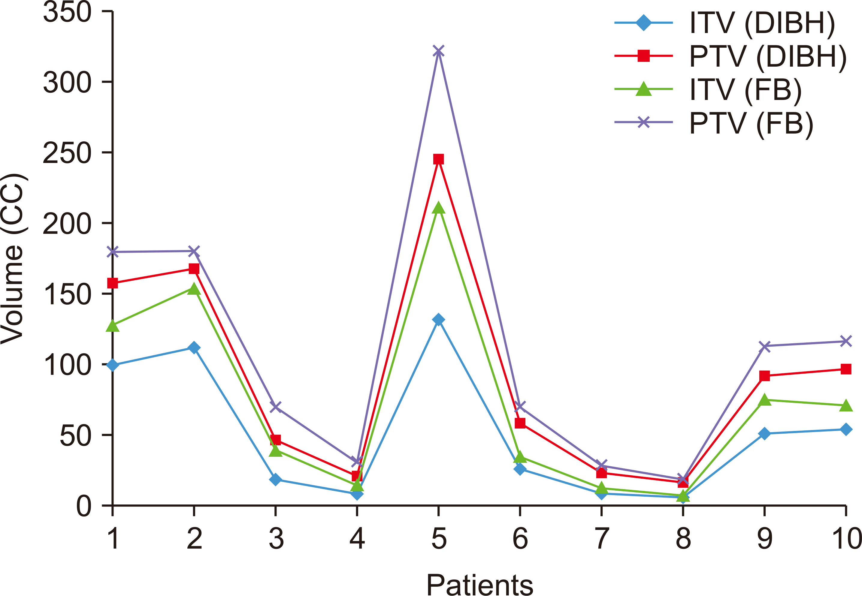
Effective respiratory training can produce more stable and predictable tumor positioning, reducing motion artifacts in imaging and treatment. Breath-holding training has been studied in patients with lung cancer and demonstrated to produce a significant difference between free breathing and deep inspiration breath-hold (DIBH) regarding overall PTV volumes and DIBH with smaller volumes (Fig. 7). In addition, significant reductions of 40% and 35% were seen in the mean and near maximum doses of the heart, one of the OARs, in lung cancer treatment. Improved control of tumor motion allows for tighter treatment margins, increasing the radiation dose conformity to the tumor and sparing healthy tissue [60].

5. Ongoing support and assessment

Respiratory patterns should be continuously monitored throughout the treatment course to ensure adherence to the trained patterns, although the respiratory training protocols may need adjustments based on the performance and comfort of the patient during the treatment course.
Patient respiratory training is a vital component of motion management in photon radiation therapy. Respiratory training enhances the consistency and predictability of tumor positioning and thereby significantly contributes to the precision and efficacy of radiation therapy. Ongoing patient support, monitoring, and adaptation of training protocols are essential for achieving optimal treatment outcomes.

Motion Management Strategies

Effective motion management in photon radiation therapy is crucial for ensuring accurate radiation dose delivery to the tumor while minimizing exposure to the surrounding healthy tissue. This section explores various strategies developed to manage and mitigate the impact of motion.

1. Motion-encompassing strategies

By expanding the margins around the target area, these strategies aim to encompass the entire range of tumor motion during the respiratory cycle. This is accomplished by expanding the clinical target volume with an appropriate margin to account for tumor movements that are independent of patient setup uncertainties, creating the so-called internal target volume (ITV) [61]. The appropriate margin, internal margin (IM) includes variation of position (daily set-up) and volume or shape during multifraction [62]. IMs can be determined by investigating the movement variation of the lesion using breath-hold CT scans performed at inspiration [63] and expiration or by using fiducial markers [64].
A 4DCT dataset is mainly used to generate an ITV. The 4DCT scanning records multiple images over time, and CT images are acquired by assessing whether these should be based on the amplitude or phase of the respiratory cycle of the patient. The maximum intensity projection (MIP) or the average intensity projection (AIP) method is a representative method of projecting dozens of images acquired in this way. MIP involves reporting the maximum intensity across all bins for each voxel to create a single three-dimensional data set. Theoretically, this should then delineate the full range of motion as higher-density tumor intensity values are painted throughout the respiratory cycle. AIP is simply an average intensity for each voxel across each bin. MIP may have image distortion due to the inhomogeneity of the body, but AIP is closer to reality in terms of treatment delivery and is the preferred data set for dose calculation. A notable advantage compared with other techniques is that this allows free breathing while acquiring CT images, making this suitable for patients with reduced physical or cognitive abilities [65,66].

2. Respiratory-gating techniques

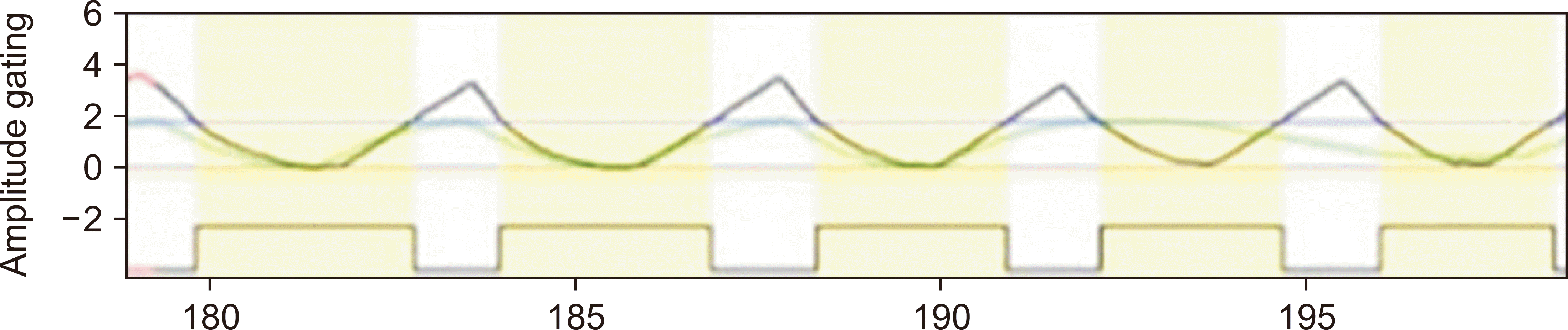
Respiratory-gating involves delivering radiation only during specific phases of the breathing cycle of the patient, typically when the tumor is in a relatively stable position. This is generally used with a surrogate, infrared light source, and charge-coupled device–tracking camera to monitor the breathing cycle. One commercially available system is referred to as the real-time position management respiratory-gating system (Varian Medical Systems). The surrogate is placed on the abdomen or chest of the patient where the breathing cycle can be easily measured, and the reference wave of the breathing phase is obtained. Based on this, the phase range is set for the portion where the patient breathes stably [67,68] by using two gaiting modes, phase and amplitude. A clinical study has shown that by performing gating treatment on patients with cancer, the mean volumes of normal tissue receiving the prescribed doses were reduced by approximately 30% compared with treatment setting ITV [69]. In addition, pulmonary function parameters, especially diffusing capacity of the lungs for carbon monoxide, although more heterogeneous, showed a tendency to reduce pulmonary toxicity in the respiratory-gated conformal radiotherapy group [70]. This technique enhances the accuracy of radiation delivery by reducing the impact of tumor motion during even normal breathing, thereby allowing for smaller margins and less radiation to nontarget tissues. A comparison study with and without gating showed that the 3%/2-mm gamma–index passing rate was improved by 6.0% [71]. The gamma–index analysis compares the delivered (evaluated) dose distribution point by point to that planned by the treatment planning system (TPS) (reference dose distribution) based on a distance to agreement criteria and a dose difference criteria. This is a helpful tool for dosimetric verification that compares the TPS plan to the measured plan and provides the metric of the agreement to dose. Fig. 8 shows the example of amplitude gaiting mode [72].

3. Breath-hold techniques

During radiation delivery, patients are instructed to hold their breath to minimize tumor movement. Two types of breath-holding techniques can be used: voluntary breath-hold (VBH) [6] and DIBH [7]. In VBH, the patient holds breathing at predetermined stages of the respiratory cycle, and, depending on their ability to hold breathing, the patient and the therapist can decide together when to start or end radiation beam-on treatment. VBH requires the patient to reproducibly hold their breath for the same time and respiratory phase during intrafraction treatment [69]. Assisted techniques, such as the ABC, may also be used. During DIBH treatment, patients must reproduce and maintain the same DIBH throughout the treatment beam-on time and treatment planning CT scan. Although delivering a therapeutic dose per beam within 10 to 15 seconds is relatively easy, it may be necessary to temporarily stop the CT scan to allow additional breathing for the patient. Therefore, patients commonly receive training or coaching sessions before their CT scan to reduce patient anxiety and improve the quality of the CT study [69]. Breath-hold methods can significantly reduce tumor motion, particularly in thoracic and upper abdominal radiation therapy, producing more precise targeting. In particular, radiation therapy of the left breast for patients with breast cancer produced a >50% reduction in radiation dose to the heart and coronary arteries compared with free breathing. In addition, by increasing the distance between the heart and PTV as shown in Fig. 9, the dose entering the heart can be reduced [52].

4. Forced shallow breathing methods

Techniques such as abdominal compression (AC) limit the depth of breathing, thereby reducing the extent of tumor motion. This is useful in cases where other motion management strategies are unfeasible or when the tumor motion is significant. As shown in Fig. 10 [73], AC is applied to the patient as much as can be tolerated. The AC technique restricts abdominal breathing and encourages lung-based surface breathing by applying pressure to the abdominal area to limit movement of the diaphragm. Therefore, this is unsuitable for using methods such as deep breathing, forced humping, or breath holding.

5. Real-time tracking

Dynamic monitoring involves advanced imaging and tracking technologies, such as optical surface monitoring or implanted fiducial markers to provide real-time information on tumor position. In the previously mentioned respiratory-gating techniques, surrogates and infrared cameras are used to monitor the respiratory phase of the patient. In addition, the fiducial marker is used to track the location of a tumor by implanting a fiducial mark around or within the tumor, which cannot be transmitted through kV X-rays and appears opaque in the image [74].
Dynamic treatment uses motion tracking for respiratory gaiting treatment, and the physician determines the window and then delivers radiation only when the tumor passes through this predefined region. This window can be determined by the amplitude and phase. For instance, some gaiting systems turn off the radiation beam when the tumor leaves the window and then reapply this when the tumor reenters the targeted treatment field. Additionally, the position of the tumor is tracked in real-time with a marker and can be used for the gating information of beam irradiation. The beam is on when the marker is within the given tolerance during beam irradiation [75]. These techniques can be used to dynamically adjust the radiation beam or pause treatment if the tumor moves outside the planned area, enhancing treatment accuracy and safety.
Motion management strategies in photon radiation therapy, including motion-encompassing techniques, respiratory-gating, breath-hold methods, forced shallow breathing, and real-time tracking, play a pivotal role in enhancing treatment delivery precision. These strategies can mitigate the impact of tumor and organ motion and significantly contribute to the efficacy and safety of radiation therapy, ultimately improving patient outcomes. These motion management strategies become more important in the case of high-dose irradiation, such as stereotactic ablative body radiotherapy, because the protection of normal tissue is critical. However, continued technological advancements and technique development are essential to refine motion management in radiation therapy further.

IGRT in Lung SBRT

IGRT has revolutionized lung SBRT, a high-precision treatment modality for lung cancer. The role of IGRT role in lung SBRT is pivotal because of the need for extreme accuracy in delivering high-dose radiation to moving lung tumors while sparing surrounding healthy tissue.

1. Importance of IGRT in lung SBRT

Lung SBRT involves delivering high doses per fraction [76,77]. However, residual tumor displacement can exist even with appropriate tumor motion management throughout patient training, [78-80]. Barnes et al. (2001) [78] reported that tumor motion under DIBH can be up to 4 mm. An investigation using a stereotactic body frame showed a variation of tumor position from 4 to 7 mm [81], and the presence of motion of not only the tumor but also the nearby OARs was also reported. Without AC, the overall motion in each organ during treatment is the largest, and as compression increases, the motion of the tumor and OAR decreases [82]. Even patients using breath holding showed tumor movement of 1.4±1.4 mm [79]. Therefore, online verification of tumor position is critical in lung SBRT.
IGRT offers image-based guidance for tumor tracking in radiotherapy delivery before and during treatment [83]. This can be implemented to reduce beam delivery error due to organ motion [77,83] or daily patient displacement by frequently matching CT images obtained before treatment to the planning CT images [84]. The IGRT ensures the radiation is accurately targeted to the tumor, accounting for any motion and changes in position between and during treatments [76,77]. Lung SBRT is challenging as the lung has large organ movement over time and should be carefully treated with exact tumor and OAR locating. By enhancing targeting accuracy, IGRT reduces unnecessary radiation exposure to the surrounding healthy lung tissue and critical organs, thus minimizing the risk of side effects.

2. Technological advances in IGRT

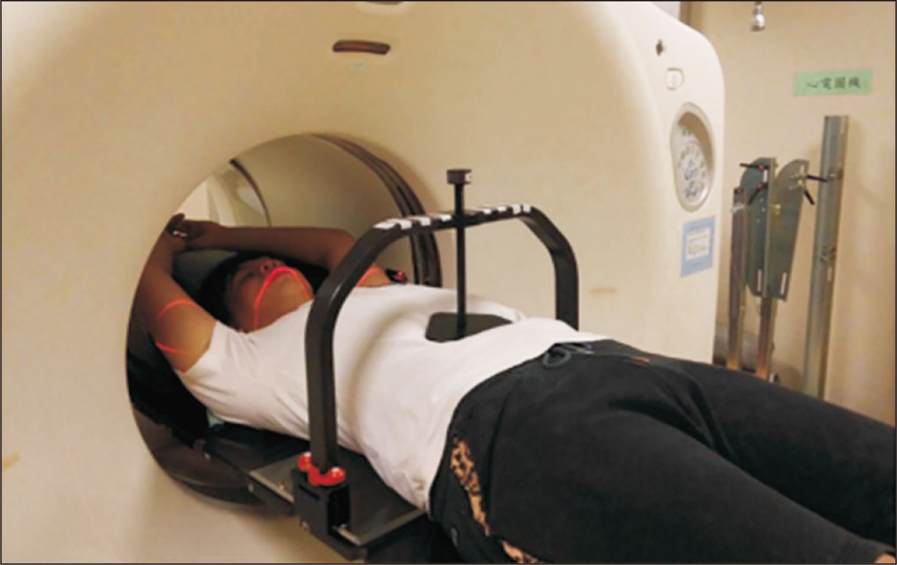
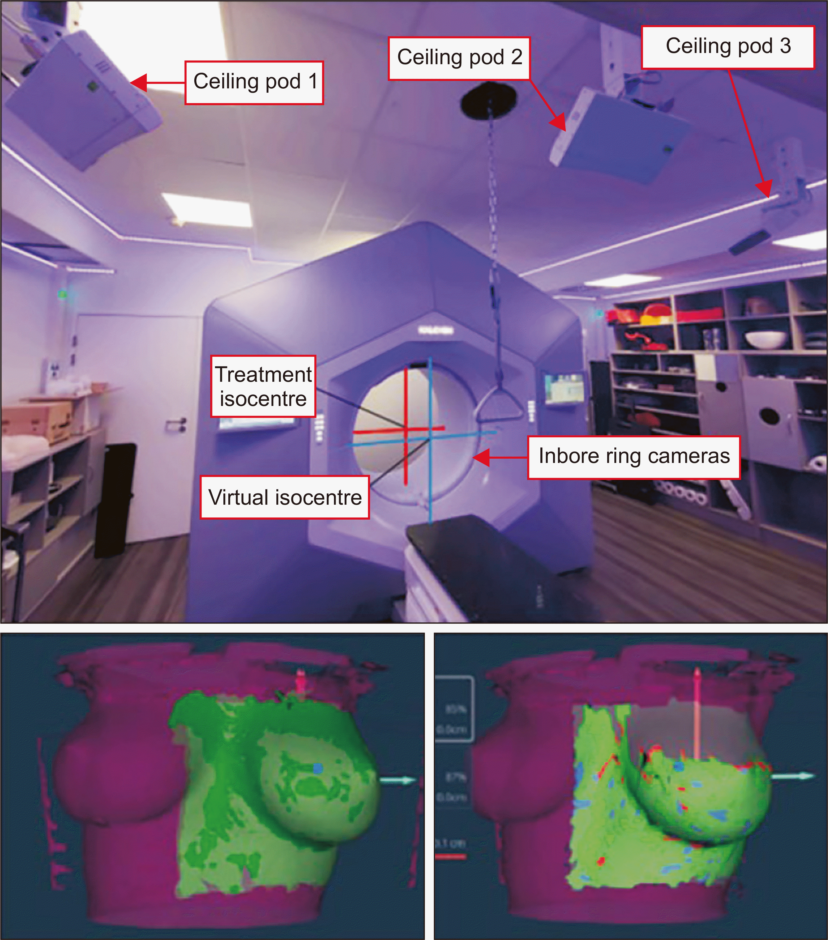
The image-guidance motion mangament device using 3 celing pods and inbore ring camera is showed in Fig. 11 [85]. Caillet classified IGRT into two groups: In-room pretreatment IGRT and intratreatment IGRT with specific motion management techniques per group [83]. Cone-beam computed tomography (CBCT) is frequently used in lung SBRT for pretreatment verification and provides 3D images of the tumor and surrounding anatomy, ensuring precise alignment before radiation delivery. With high dependence of anatomical visibilities in kV radiographic images based on using Compton scattering cross-sectioning, bone (and sometimes metal as an implanted marker) can be used as a reference for the verification of the position of the patient [83]. However, displaying the lung anatomy is limited because of the large range of motion, which can induce motion artifacts such as blurring and double images or streaks on images. This can be relieved by the application of motion management strategies such as breath-holding and respiratory-gating.
Advanced IGRT systems can track the real-time movement of tumors, adjusting the radiation beam dynamically to maintain accurate targeting despite respiratory motion. IGRT technologies are often combined with respiratory-gating or breath-hold techniques, synchronizing radiation delivery with specific phases of the breathing cycle of the patient for improved precision [39,86,87]. For intrafractional motion monitoring, the combination of IGRT and SGRT devices has been suggested [88]. In addition, the kV-imaging device itself can be a solution for real-time image-guidance for tumor tracking. One device used for real-time image-guided radiotherapy for lung tumors is the CyberKnife [83] while another is the Vero tracking system [89]. IGRT allows for adaptive radiation therapy, where treatment plans are modified based on changes in tumor size, shape, or position displayed in the radiographic images over the course of treatment.

3. Challenges and considerations

The precision offered by IGRT in lung SBRT produced better local control of the tumor [75,76]. By accurately targeting the tumor and sparing healthy tissue, IGRT contributes to reduced toxicity and improved patient quality of life. The high level of precision required in lung SBRT with IGRT demands meticulous treatment planning and execution. Variations in tumor location, size, and patient anatomy necessitate individualized treatment planning and execution [90-92]. Ongoing advancements in imaging and radiation delivery technologies promise further improvements in the precision and efficacy of lung SBRT. Continued research is essential to optimize IGRT protocols and further understand the long-term outcomes of lung cancer treatment [93-95]. Clinical outcomes of motion management can be reportedly hard to interpret due to the difficulty of defining the exact effect of the absence or presence of the image guidance produced by random variation between patients [83]. A solution for this is by using a sufficiently large enough cohort of patients to support the benefits of image guidance.
Challenges also exist for IGRT in lung SBRT, such as the difficulty in managing motion and respiration-induced artifacts that decrease the accuracy of tumor delineation [96]. Several attempts to reduce motion-induced artifacts in CBCT images have been reported [97,98]. Furthermore, while the CT image, which is the most frequently used in the lung SBRT, has good visibility for high-density materials (e.g., bone), soft tissues have a low visibility contrast. The application of magnetic resonance imaging (MRI), which can visualize soft tissues, to IGRT has been suggested through the studies on the availability and potential of the MRI-guided IGRT [99-101]. The MRI-based guidance of tumor locations in soft tissues such as the lung is superior to that of kV-based imaging with improved delineation of target and nearby organs [101,102]. As technology advances, a linear accelerator (LINAC) has been combined directly with an MRI scanner, which can perform adaptive radiation therapy (RT), for MRI-guided LINAC [103]. This acquires MRI images on the day of treatment, and the oncologist confirms the positions of the target and OAR through comparison with the reference image used in treatment planning. If discrepancies between images are out of the target margin or tolerance, oncologists adjust the radiation treatment plan by redrawing the target and OAR. However, the treatment time required by MRI-guide LINAC may be longer than general treatment, although uncertainties, such as inter- and intrafraction setup error and variability, can be reduced and highly accurate RT can be performed. IGRT has become an integral component of the lung SBRT, significantly enhancing the accuracy of treatment delivery. This advancement allows for the safe administration of high radiation doses to lung tumors, improving tumor control and patient outcomes while minimizing the risk of toxicity. As technology advances, IGRT will continue to play a crucial role in refining lung cancer treatment, potentially extending the benefits of SBRT to a broader range of patients [75,76,99].

Conclusion

Integrating motion management in photon radiation therapy, real-time control of respiratory motion, and applying IGRT in lung SBRT are vital advancements. They significantly improve the accuracy, safety, and effectiveness of radiation therapy, producing better patient outcomes. Continued technological advancements and clinical research in these areas are crucial for the ongoing refinement of cancer treatment strategies.

Notes

Funding

This research was supported by Basic Science Research Program through the National Research Foundation of Korea (NRF) funded by the Ministry of Education (NRF-2021R1|1A1A01059845).

Conflicts of Interest

The authors have nothing to disclose.

Availability of Data and Materials

All relevant data are within the paper and its Supporting Information files.

Author Contributions

Conceptualization: Jung-in Kim. Data curation: Jin Jegal, Hyojun Park, Jung-in Kim. Formal analysis: Seonghee Kang, Chang Heon Choi, Jung-in Kim. Funding acquisition: Jung-in Kim. Investigation: Jin Jegal, Hyojun Park, Jung-in Kim Project administration: Seonghee Kang, Chang Heon Choi, Jung-in Kim. Visualization: Jin Jegal,. Writing – original draft: Jin Jegal, Hyojun Park. Writing – review & editing: Seonghee Kang, Chang Heon Choi, Jung-in Kim.

References

1. Wang JZ, Li JB, Wang W, Qi HP, Ma ZF, Zhang YJ, et al. 2015; Changes in tumour volume and motion during radiotherapy for thoracic oesophageal cancer. Radiother Oncol. 114:201–205. DOI: 10.1016/j.radonc.2014.12.010. PMID: 25595652.
crossref
2. Botticella A, Levy A, Auzac G, Chabert I, Berthold C, Le Pechoux C. 2021; Tumour motion management in lung cancer: a narrative review. Transl Lung Cancer Res. 10:2011–2017. DOI: 10.21037/tlcr-20-856. PMID: 34012810. PMCID: PMC8107759.
crossref
3. Eminowicz G, Motlib J, Khan S, Perna C, McCormack M. 2016; Pelvic organ motion during radiotherapy for cervical cancer: understanding patterns and recommended patient preparation. Clin Oncol (R Coll Radiol). 28:e85–e91. DOI: 10.1016/j.clon.2016.04.044. PMID: 27178706.
crossref
4. Langen KM, Jones DT. 2001; Organ motion and its management. Int J Radiat Oncol Biol Phys. 50:265–278. DOI: 10.1016/S0360-3016(01)01453-5. PMID: 11316572.
crossref
5. Keall P. 2004; 4-dimensional computed tomography imaging and treatment planning. Semin Radiat Oncol. 14:81–90. DOI: 10.1053/j.semradonc.2003.10.006. PMID: 14752736.
crossref
6. Keall PJ, Joshi S, Vedam SS, Siebers JV, Kini VR, Mohan R. 2005; Four-dimensional radiotherapy planning for DMLC-based respiratory motion tracking. Med Phys. 32:942–951. DOI: 10.1118/1.1879152. PMID: 15895577.
crossref
7. Cho B, Poulsen PR, Keall PJ. 2010; Real-time tumor tracking using sequential kV imaging combined with respiratory monitoring: a general framework applicable to commonly used IGRT systems. Phys Med Biol. 55:3299–3316. DOI: 10.1088/0031-9155/55/12/003. PMID: 20484777. PMCID: PMC2974817.
crossref
8. Bertholet J, Knopf A, Eiben B, McClelland J, Grimwood A, Harris E, et al. 2019; Real-time intrafraction motion monitoring in external beam radiotherapy. Phys Med Biol. 64:15TR01. DOI: 10.1088/1361-6560/ab2ba8. PMID: 31226704. PMCID: PMC7655120.
crossref
9. Kang SH, Kim S, Kim DS, Kim TH, Park SH, Shin DS, et al. 2017; A method of respiratory phase optimization for better dose sparing of organs at risks: a validation study in patients with lung cancer. Oncotarget. 9:205–216. DOI: 10.18632/oncotarget.23353. PMID: 29416607. PMCID: PMC5787457.
crossref
10. Jadon R, Pembroke CA, Hanna CL, Palaniappan N, Evans M, Cleves AE, et al. 2014; A systematic review of organ motion and image-guided strategies in external beam radiotherapy for cervical cancer. Clin Oncol (R Coll Radiol). 26:185–196. DOI: 10.1016/j.clon.2013.11.031. PMID: 24566332.
crossref
11. Chan P, Dinniwell R, Haider MA, Cho YB, Jaffray D, Lockwood G, et al. 2008; Inter- and intrafractional tumor and organ movement in patients with cervical cancer undergoing radiotherapy: a cinematic-MRI point-of-interest study. Int J Radiat Oncol Biol Phys. 70:1507–1515. DOI: 10.1016/j.ijrobp.2007.08.055. PMID: 18164850.
crossref
12. Balter JM, Lam KL, Sandler HM, Littles JF, Bree RL, Ten Haken RK. 1995; Automated localization of the prostate at the time of treatment using implanted radiopaque markers: technical feasibility. Int J Radiat Oncol Biol Phys. 33:1281–1286. DOI: 10.1016/0360-3016(95)02083-7. PMID: 7493853.
crossref
13. Nederveen A, Lagendijk J, Hofman P. 2000; Detection of fiducial gold markers for automatic on-line megavoltage position verification using a marker extraction kernel (MEK). Int J Radiat Oncol Biol Phys. 47:1435–1442. DOI: 10.1016/S0360-3016(00)00523-X. PMID: 10889399.
crossref
14. Roach M 3rd, Pickett B, Rosenthal SA, Verhey L, Phillips TL. 1994; Defining treatment margins for six field conformal irradiation of localized prostate cancer. Int J Radiat Oncol Biol Phys. 28:267–275. DOI: 10.1016/0360-3016(94)90167-8. PMID: 8270451.
crossref
15. Yan D, Lockman D, Brabbins D, Tyburski L, Martinez A. 2000; An off-line strategy for constructing a patient-specific planning target volume in adaptive treatment process for prostate cancer. Int J Radiat Oncol Biol Phys. 48:289–302. DOI: 10.1016/S0360-3016(00)00608-8. PMID: 10925000.
crossref
16. Maleike D, Unkelbach J, Oelfke U. 2006; Simulation and visualization of dose uncertainties due to interfractional organ motion. Phys Med Biol. 51:2237–2252. DOI: 10.1088/0031-9155/51/9/009. PMID: 16625039.
crossref
17. Liu G, Hu F, Ding X, Li X, Shao Q, Wang Y, et al. 2019; Simulation of dosimetry impact of 4DCT uncertainty in 4D dose calculation for lung SBRT. Radiat Oncol. 14:1. DOI: 10.1186/s13014-018-1191-y. PMID: 30621744. PMCID: PMC6323842.
crossref
18. Duan J, Shen S, Fiveash JB, Popple RA, Brezovich IA. 2006; Dosimetric and radiobiological impact of dose fractionation on respiratory motion induced IMRT delivery errors: a volumetric dose measurement study. Med Phys. 33:1380–1387. DOI: 10.1118/1.2192908. PMID: 16752574.
crossref
19. Jiang SB, Pope C, Al Jarrah KM, Kung JH, Bortfeld T, Chen GT. 2003; An experimental investigation on intra-fractional organ motion effects in lung IMRT treatments. Phys Med Biol. 48:1773–1784. DOI: 10.1088/0031-9155/48/12/307. PMID: 12870582.
crossref
20. Ong CL, Dahele M, Slotman BJ, Verbakel WF. 2013; Dosimetric impact of the interplay effect during stereotactic lung radiation therapy delivery using flattening filter-free beams and volumetric modulated arc therapy. Int J Radiat Oncol Biol Phys. 86:743–748. DOI: 10.1016/j.ijrobp.2013.03.038. PMID: 23773394.
crossref
21. Berbeco RI, Pope CJ, Jiang SB. 2006; Measurement of the interplay effect in lung IMRT treatment using EDR2 films. J Appl Clin Med Phys. 7:33–42. DOI: 10.1120/jacmp.v7i4.2222. PMID: 17533350. PMCID: PMC5722391.
crossref
22. Peng C, Ahunbay E, Chen G, Anderson S, Lawton C, Li XA. 2011; Characterizing interfraction variations and their dosimetric effects in prostate cancer radiotherapy. Int J Radiat Oncol Biol Phys. 79:909–914. DOI: 10.1016/j.ijrobp.2010.05.008. PMID: 20732764.
crossref
23. Huang TC, Liang JA, Dilling T, Wu TH, Zhang G. 2010; Four-dimensional dosimetry validation and study in lung radiotherapy using deformable image registration and Monte Carlo techniques. Radiat Oncol. 5:45. DOI: 10.1186/1748-717X-5-45. PMID: 20509955. PMCID: PMC2890615.
crossref
24. Selvaraj JK. 2013. Modelling the effect of geometric uncertainties, clonogen distribution and IMRT interplay effect on tumour control probability. [thesis]. University of Liverpool;Liverpool:
25. Zhang Q, Hu YC, Liu F, Goodman K, Rosenzweig KE, Mageras GS. 2010; Correction of motion artifacts in cone-beam CT using a patient-specific respiratory motion model. Med Phys. 37:2901–2909. DOI: 10.1118/1.3397460. PMID: 20632601. PMCID: PMC2887907.
crossref
26. Kong VC, Marshall A, Chan HB. 2016; Cone beam computed tomography: the challenges and strategies in its application for dose accumulation. J Med Imaging Radiat Sci. 47:92–97. DOI: 10.1016/j.jmir.2015.09.012. PMID: 31047170.
crossref
27. Jiang SB, Wolfgang J, Mageras GS. 2008; Quality assurance challenges for motion-adaptive radiation therapy: gating, breath holding, and four-dimensional computed tomography. Int J Radiat Oncol Biol Phys. 71:S103–S107. DOI: 10.1016/j.ijrobp.2007.07.2386. PMID: 18406905.
crossref
28. Lim-Reinders S, Keller BM, Al-Ward S, Sahgal A, Kim A. 2017; Online adaptive radiation therapy. Int J Radiat Oncol Biol Phys. 99:994–1003. DOI: 10.1016/j.ijrobp.2017.04.023. PMID: 28916139.
crossref
29. Pos F, Remeijer P. 2010; Adaptive management of bladder cancer radiotherapy. Semin Radiat Oncol. 20:116–120. DOI: 10.1016/j.semradonc.2009.11.005. PMID: 20219549.
crossref
30. Sonke JJ, Belderbos J. 2010; Adaptive radiotherapy for lung cancer. Semin Radiat Oncol. 20:94–106. DOI: 10.1016/j.semradonc.2009.11.003. PMID: 20219547.
crossref
31. Ketting CH, Austin-Seymour M, Kalet I, Jacky J, Kromhout-Schiro S, Hummel S, et al. 1997; Automated planning target volume generation: an evaluation pitting a computer-based tool against human experts. Int J Radiat Oncol Biol Phys. 37:697–704. DOI: 10.1016/S0360-3016(96)00562-7. PMID: 9112469.
crossref
32. Nugent K, O'Neill B, Brennan V, Lynch J, Higgins M, Dunne M, et al. 2023; Quantification of organ motion in male and female patients undergoing long-course radiation therapy for rectal cancer in the supine position. Adv Radiat Oncol. 8:101109. DOI: 10.1016/j.adro.2022.101109. PMID: 36483059. PMCID: PMC9723314.
crossref
33. Elkin EB, Hudis C, Begg CB, Schrag D. 2005; The effect of changes in tumor size on breast carcinoma survival in the U.S.: 1975-1999. Cancer. 104:1149–1157. DOI: 10.1002/cncr.21285. PMID: 16088887.
crossref
34. Dykstra BJ, Scanlon PD, Kester MM, Beck KC, Enright PL. 1999; Lung volumes in 4,774 patients with obstructive lung disease. Chest. 115:68–74. DOI: 10.1378/chest.115.1.68. PMID: 9925064.
crossref
35. Bhatti U, Rani K, Memon MQ. 2014; Variation in lung volumes and capacities among young males in relation to height. J Ayub Med Coll Abbottabad. 26:200–202.
36. Hepper NG, Fowler WS, Helmholz HF Jr. 1960; Relationship of height to lung volume in healthy men. Dis Chest. 37:314–320. DOI: 10.1378/chest.37.3.314. PMID: 14401182.
37. Um EH, Hwang S, Song GW, Jung DH, Ahn CS, Kim KH, et al. 2015; Calculation of standard liver volume in Korean adults with analysis of confounding variables. Korean J Hepatobiliary Pancreat Surg. 19:133–138. DOI: 10.14701/kjhbps.2015.19.4.133. PMID: 26693231. PMCID: PMC4683924.
crossref
38. Ragnarsdóttir M, Kristinsdóttir EK. 2006; Breathing movements and breathing patterns among healthy men and women 20-69 years of age: reference values. Respiration. 73:48–54. DOI: 10.1159/000087456. PMID: 16106113.
crossref
39. Massaccesi M. 2020. CT in room gating during radiotherapy. Imaging and interventional radiology for radiation oncology. Springer;p. 91–106. DOI: 10.1007/978-3-030-38261-2_7.
40. Knybel L, Cvek J, Molenda L, Stieberova N, Feltl D. 2016; Analysis of lung tumor motion in a large sample: patterns and factors influencing precise delineation of internal target volume. Int J Radiat Oncol Biol Phys. 96:751–758. DOI: 10.1016/j.ijrobp.2016.08.008. PMID: 27788948.
crossref
41. Savanović M, Štrbac B, Mihajlović B, Trokić D, Jaroš D, Kolarević G, et al. 2021; Evaluation of thoracic surface motion during the free breathing and deep inspiration breath hold methods. Med Dosim. 46:274–278. DOI: 10.1016/j.meddos.2021.02.006. PMID: 33766492.
crossref
42. Li XA, Stepaniak C, Gore E. 2006; Technical and dosimetric aspects of respiratory gating using a pressure-sensor motion monitoring system. Med Phys. 33:145–154. DOI: 10.1118/1.2147743. PMID: 16485421.
crossref
43. Purdie TG, Moseley DJ, Bissonnette JP, Sharpe MB, Franks K, Bezjak A, et al. 2006; Respiration correlated cone-beam computed tomography and 4DCT for evaluating target motion in stereotactic lung radiation therapy. Acta Oncol. 45:915–922. DOI: 10.1080/02841860600907345. PMID: 16982558.
crossref
44. Bortfeld T, Jiang SB, Rietzel E. 2004; Effects of motion on the total dose distribution. Semin Radiat Oncol. 14:41–51. DOI: 10.1053/j.semradonc.2003.10.011. PMID: 14752732.
crossref
45. Mohn S, Wasbø E. 2011; Simulation of respiratory motion during IMRT dose delivery. Acta Oncol. 50:935–943. DOI: 10.3109/0284186X.2011.580002. PMID: 21767194.
crossref
46. Qi XS, White J, Rabinovitch R, Merrell K, Sood A, Bauer A, et al. 2010; Respiratory organ motion and dosimetric impact on breast and nodal irradiation. Int J Radiat Oncol Biol Phys. 78:609–617. DOI: 10.1016/j.ijrobp.2009.11.053. PMID: 20472366.
crossref
47. Shin E, Park HC, Han T, Ju SG, Shin JS, Ahn YC. 2008; Efficacy of a respiratory training system on the regularity of breathing. J Korean Soc Ther Radiol Oncol. 26:181–188. DOI: 10.3857/jkstro.2008.26.3.181.
crossref
48. Kang SH, Yoon JW, Kim TH, Suh TS. 2012. May. 26-31. Development of respiratory training system using individual characteristic guiding waveform. Paper presented at: World Congress on Medical Physics and Biomedical Engineering. Beijing, China: p. 2123–2125. DOI: 10.1007/978-3-642-29305-4_557.
49. Pépin A, Daouk J, Bailly P, Hapdey S, Meyer ME. 2014; Management of respiratory motion in PET/computed tomography: the state of the art. Nucl Med Commun. 35:113–122. DOI: 10.1097/MNM.0000000000000048. PMID: 24352107. PMCID: PMC3868022.
50. Chopra S, Dinshaw KA, Kamble R, Sarin R. 2006; Breast movement during normal and deep breathing, respiratory training and set up errors: implications for external beam partial breast irradiation. Br J Radiol. 79:766–773. DOI: 10.1259/bjr/98024704. PMID: 16940376.
crossref
51. Bruzzaniti V, Abate A, Pinnarò P, D'Andrea M, Infusino E, Landoni V, et al. 2013; Dosimetric and clinical advantages of deep inspiration breath-hold (DIBH) during radiotherapy of breast cancer. J Exp Clin Cancer Res. 32:88. DOI: 10.1186/1756-9966-32-88. PMID: 24423396. PMCID: PMC3826503.
crossref
52. Wolf J, Stoller S, Lübke J, Rothe T, Serpa M, Scholber J, et al. 2023; Deep inspiration breath-hold radiation therapy in left-sided breast cancer patients: a single-institution retrospective dosimetric analysis of organs at risk doses. Strahlenther Onkol. 199:379–388. DOI: 10.1007/s00066-022-01998-z. PMID: 36074138. PMCID: PMC10033469.
crossref
53. Romera-Martínez I, Muñoz-Montplet C, Jurado-Bruggeman D, Onsès-Segarra A, Fuentes-Raspall R, Buxó M, et al. 2020; A novel device for deep-inspiration breath hold (DIBH): results from a single-institution phase 2 clinical trial for patients with left-sided breast cancer. Pract Radiat Oncol. 10:e290–e297. DOI: 10.1016/j.prro.2020.02.004. PMID: 32068155.
crossref
54. Mittauer KE, Deraniyagala R, Li JG, Lu B, Liu C, Samant SS, et al. 2015; Monitoring ABC-assisted deep inspiration breath hold for left-sided breast radiotherapy with an optical tracking system. Med Phys. 42:134–143. DOI: 10.1118/1.4903511. PMID: 25563254.
crossref
55. Kaza E, Dunlop A, Panek R, Collins DJ, Orton M, Symonds-Tayler R, et al. 2017; Lung volume reproducibility under ABC control and self-sustained breath-holding. J Appl Clin Med Phys. 18:154–162. DOI: 10.1002/acm2.12034. PMID: 28300372. PMCID: PMC5689958.
crossref
56. Parasuramar A, DeSmit A, Borg M. 2017; Deep inspiration breath hold techniques in the radiotherapeutic management of left-sided breast cancer- active breathing coordinator vs. voluntary breath hold. Int J Gynecol Clin Pract. 4:133. DOI: 10.15344/2394-4986/2017/133.
57. Yamauchi R, Mizuno N, Itazawa T, Masuda T, Akiyama S, Kawamori J. 2021; Assessment of visual feedback system for reproducibility of voluntary deep inspiration breath hold in left-sided breast radiotherapy. J Med Imaging Radiat Sci. 52:544–551. DOI: 10.1016/j.jmir.2021.08.018. PMID: 34538757.
crossref
58. Heerink WJ, Dorrius MD, Groen HJM, Van Ooijen PMA, Vliegenthart R, Oudkerk M. 2018; Respiratory level tracking with visual biofeedback for consistent breath-hold level with potential application in image-guided interventions. Eur Radiol Exp. 2:22. DOI: 10.1186/s41747-018-0052-7. PMID: 30238087. PMCID: PMC6123338.
crossref
59. Masaoka Y, Homma I. 1997; Anxiety and respiratory patterns: their relationship during mental stress and physical load. Int J Psychophysiol. 27:153–159. DOI: 10.1016/S0167-8760(97)00052-4. PMID: 9342646.
crossref
60. Mani KR, Bhuiyan A, Alam M, Ahmed S, Sumon MA, Sengupta AK, et al. 2018; Dosimetric comparison of deep inspiration breath hold and free breathing technique in stereotactic body radiotherapy for localized lung tumor using Flattening Filter Free beam. Polish J Med Phys Eng. 24:15–24. DOI: 10.2478/pjmpe-2018-0003.
crossref
61. Abbas H, Chang B, Chen ZJ. 2014; Motion management in gastrointestinal cancers. J Gastrointest Oncol. 5:223–235.
62. Wambersie A. 1999. ICRU report 62: prescribing, recording and reporting photon beam therapy (supplement to ICRU report 50). International Commission on Radiation Units & Measurements (ICRU). 62.
63. Cohen RJ, Paskalev K, Litwin S, Price RA Jr, Feigenberg SJ, Konski AA. 2010; Esophageal motion during radiotherapy: quantification and margin implications. Dis Esophagus. 23:473–479. DOI: 10.1111/j.1442-2050.2009.01037.x. PMID: 20095993. PMCID: PMC2933373.
crossref
64. Akasaka H, Mizonobe K, Oki Y, Uehara K, Nakayama M, Tamura S, et al. 2022; Fiducial marker position affects target volume in stereotactic lung irradiation. J Appl Clin Med Phys. 23:e13596. DOI: 10.1002/acm2.13596. PMID: 35377962. PMCID: PMC9195037.
crossref
65. Kissick MW, Mackie TR. 2009; Task group 76 report on 'the management of respiratory motion in radiation oncology' [med. phys. 33, 3874-3900 (2006)]. Med Phys. 36:5721–5722. DOI: 10.1118/1.3260838. PMID: 20095285. PMCID: PMC2797047.
crossref
66. Dieterich S, Ford E, Pavord D, Zeng J. Chapter 19 - respiratory motion management for external beam radiotherapy. 2016. Practical radiation oncology physics: a companion to gunderson and tepper's clinical radiation oncology. Elsevier;p. 252–263. DOI: 10.1016/B978-0-323-26209-5.00019-5.
67. Berson AM, Emery R, Rodriguez L, Richards GM, Ng T, Sanghavi S, et al. 2004; Clinical experience using respiratory gated radiation therapy: comparison of free-breathing and breath-hold techniques. Int J Radiat Oncol Biol Phys. 60:419–426. DOI: 10.1016/j.ijrobp.2004.03.037. PMID: 15380575.
crossref
68. Cardenas A, Fontenot J, Forster KM, Stevens CW, Starkschall G. 2004; Quality assurance evaluation of delivery of respiratory-gated treatments. J Appl Clin Med Phys. 5:55–61. DOI: 10.1120/jacmp.2021.25276. PMID: 15753939. PMCID: PMC5723481.
crossref
69. Giraud P, Houle A. 2013; Respiratory gating for radiotherapy: main technical aspects and clinical benefits. ISRN Pulmonology. 2013:1–13. DOI: 10.1155/2013/519602.
crossref
70. Giraud P, Morvan E, Claude L, Mornex F, Le Pechoux C, Bachaud JM, et al. STIC Study Centers. 2011; Respiratory gating techniques for optimization of lung cancer radiotherapy. J Thorac Oncol. 6:2058–2068. DOI: 10.1097/JTO.0b013e3182307ec2. PMID: 22052228.
crossref
71. De Roover R, Hansen R, Crijns W, Muurholm CG, Poels K, Skouboe S, et al. 2021; Dosimetric impact of intrafraction prostate rotation and accuracy of gating, multi-leaf collimator tracking and couch tracking to manage rotation: an end-to-end validation using volumetric film measurements. Radiother Oncol. 156:10–18. DOI: 10.1016/j.radonc.2020.11.031. PMID: 33264640.
crossref
72. Cetnar AJ, James J, Wang B. 2016; Commissioning of a motion system to investigate dosimetric consequences due to variability of respiratory waveforms. J Appl Clin Med Phys. 17:283–292. DOI: 10.1120/jacmp.v17i1.5921. PMID: 26894366. PMCID: PMC5690223.
crossref
73. Huang TC, Wang YC, Chiou YR, Kao CH. 2014; Respiratory motion reduction in PET/CT using abdominal compression for lung cancer patients. PLoS One. 9:e98033. DOI: 10.1371/journal.pone.0098033. PMID: 24837352. PMCID: PMC4024027.
crossref
74. Scher N, Bollet M, Bouilhol G, Tannouri R, Khemiri I, Vouillaume A, et al. 2019; Safety and efficacy of fiducial marker implantation for robotic stereotactic body radiation therapy with fiducial tracking. Radiat Oncol. 14:167. DOI: 10.1186/s13014-019-1373-2. PMID: 31519194. PMCID: PMC6743112.
crossref
75. De Los Santos J, Popple R, Agazaryan N, Bayouth JE, Bissonnette JP, Bucci MK, et al. 2013; Image guided radiation therapy (IGRT) technologies for radiation therapy localization and delivery. Int J Radiat Oncol Biol Phys. 87:33–45. DOI: 10.1016/j.ijrobp.2013.02.021. PMID: 23664076.
crossref
76. Martin A, Gaya A. 2010; Stereotactic body radiotherapy: a review. Clin Oncol (R Coll Radiol). 22:157–172. DOI: 10.1016/j.clon.2009.12.003. PMID: 20092981.
crossref
77. Tsang MW. 2016; Stereotactic body radiotherapy: current strategies and future development. J Thorac Dis. 8(Suppl 6):S517–S527. DOI: 10.21037/jtd.2016.03.14. PMID: 27606082. PMCID: PMC4990666.
crossref
78. Barnes EA, Murray BR, Robinson DM, Underwood LJ, Hanson J, Roa WH. 2001; Dosimetric evaluation of lung tumor immobilization using breath hold at deep inspiration. Int J Radiat Oncol Biol Phys. 50:1091–1098. DOI: 10.1016/S0360-3016(01)01592-9. PMID: 11429237.
crossref
79. Baba F, Shibamoto Y, Tomita N, Ikeya-Hashizume C, Oda K, Ayakawa S, et al. 2009; Stereotactic body radiotherapy for stage I lung cancer and small lung metastasis: evaluation of an immobilization system for suppression of respiratory tumor movement and preliminary results. Radiat Oncol. 4:15. DOI: 10.1186/1748-717X-4-15. PMID: 19476628. PMCID: PMC2694202.
crossref
80. Nelson C, Starkschall G, Balter P, Morice RC, Stevens CW, Chang JY. 2007; Assessment of lung tumor motion and setup uncertainties using implanted fiducials. Int J Radiat Oncol Biol Phys. 67:915–923. DOI: 10.1016/j.ijrobp.2006.10.033. PMID: 17293241.
crossref
81. Negoro Y, Nagata Y, Aoki T, Mizowaki T, Araki N, Takayama K, et al. 2001; The effectiveness of an immobilization device in conformal radiotherapy for lung tumor: reduction of respiratory tumor movement and evaluation of the daily setup accuracy. Int J Radiat Oncol Biol Phys. 50:889–898. DOI: 10.1016/S0360-3016(01)01516-4. PMID: 11429216.
crossref
82. Heinzerling JH, Anderson JF, Papiez L, Boike T, Chien S, Zhang G, et al. 2008; Four-dimensional computed tomography scan analysis of tumor and organ motion at varying levels of abdominal compression during stereotactic treatment of lung and liver. Int J Radiat Oncol Biol Phys. 70:1571–1578. DOI: 10.1016/j.ijrobp.2007.12.023. PMID: 18374231.
crossref
83. Caillet V, Booth JT, Keall P. 2017; IGRT and motion management during lung SBRT delivery. Phys Med. 44:113–122. DOI: 10.1016/j.ejmp.2017.06.006. PMID: 28647449.
crossref
84. Kim B, Kirkby C, Semaka A, Debenham B, Campbell T. 2021; Assessment of IGRT variability for lung SBRT. J Med Imaging Radiat Sci. 52:191–197. DOI: 10.1016/j.jmir.2021.02.004. PMID: 33707110.
crossref
85. Nguyen D, Khodri M, Sporea C, Reinoso R, Jacob Y, Farah J. 2023; Investigating the robustness of the AlignRT InBore™ co-calibration process and determining the overall tracking errors. Phys Med. 108:102567. DOI: 10.1016/j.ejmp.2023.102567. PMID: 36996575.
crossref
86. Boda-Heggemann J, Knopf AC, Simeonova-Chergou A, Wertz H, Stieler F, Jahnke A, et al. 2016; Deep inspiration breath hold-based radiation therapy: a clinical review. Int J Radiat Oncol Biol Phys. 94:478–492. DOI: 10.1016/j.ijrobp.2015.11.049. PMID: 26867877.
crossref
87. Moseley DJ, Hawkins M, Eccles C, Euler C, White EA, Bissonnette J, et al. 2005; Respiratory gated cone-beam CT volumetric imaging for external beam radiotherapy. Int J Radiat Oncol Biol Phys. 63(Suppl 1):S27–S28. DOI: 10.1016/j.ijrobp.2005.07.054.
crossref
88. Heinzerling JH, Hampton CJ, Robinson M, Bright M, Moeller BJ, Ruiz J, et al. 2020; Use of surface-guided radiation therapy in combination with IGRT for setup and intrafraction motion monitoring during stereotactic body radiation therapy treatments of the lung and abdomen. J Appl Clin Med Phys. 21:48–55. DOI: 10.1002/acm2.12852. PMID: 32196944. PMCID: PMC7286017.
crossref
89. Depuydt T, Verellen D, Haas O, Gevaert T, Linthout N, Duchateau M, et al. 2011; Geometric accuracy of a novel gimbals based radiation therapy tumor tracking system. Radiother Oncol. 98:365–372. DOI: 10.1016/j.radonc.2011.01.015. PMID: 21316786.
crossref
90. Giaquinto AN, Sung H, Miller KD, Kramer JL, Newman LA, Minihan A, et al. 2022; Breast cancer statistics, 2022. CA Cancer J Clin. 72:524–541. DOI: 10.3322/caac.21754. PMID: 36190501.
crossref
91. Mubenga LE, Hermans MP, Chimanuka D, Muhindo L, Bwenge E, Tombal B. 2020; Prostate volume and its relationship with anthropometric variables among different ethnic groups of South-Kivu, DR Congo. Afr J Urol. 26:32. DOI: 10.1186/s12301-020-00040-x.
crossref
92. Bosch JL, Tilling K, Bohnen AM, Bangma CH, Donovan JL. 2007; Establishing normal reference ranges for prostate volume change with age in the population-based Krimpen-study: prediction of future prostate volume in individual men. Prostate. 67:1816–1824. DOI: 10.1002/pros.20663. PMID: 17935157.
crossref
93. Smit C, Shaw W. 2015; An investigation of dose and quality in clinical IGRT imaging protocols. Phys Med. 31(Suppl 1):S12. DOI: 10.1016/j.ejmp.2015.07.127.
crossref
94. Mayyas E, Chetty IJ, Chetvertkov M, Wen N, Neicu T, Nurushev T, et al. 2013; Evaluation of multiple image-based modalities for image-guided radiation therapy (IGRT) of prostate carcinoma: a prospective study. Med Phys. 40:041707. DOI: 10.1118/1.4794502. PMID: 23556877.
crossref
95. Stock M, Palm A, Altendorfer A, Steiner E, Georg D. 2012; IGRT induced dose burden for a variety of imaging protocols at two different anatomical sites. Radiother Oncol. 102:355–363. DOI: 10.1016/j.radonc.2011.10.005. PMID: 22098793.
crossref
96. Balter JM, Ten Haken RK, Lawrence TS, Lam KL, Robertson JM. 1996; Uncertainties in CT-based radiation therapy treatment planning associated with patient breathing. Int J Radiat Oncol Biol Phys. 36:167–174. DOI: 10.1016/S0360-3016(96)00275-1. PMID: 8823272.
crossref
97. Peterlik I, Strzelecki A, Lehmann M, Messmer P, Munro P, Paysan P, et al. 2021; Reducing residual-motion artifacts in iterative 3D CBCT reconstruction in image-guided radiation therapy. Med Phys. 48:6497–6507. DOI: 10.1002/mp.15236. PMID: 34529270.
crossref
98. Lewis JH, Li R, Jia X, Watkins WT, Lou Y, Song WY, et al. 2011; Mitigation of motion artifacts in CBCT of lung tumors based on tracked tumor motion during CBCT acquisition. Phys Med Biol. 56:5485–5502. DOI: 10.1088/0031-9155/56/17/003. PMID: 21813959.
crossref
99. Fischer-Valuck BW, Henke L, Green O, Kashani R, Acharya S, Bradley JD, et al. 2017; Two-and-a-half-year clinical experience with the world's first magnetic resonance image guided radiation therapy system. Adv Radiat Oncol. 2:485–493. DOI: 10.1016/j.adro.2017.05.006. PMID: 29114617. PMCID: PMC5605309.
crossref

Fig. 1
The changes of geometric relationships between the planning target volume (PTV) and organs at risk (OARs) (esophagus, heart and spinal cord) in difference respiratory phases. (a) Phase-wise PTVs in a coronal computed tomography (CT) image, (b) phase-wise expanded PTVs and OARs in an axial CT image, and (c) example of overlap volume historgram curves for esophagus. Reused from the article of Kang et al. (Oncotarget. 2017;9:205-216) [9].
pmp-35-2-21-f1.tif
Fig. 2
Dose-volumetric histograms of the gross tumor volume from various respiratory phases as well as the four-dimensional (4D) cumulative dose DVH (average). Reused from the article of Huang et al. (Radiat Oncol. 2010;5:45) [23].
pmp-35-2-21-f2.tif
Fig. 3
Axial CBCT section of phantom with simulated respiratory motion (a) with phase-based motion correction and (b) with principal component analysis-model-based correction. Display window settings are the same for all images. Reused from the article of Zhang et al. (Med Phys. 2010;37:2901-2909) [25].
pmp-35-2-21-f3.tif
Fig. 4
A patient using the active breathing coordinators (ABC) deep inspiration breath hold. Reused from the article of Parasuramar et al. (Int J Gynecol Clin Pract. 2017;4:133) [56].
pmp-35-2-21-f4.tif
Fig. 5
Gating apparatus. The real-time position management (RPM) system (Varian Medical Systems). Reused from the article of Pépin et al. (Nucl Med Commun. 2014;35:113-122) [49].
pmp-35-2-21-f5.tif
Fig. 6
The exeperimental setup of visual biofeedback (BFB) (a) and screenshot of BFB provided to the volunteers.
pmp-35-2-21-f6.tif
Fig. 7
Planning target volume (PTV) and internal target volume (ITV) volume in free breathing and deep inspiration breath-hold (DIBH). FB, free breathing. Reused from the article of Mani et al. (Polish J Med Phys Eng. 2018;24:15-24) [60].
pmp-35-2-21-f7.tif
Fig. 8
Example of waveforms for closed limits to interrupt beam if treatment amplitude is greater than simulation amplitude. Horizontal lines represent the upper (blue) and lower (orange) limits. The black curve is an example of a respiratory waveform during treatment greater than the original, and the green curve is the waveform from computed tomography (CT) simulation. The yellow shaded area indicates that the beam is on. Reused from the article of Cetnar et al. (J Appl Clin Med Phys. 2016;17:283-292) [72].
pmp-35-2-21-f8.tif
Fig. 9
Isodose distribution in free breathing (FB) and deep inspiration breath-hold (DIBH) plans for the same patient. (a) Axial scan and (b) oblique reconstruction with visualization of tangential irradiation fields including the adaptations of the multileaf collimators “beam’s eye view,” in FB; (c) axial scan and (d) oblique reconstruction with visualization of tangential irradiation fields including the adapted of the multileaf collimators “beam’s eye view,” in DIBH. Reused from the article of Wolf et al. (Strahlenther Onkol. 2023;199:379-388) [52].
pmp-35-2-21-f9.tif
Fig. 10
Photographs of a patient in the simulation procedure with the abdominal compression device equipment. Reused from the article of Huang et al. (PLoS One. 2014;9:e98033) [73].
pmp-35-2-21-f10.tif
Fig. 11
Image-guidance motion management devices for pretreatment image-guided radiation therapy (IGRT) (AlignRT). Note that, these devices are also available for the IGRT during treatment in combination with real-time tracking technologies. Reused from the article of Nguyen et al. (Phys Med. 2023;108:102567) [85]
pmp-35-2-21-f11.tif
TOOLS
Similar articles