Journal List > J Rheum Dis > v.31(3) > 1516087679

Ha, Ahn, Song, Park, and Lee: The monocyte-to-high-density lipoprotein-cholesterol ratio at diagnosis is associated with cerebrovascular accident during follow-up in patients with antineutrophil cytoplasmic antibody-associated vasculitis

Abstract

Objective

In this study, the association between the monocyte-to-high-density lipoprotein cholesterol ratio (MHR) at diagnosis and poor outcomes of atherosclerosis-related antineutrophil cytoplasmic antibody-associated vasculitis (AAV) during follow-up in patients with AAV was investigated.

Methods

This retrospective study included 138 patients diagnosed with AAV. Their comprehensive medical records were meticulously reviewed. All-cause mortality, cerebrovascular accident (CVA), and acute coronary syndrome (ACS) were evaluated as atherosclerosis-related poor outcomes of AAV. MHR was obtained by dividing monocyte counts (/mm3) by high-density lipoprotein cholesterol (mg/dL) levels.

Results

The median age of the 138 patients was 58.3 years with 44 being male (31.9%). Among the 138 patients, 11 (8.0%) died, and 11 (8.0%) and 9 (6.5%) had CVA, and ACS, respectively. MHR at diagnosis was significantly correlated with the Birmingham vasculitis activity score, erythrocyte sedimentation rate, and C-reactive protein at diagnosis. Among the three poor outcomes of AAV, only CVA during follow-up was significantly associated with MHR at diagnosis, and thus, only CVA was considered an atherosclerosis-related poor outcome of AAV. In the multivariable Cox hazards model analysis, MHR (hazard ratio [HR] 1.195) and serum albumin (HR 0.203) at diagnosis were independently associated with CVA during follow-up. Additionally, patients with MHR at diagnosis ≥3.0 exhibited a significantly higher risk for CVA and lower cumulative CVA-free survival rate than those with MHR at diagnosis <3.0.

Conclusion

This study is the first to demonstrate clinical implications of MHR suggesting that MHR at diagnosis is significantly and independently associated with CVA during follow-up in patients with AAV.

INTRODUCTION

Antineutrophil cytoplasmic antibody-associated vasculitis (AAV) is a small vessel vasculitis that affects the capillaries, arterioles, venules, and occasionally medium-sized arteries [1]. Based on the clinical features, AAV is categorized into three subtypes, microscopic polyangiitis (MPA), granulomatosis with polyangiitis (GPA), and eosinophilic granulomatosis with polyangiitis (EGPA) [1,2]. Although clinical symptoms vary depending on AAV subtypes, theoretically, AAV can induce inflammation in almost any organ, leading to atherosclerosis-related poor outcomes of AAV including all-cause mortality, and cerebrovascular and cardiovascular diseases [3,4]. Therefore, discovering initial risk factors to predict poor outcomes of AAV could have clinical implications.
Recently, the monocyte-to-high-density lipoprotein-cholesterol ratio (MHR) was introduced, and MHR at diagnosis or study entry was reported to be associated with all-cause mortality, acute coronary syndrome (ACS), and atherosclerosis [5-7]. However, the predictive ability of MHR at diagnosis for atherosclerosis-related poor outcomes of AAV in patients with AAV has not been reported to date. Given the need to discover various predictors for poor outcomes of AAV [8], in this study, we investigated whether MHR at diagnosis might be associated with atherosclerosis-related poor outcomes of AAV during follow-up in patients with AAV.

MATERIALS AND METHODS

Patients

This study included 138 patients with AAV according to the following inclusion criteria: i) patients for whom diagnosed with MPA, GPA, and EGPA at the tertiary university hospital from November 2016 to March 2023; ii) patients who fulfilled the 2007 European Medicine Agency algorithm for AAV, the 2012 revised Chapel Hill Consensus Conference nomenclature of vasculitides, and the 2022 American College of Rheumatology/European Alliance of Associations for Rheumatology classification criteria for MPA, GPA, and EGPA [1,2,9-12]; iii) patients for whom medical records included clinical, laboratory, radiological, and histological data sufficient for confirming the classification of AAV as well as collecting information on AAV-specific indices and acute-phase reactants such as the Birmingham vasculitis activity score (BVAS), the five-factor score (FFS), erythrocyte sedimentation rate (ESR), and C-reactive protein (CRP) levels at diagnosis; iv) patients for whom medical records included monocyte counts and high-density lipoprotein cholesterol (HDL-cholesterol) levels at diagnosis; v) patients who were followed up for ≥6 months after AAV diagnosis and for whom atherosclerosis-related poor outcomes of AAV were clearly recorded; vi) patients who did not have concomitant malignancies or serious infectious diseases at diagnosis; vii) patients who had not received glucocorticoids at a dose of ≥10 mg/day equivalent to prednisolone, or immunosuppressive drugs within at least 4 weeks before AAV diagnosis.

Ethical disclosure

This study was approved by the Institutional Review Board (IRB) of Severance Hospital (Seoul, Korea; IRB No. 4-2020-1071). The requirement for additional written informed consent was waived by the IRB owing to the retrospective nature of this study and the use of anonymized patient data.

Data at diagnosis

Demographic data included age, sex, body mass index, and smoking history. The AAV subtype, antineutrophil cytoplasmic antibody (ANCA) type and positivity, AAV-specific indices, and laboratory results, including ESR, CRP, monocyte counts, and HDL-cholesterol were collected. Hypertension and type 2 diabetes mellitus were also recorded as initial comorbidities.

Data during follow-up

Atherosclerosis-related poor outcomes of AAV included three atherosclerosis-related systemic complications, namely, all-cause mortality, cerebrovascular accident (CVA), and ACS. CVA and ACS that had occurred before AAV diagnosis were not considered the poor outcomes of AAV in this study and were not counted. For patients with each poor outcome, the follow-up duration based on the corresponding poor outcome was defined as the period between AAV diagnosis and the time of the corresponding poor outcome occurrence. Whereas, for patients not having each poor outcome, the follow-up duration was defined as the time between AAV diagnosis and the last visit. Additionally, lipid-lowering agents, aspirin, antihypertensive drugs, and immunosuppressive drugs administered during follow-up were recorded.

Monocyte-to-high-density lipoprotein-cholesterol ratio

MHR was obtained by dividing monocyte counts (/mm3) by HDL-cholesterol (mg/dL) levels [6].

Statistical analysis

All statistical analyses were performed using SPSS Statistics for Windows, version 26 (IBM Corp., Armonk, NY, USA). Continuous and categorical variables are expressed as medians with 25th to 75th quartiles and numbers (percentages). The correlation coefficient (r) between the two variables was obtained by performing the Pearson correlation analysis. The multivariable Cox hazard model analysis using variables with statistical significance in the univariable Cox hazard model analysis was conducted to appropriately obtain the hazard ratios (HRs) during the considerable follow-up duration. Significant differences between the two categorical and continuous variables were compared using the chi-square and Fisher’s exact tests, and the Mann–Whitney U test, respectively. The cut-off value of MHR for a poor outcome was extrapolated by performing receiver operating characteristic (ROC) curve analysis. The relative risk (RR) of the cut-off value of MHR at diagnosis for CVA during follow-up was analyzed using contingency tables and the chi-square test. The cumulative survival rates between the two groups were compared using the Kaplan–Meier survival analysis with the log-rank test. Statistical significance was set at p-values <0.05.

RESULTS

Data at diagnosis

The median age of the 138 patients was 58.3 years, with 44 (31.9%) and 94 (68.1%) male and female patients, respectively. Ten patients had ever smoked cigarettes; however, none were current smokers.
Seventy-six, 30, and 32 patients were diagnosed with MPA, GPA, and EGPA, respectively. Myeloperoxidase-ANCA (or perinuclear-ANCA), and proteinase 3-ANCA (or cytoplasmic-ANCA) were detected in 91 (65.9%) and 22 (15.9%) patients, respectively. The median BVAS, FFS, ESR, and CRP levels were 13.0, 1.0, 61.0 mm/h, and 7.1 mg/L, respectively. The median MHR was calculated as 0.87 (Table 1).

Data during follow-up

Eleven (8.0%) patients died during the median follow-up duration based on all-cause mortality of 34.0 months. Additionally, among the 138 patients, 11 (8.0%) and 9 (6.5%) had CVA and ACS during the follow-up durations based on the corresponding atherosclerosis-related poor outcomes (31.1 and 32.7 months), respectively (Table 1).

Correlation analysis

Among AAV-specific indices and acute-phase reactants, MHR at diagnosis was significantly correlated with BVAS (r=0.198, p=0.021), ESR (r=0.242, p=0.005), and CRP levels (r=0.236, p=0.006) at diagnosis (Figure 1).

Univariable Cox analysis of MHR at diagnosis for CVA during follow-up

Among the three atherosclerosis-related poor outcomes of AAV, MHR at diagnosis was significantly associated with only CVA during follow-up (HR: 1.246; 95% confidence interval [CI]: 1.096, 1.417) in the univariable Cox analysis (Supplementary Table 1). Therefore, we investigated the independent association between MHR at diagnosis and CVA during follow-up in patients with AAV. In the univariable Cox analysis, together with MHR, CRP (HR: 1.008; 95% CI: 1.002, 1.015), and serum albumin (HR: 0.210; 95% CI: 0.084, 0.525) at diagnosis were proportionally and inversely associated with CVA during follow-up in patients with AAV. Moreover, age, BVAS, ESR, and hemoglobin at diagnosis tended to be associated with CVA during follow-up in patients with AAV; however, no statistical significance was observed (Table 2).

Multivariable Cox analysis of MHR at diagnosis for CVA during follow-up

In the multivariable Cox analysis that included three variables with p-values <0.05 in the univariable analysis, MHR (HR: 1.179; 95% CI: 1.024, 1.359) and serum albumin (HR: 0.235; 95% CI: 0.064, 0.863) at diagnosis were independently associated with CVA during follow-up in patients with AAV. Furthermore, in the multivariable Cox analysis that included six variables with p-values <0.01 in the univariable analysis, similarly, only MHR (HR: 1.195; 95% CI: 1.030, 1.386) and serum albumin (HR: 0.203; 95% CI: 0.042, 0.987) at diagnosis were independently associated with CVA during follow-up in patients with AAV (Table 2).

Comparison of medications administered during follow-up between patients with and without CVA

Given the effects of lipid-lowering agents, aspirin, and antihypertensive drugs on CVA development and progression, we compared the numbers of patients receiving those medications together with immunosuppressive drugs between AAV patients with CVA and those without and found no significant differences between the two groups. Additionally, the cumulative dose of glucocorticoids (equivalent to prednisolone) did not significantly differ between the two groups (Table 3).

Optimal cut-off of MHR for CVA and relative risk

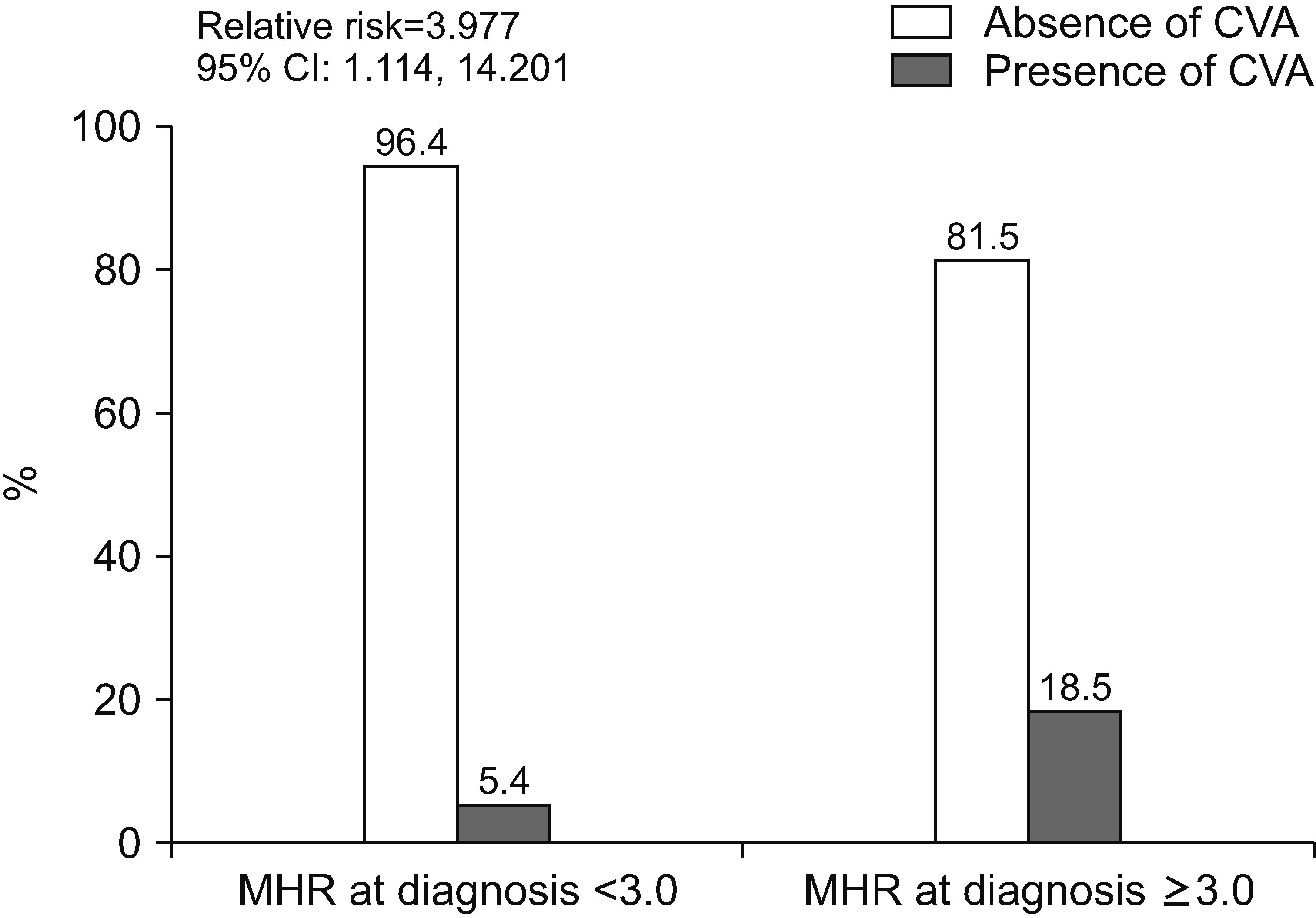
The optimal cut-off of MHR at diagnosis for CVA during follow-up in patients with AAV was determined to be 3.0 using the ROC curve analysis (the area under the curve 0.585). The sensitivity and specificity of the cut-off were 45.5% and 82.7%, respectively. When patients with AAV were divided into two groups according to MHR at diagnosis ≥3.0, 27 of the 138 patients were assigned to the group with MHR at diagnosis ≥3.0. CVA during follow-up was identified more frequently in patients with MHR at diagnosis ≥3.0 than in those with MHR at diagnosis <3.0 (18.5% vs. 5.4%, p=0.024). Furthermore, patients with MHR at diagnosis ≥3.0 exhibited a significantly higher risk of CVA than those with MHR at diagnosis <3.0 (RR: 3.977; 95% CI: 1.114, 14.201) (Figure 2).

Comparison of cumulative survival rates

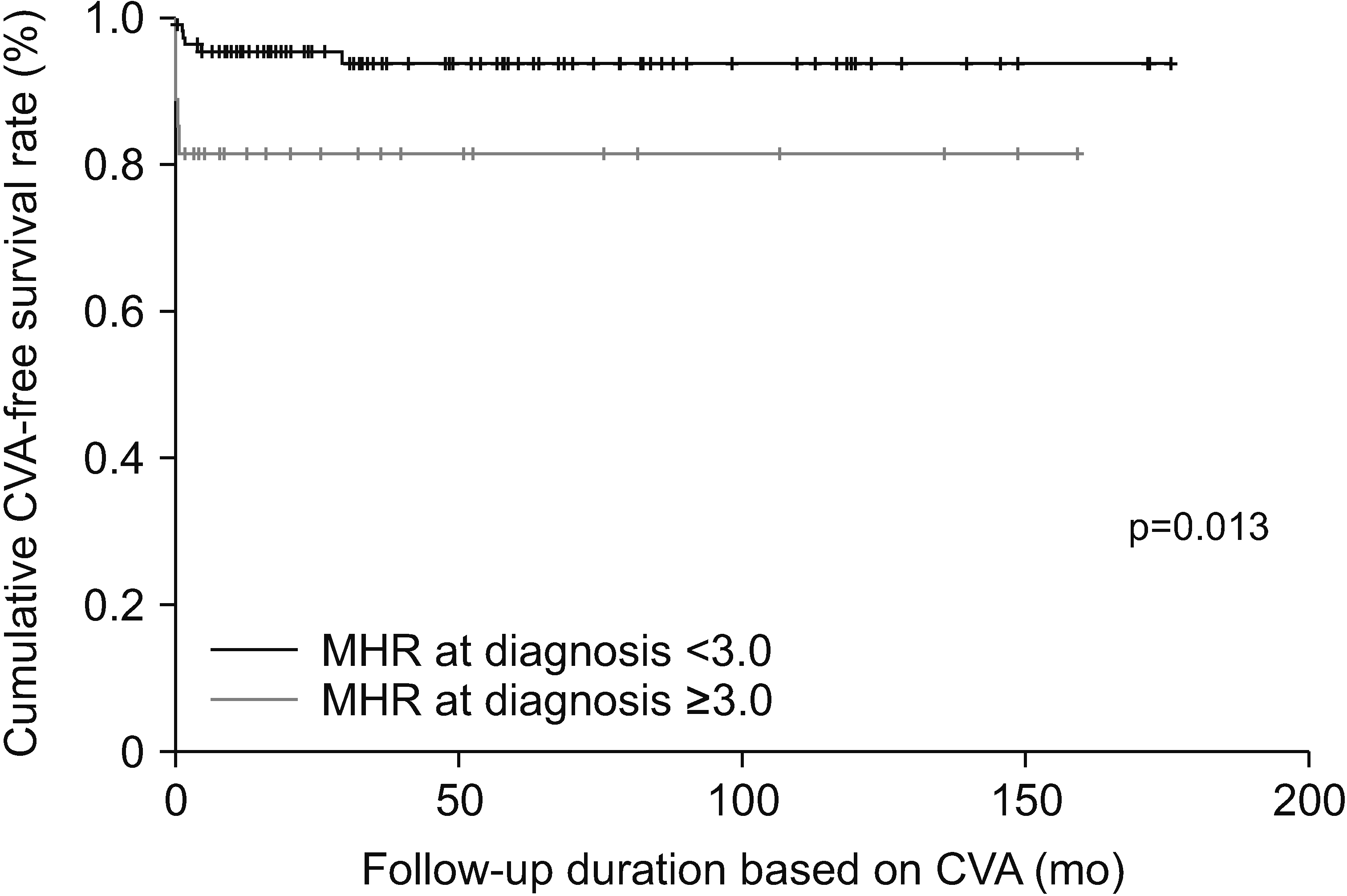
Patients with MHR at diagnosis ≥3.0 exhibited a significantly lower cumulative CVA-free survival rate than those with MHR at diagnosis <3.0 (p=0.013) (Figure 3).

DISCUSSION

In this study, the association between MHR at diagnosis and atherosclerosis-related poor outcomes of AAV during follow-up in patients with AAV was investigated, with several interesting findings. First, MHR at diagnosis reflected the cross-sectional activity of AAV and inflammatory burden. Second, among the three atherosclerosis-related poor outcomes of AAV, a significant and independent association between MHR at diagnosis and CVA during follow-up was identified using the univariable Cox analysis. Third, patients with MHR at diagnosis >3.0 had a significantly higher risk of CVA during follow-up and exhibited a significantly lower cumulative CVA-free survival rate than those without. Therefore, we conclude that MHR at diagnosis can be a useful index for predicting CVA during follow-up in patients with AAV.
Some reports suggested that MHR could be predictive of the development and progression of atherosclerosis, leading to the subsequent occurrence of cardiovascular and cerebrovascular diseases. Particularly, increased monocyte counts were associated with an elevated risk of cardiovascular disease [13]. Additionally, MHR could be a significant indicator of the extent of carotid arterial plaques [7] and might have predictive capabilities for ischemic stroke and related complications [14,15]. Therefore, previous studies reporting the association between MHR and the occurrence of atherosclerosis might support our findings, suggesting that MHR at diagnosis has an independent association with CVA during follow-up in patients with AAV. However, acknowledging that AAV-specific situations may introduce factors beyond typical causality, we present several following hypotheses.
First, MHR at diagnosis was significantly correlated with the cross-sectional BVAS, ESR, and CRP levels (Figure 1). Systemic inflammation might lead to the initiation and acceleration of atherosclerosis, which could provoke the development of CVA and ACS [16]. The link between CRP and atherosclerosis has also been established [17], and in this study, BVAS at diagnosis was also correlated with the cross-sectional ESR (r=0.242, p=0.005), and CRP (r=0.226, p=0.008). Additionally, compared to that in the general population, patients with AAV are known to have a higher risk of stroke [18]. Therefore, we hypothesize that MHR at diagnosis might reflect the cross-sectional AAV activity and inflammatory burden, which could sequentially augment the likelihood of CVA during follow-up through the onset and progression of atherosclerosis in patients with AAV.
Second, MHR at diagnosis was significantly correlated with age at diagnosis (r=0.176, p=0.039); however, neither monocyte counts nor HDL-cholesterol levels were correlated with age at diagnosis. Therefore, although MHR might not account for a large proportion, given that age is a significant risk factor for stroke [19], we hypothesize that MHR at diagnosis could be used to predict CVA during follow-up in patients with AAV by reflecting the age at diagnosis.
Third, in the multivariable Cox analysis, MHR at diagnosis rather than BVAS, ESR, CRP, or age at diagnosis was a significant and independent indicator that was predictive of CVA during follow-up in patients with AAV (Table 2). This suggests that another associated mechanism could exist other than those suggested in the first and second hypotheses. Given that serum albumin at diagnosis was also independently associated with CVA during follow-up and that monocyte recruitment can be promoted during inflammation [20], we hypothesize that MHR at diagnosis might reflect the total amount of inflammation, which is more closely related to the development of CVA during follow-up in patients with AAV than BVAS, ESR, or CRP levels at diagnosis.
On the other hand, to investigate the statistical significance of the four cholesterol-related variables at diagnosis for predicting CVA during follow-up, we performed the univariable Cox analysis using these variables. We found that total cholesterol (HR: 0.991; 95% CI: 0.976, 1.006), low-density lipoprotein cholesterol (HR: 0.008; 95% CI: 0.983, 1.014), and triglyceride (HR: 1.000; 95% CI: 0.990, 1.010) were not associated with CVA, whereas HDL-cholesterol (HR: 0.948; 95% CI: 0.909, 0.989) exhibited an inverse predictive potential for CVA. When we, however, included HDL-cholesterol in the multivariable Cox analysis, we witnessed the statistically independent association of MHR (HR: 1.147; 95% CI: 0.973, 1.351) with CVA disappeared along with HDL-cholesterol (HR: 0.986; 95% CI: 0.944, 1.030), CRP (HR: 0.999; 95% CI: 0.987, 1.011), and serum albumin (HR: 0.261; 95% CI: 0.067, 1.013) at all. Nevertheless, given the well-known protective effect of HDL-cholesterol on CVA as well as the multicollinearity between HDL-cholesterol and MHR due to their high correlation, we conclude that it would be better not to include HDL-cholesterol in the Cox analyses to evaluate the clinical implication of MHR independently in the present study.
This study had certain limitations. The number of patients was not sufficient to represent all Korean patients with AAV, and thus, the results of this study cannot be immediately applied to patients in real clinical settings. The retrospective study design did not enable the serial measurements of MHR or the detection of subclinical CVA. Furthermore, the accurate incidence of vascular complications other than death, CVA, and ACS, such as deep vein thrombosis or thrombotic microangiopathy could not be investigated and provided because of the limitation of a retrospective study design. Nevertheless, this study had an advantage in that it was the first to demonstrate that MHR at diagnosis is independently associated with CVA during follow-up in patients with AAV. This study had another advantage in that it did not propose this cut-off of MHR at diagnosis as an absolute value but suggested a method to obtain the cut-off using the ROC curve for each cohort. A future prospective study with more patients will reliably reveal the dynamic clinical implications of MHR at diagnosis for predicting CVA during follow-up in patients with AAV.
CONCLUSION

This study was the first to demonstrate the clinical implications of MHR. In particular, MHR at diagnosis was identified to be significantly and independently associated with CVA during follow-up in patients with AAV.
SUPPLEMENTARY DATA

Supplementary data can be found with this article online at https://doi.org/10.4078/jrd.2024.0001

ACKNOWLEDGMENTS

None.

Notes

FUNDING

This study received funding from CELLTRION PHARM, Inc., Chungcheongbuk-do, Republic of Korea (NCR 2019-6), and Chong Kun Dang Pharmaceutical Corp., Seoul, Republic of Korea. The funder was not involved in the study design, collection, analysis, interpretation of data, the writing of this article or the decision to submit it for publication.

CONFLICT OF INTEREST

No potential conflict of interest relevant to this article was reported.

AUTHOR CONTRIBUTIONS

Conceptualization: All authors. Data curation: JWH, SSA, SWL. Validation: JJS, YBP. Writing - original draft: JWH, SSA, SWL. Writing - review & editing: All authors.

REFERENCES

1. Jennette JC, Falk RJ, Bacon PA, Basu N, Cid MC, Ferrario F, et al. 2013; 2012 revised International Chapel Hill Consensus Conference Nomenclature of Vasculitides. Arthritis Rheum. 65:1–11. DOI: 10.1002/art.37715. PMID: 23045170.
crossref
2. Watts R, Lane S, Hanslik T, Hauser T, Hellmich B, Koldingsnes W, et al. 2007; Development and validation of a consensus methodology for the classification of the ANCA-associated vasculitides and polyarteritis nodosa for epidemiological studies. Ann Rheum Dis. 66:222–7. DOI: 10.1136/ard.2006.054593. PMID: 16901958. PMCID: PMC1798520.
crossref
3. Millet A, Pederzoli-Ribeil M, Guillevin L, Witko-Sarsat V, Mouthon L. 2013; Antineutrophil cytoplasmic antibody-associated vasculitides: is it time to split up the group? Ann Rheum Dis. 72:1273–9. DOI: 10.1136/annrheumdis-2013-203255. PMID: 23606701.
crossref
4. Mukhtyar C, Lee R, Brown D, Carruthers D, Dasgupta B, Dubey S, et al. 2009; Modification and validation of the Birmingham Vasculitis Activity Score (version 3). Ann Rheum Dis. 68:1827–32. DOI: 10.1136/ard.2008.101279. PMID: 19054820.
crossref
5. Jiang M, Yang J, Zou H, Li M, Sun W, Kong X. 2022; Monocyte-to-high-density lipoprotein-cholesterol ratio (MHR) and the risk of all-cause and cardiovascular mortality: a nationwide cohort study in the United States. Lipids Health Dis. 21:30. DOI: 10.1186/s12944-022-01638-6. PMID: 35300686. PMCID: PMC8931976.
crossref
6. Chen JW, Li C, Liu ZH, Shen Y, Ding FH, Shu XY, et al. 2019; The role of monocyte to high-density lipoprotein cholesterol ratio in prediction of carotid intima-media thickness in patients with type 2 diabetes. Front Endocrinol (Lausanne). 10:191. DOI: 10.3389/fendo.2019.00191. PMID: 31019490. PMCID: PMC6458254.
crossref
7. Xi J, Men S, Nan J, Yang Q, Dong J. 2022; The blood monocyte to high density lipoprotein cholesterol ratio (MHR) is a possible marker of carotid artery plaque. Lipids Health Dis. 21:130. Erratum in: Lipids Health Dis 2022;21:144. DOI: 10.1186/s12944-022-01741-8. PMID: 36463176. PMCID: PMC9719628.
crossref
8. Lee SW, Park YB. 2019; Classification of antineutrophil cytoplasmic antibody-associated vasculitis. J Rheum Dis. 26:156–64. DOI: 10.4078/jrd.2019.26.3.156.
crossref
9. Suppiah R, Robson JC, Grayson PC, Ponte C, Craven A, Khalid S, et al. 2022; 2022 American College of Rheumatology/European Alliance of Associations for Rheumatology classification criteria for microscopic polyangiitis. Ann Rheum Dis. 81:321–6. DOI: 10.1136/annrheumdis-2021-221796. PMID: 35110332.
crossref
10. Robson JC, Grayson PC, Ponte C, Suppiah R, Craven A, Judge A, et al. 2022; 2022 American College of Rheumatology/European Alliance of Associations for Rheumatology classification criteria for granulomatosis with polyangiitis. Ann Rheum Dis. 81:315–20. DOI: 10.1136/annrheumdis-2021-221795. PMID: 35110333.
crossref
11. Grayson PC, Ponte C, Suppiah R, Robson JC, Craven A, Judge A, et al. 2022; 2022 American College of Rheumatology/European Alliance of Associations for Rheumatology classification criteria for eosinophilic granulomatosis with polyangiitis. Ann Rheum Dis. 81:309–14. DOI: 10.1136/annrheumdis-2021-221794. PMID: 35110334.
crossref
12. Pyo JY, Lee LE, Park YB, Lee SW. 2023; Comparison of the 2022 ACR/EULAR classification criteria for antineutrophil cytoplasmic antibody-associated vasculitis with previous criteria. Yonsei Med J. 64:11–7. DOI: 10.3349/ymj.2022.0435. PMID: 36579374. PMCID: PMC9826961.
crossref
13. Kim JH, Lim S, Park KS, Jang HC, Choi SH. 2017; Total and differential WBC counts are related with coronary artery atherosclerosis and increase the risk for cardiovascular disease in Koreans. PLoS One. 12:e0180332. DOI: 10.1371/journal.pone.0180332. PMID: 28753607. PMCID: PMC5533311.
crossref
14. Li Y, Zhang M, Xue M, Liu D, Sun J. 2022; Elevated monocyte-to-HDL cholesterol ratio predicts post-stroke depression. Front Psychiatry. 13:902022. DOI: 10.3389/fpsyt.2022.902022. PMID: 35935403. PMCID: PMC9354071.
crossref
15. Liu Z, Fan Q, Wu S, Lei Y. 2021; Associations of monocytes and the monocyte/high-density lipoprotein ratio with extracranial and intracranial atherosclerotic stenosis. Front Neurol. 12:756496. DOI: 10.3389/fneur.2021.756496. PMID: 34925216. PMCID: PMC8678066.
crossref
16. Geovanini GR, Libby P. 2018; Atherosclerosis and inflammation: overview and updates. Clin Sci (Lond). 132:1243–52. DOI: 10.1042/CS20180306. PMID: 29930142.
crossref
17. Singh SK, Suresh MV, Voleti B, Agrawal A. 2008; The connection between C-reactive protein and atherosclerosis. Ann Med. 40:110–20. DOI: 10.1080/07853890701749225. PMID: 18293141. PMCID: PMC3364506.
crossref
18. Tabakovic D, Smith R, Jayne D, Mohammad AJ. 2023; High risk of stroke in ANCA-associated vasculitis-a population-based study. Rheumatology (Oxford). 62:2806–12. DOI: 10.1093/rheumatology/keac669. PMID: 36440920. PMCID: PMC10393431.
crossref
19. Kelly-Hayes M. 2010; Influence of age and health behaviors on stroke risk: lessons from longitudinal studies. J Am Geriatr Soc. 58(Suppl 2):S325–8. DOI: 10.1111/j.1532-5415.2010.02915.x. PMID: 21029062. PMCID: PMC3006180.
20. Shi C, Pamer EG. 2011; Monocyte recruitment during infection and inflammation. Nat Rev Immunol. 11:762–74. DOI: 10.1038/nri3070. PMID: 21984070. PMCID: PMC3947780.
crossref

Figure 1
Correlation analysis. MHR at diagnosis is significantly correlated with the cross-sectional BVAS, ESR, and CRP levels. MHR: monocyte-to-high-density lipoprotein-cholesterol ratio, BVAS: Birmingham vasculitis activity score, ESR: erythrocyte sedimentation rate, CRP: C-reactive protein.
jrd-31-3-151-f1.tif
Figure 2
Relative risk. Patients with MHR at diagnosis ≥3.0 exhibits a significantly higher risk of CVA than those with MHR at diagnosis <3.0. MHR: monocyte-to-high-density lipoprotein-cholesterol ratio, CVA: cerebrovascular accident, CI: confidence interval.
jrd-31-3-151-f2.tif
Figure 3
Comparison of cumulative survival rates. Patients with MHR at diagnosis ≥3.0 exhibits a significantly lower cumulative CVA-free survival rate than those with MHR at diagnosis <3.0. MHR: monocyte-to-high-density lipoprotein-cholesterol ratio, CVA: cerebrovascular accident.
jrd-31-3-151-f3.tif
Table 1
Characteristics of patients with AAV
Variable Value (n=138)
At diagnosis
Demographic data
Age (yr) 58.3 (46.5~69.0)
Male sex 44 (31.9)
Female sex 94 (68.1)
BMI (kg/m2) 22.0 (19.6~24.0)
Ex-smoker 10 (7.2)
AAV subtype
MPA 76 (55.1)
GPA 30 (21.7)
EGPA 32 (23.2)
ANCA type and positivity
MPO-ANCA (or P-ANCA) positivity 91 (65.9)
PR3-ANCA (or C-ANCA) positivity 22 (15.9)
AAV-specific indices
BVAS 13.0 (7.0~18.0)
FFS 1.0 (1.0~2.0)
Acute phase reactants
ESR (mm/h) 61.0 (24.0~91.0)
CRP (mg/L) 7.1 (1.4~71.0)
Routine laboratory results
White blood cell count (/mm3) 8,470.0 (6,220.0~13,090.0)
Neutrophil count 450.0 (310.0~602.5)
Lymphocytes count 20.0 (10.0~40.0)
Monocyte count 50.0 (0~110.0)
Hemoglobin (g/dL) 11.1 (9.1~13.1)
Platelet count (×1,000/mm3) 286.0 (207.0~378.0)
Blood urea nitrogen (mg/dL) 19.0 (12.3~35.2)
Serum creatinine (mg/dL) 1.0 (0.7~2.1)
Serum albumin (g/dL) 3.6 (3.1~4.0)
Lipid profile (mg/dL)
Total cholesterol 171.0 (144.8~202.3)
HDL-cholesterol 47.5 (36.0~63.0)
TG 108.0 (86.8~155.0)
LDL-cholesterol 97.2 (78.7~121.3)
MHR
Comorbidities 0.87 (0~2.36)
Hypertension 39 (28.3)
T2DM 41 (29.7)
During the follow-up duration
Typical poor outcomes of AAV
All-cause mortality 11 (8.0)
Follow-up duration based on all-cause mortality (mo) 34.0 (11.5~76.3)
CVA 11 (8.0)
Follow-up duration based on stroke (mo) 31.1 (9.1~71.0)
ACS 9 (6.5)
Follow-up duration based on ACS (mo) 32.7 (9.8~72.4)

Values are expressed as a median (25~75 percentiles) or number (%). AAV: antineutrophil cytoplasmic antibody-associated vasculitis, ANCA: antineutrophil cytoplasmic antibody, BMI: body mass index, MPA: microscopic polyangiitis, GPA: granulomatosis with polyangiitis, EGPA: eosinophilic granulomatosis with polyangiitis, MPO: myeloperoxidase, P: perinuclear, PR3: proteinase 3, C: cytoplasmic, BVAS: Birmingham vasculitis activity score, FFS: five-factor score, ESR: erythrocyte sedimentation rate, CRP: C-reactive protein, HDL-cholesterol: high-density lipoprotein cholesterol, TG: triglyceride, LDL-cholesterol: low-density lipoprotein cholesterol, MHR: monocyte-to-high-density lipoprotein-cholesterol ratio, T2DM: type 2 diabetes mellitus, CVA: cerebrovascular accident, ACS: acute coronary syndrome.

Table 2
Cox hazards model analyses of MHR and variables at diagnosis for CVA during follow-up in patients with AAV
Variable Univariable Multivariable
(variables with p<0.05)
Multivariable
(variables with p<0.1)
HR 95% CI p-value HR 95% CI p-value HR 95% CI p-value
Age (yr) 1.046 0.994, 1.100 0.087 1.010 0.955, 1.068 0.736
Male sex 1.274 0.372, 4.361 0.699
BMI (kg/m2) 1.145 0.942, 1.392 0.174
Ex-smoker 1.333 0.170, 10.432 0.784
MPO-ANCA (or P-ANCA) positivity 2.405 0.519, 11.138 0.262
PR3-ANCA (or C-ANCA) positivity 0.515 0.066, 4.023 0.527
BVAS 1.086 0.999, 1.180 0.053 1.065 0.954, 1.188 0.260
FFS 1.533 0.880, 2.670 0.131
ESR (mm/h) 1.016 0.999, 1.032 0.068 0.994 0.970, 1.018 0.618
CRP (mg/L) 1.008 1.002, 1.015 0.013 0.999 0.987, 1.011 0.857 1.000 0.987, 1.014 0.953
White blood cell count (/mm3) 1.000 1.000, 1.000 0.156
Hemoglobin (g/dL) 0.770 0.583, 1.016 0.065 1.126 0.774, 1.637 0.535
Platelet count (×1,000/mm3) 1.002 0.999, 1.005 0.189
Blood urea nitrogen (mg/dL) 1.006 0.988, 1.025 0.501
Serum creatinine (mg/dL) 1.058 0.811, 1.381 0.677
Serum albumin (g/dL) 0.210 0.084, 0.525 0.001 0.235 0.064, 0.863 0.029 0.203 0.042, 0.987 0.048
T2DM 0.530 0.115, 2.455 0.417
Hypertension 1.975 0.602, 6.477 0.261
MHR 1.246 1.096, 1.417 0.001 1.179 1.024, 1.359 0.022 1.195 1.030, 1.386 0.018

MHR: monocyte-to-high-density lipoprotein-cholesterol ratio, AAV: antineutrophil cytoplasmic antibody-associated vasculitis, CVA: cerebrovascular accident, ANCA: antineutrophil cytoplasmic antibody; HR: hazard ratio, CI: confidence interval, BMI: body mass index, MPO: myeloperoxidase, P: perinuclear, PR3: proteinase 3, C: cytoplasmic, BVAS: Birmingham vasculitis activity score, FFS: five-factor score, ESR: erythrocyte sedimentation rate, CRP: C-reactive protein, T2DM: type 2 diabetes mellitus.

Table 3
Comparison of medications administered during follow-up between AAV patients with CVA and those without
Variable Patients without CVA (n=127) Patients with CVA (n=11) p-value
Lipid-lowering agent
Statins 69 (54.3) 8 (72.7) 0.346
Ezetimibe 15 (11.8) 2 (18.2) 0.626
Aspirin 22 (17.3) 4 (36.4) 0.219
Antihypertensive drug
Angiotensin converting enzyme inhibitor or angiotensin receptor II blocker 72 (56.7) 9 (81.8) 0.123
Calcium channel blocker 79 (62.2) 10 (90.9) 0.097
Beta-adrenergic blocker 46 (36.2) 4 (36.4) 1.000
Diuretics 51 (40.2) 5 (45.5) 0.731
Other antihypertensive agents 17 (13.4) 3 (27.3) 0.200
Immunosuppressive drug
Glucocorticoids 121 (95.3) 11 (100) 1.000
Cumulative dose of glucocorticoids (equivalent to prednisolone, mg) 11,852.2 (6,085.0~17,525.0) 11,203.1 (7,810.0~18,728.3) 0.878
Cyclophosphamide 47 (37.0) 5 (45.5) 0.579
Rituximab 23 (18.1) 3 (27.3) 0.434
Azathioprine 44 (34.6) 2 (18.2) 0.336
Mycophenolate mofetil 27 (21.3) 2 (18.2) 1.000
Tacrolimus 13 (10.2) 1 (9.1) 1.000
Methotrexate 18 (14.2) 1 (9.1) 1.000

Values are expressed as number (%) or median (interquartile range). AAV: antineutrophil cytoplasmic antibody-associated vasculitis, CVA: cerebrovascular accident.

TOOLS
Similar articles