This article has been
cited by other articles in ScienceCentral.
Abstract
This case report presents the successful endoscopic submucosal dissection (ESD) of a well-differentiated esophageal liposarcoma in a 51-year-old male with persistent dysphagia. The cause was initially diagnosed as a 10 cm pedunculated lesion extending from the upper esophageal sphincter to the mid-esophagus. An ESD was chosen over traditional surgery because it is less invasive. The procedure involved a precise submucosal injection and excision with special techniques to manage bleeding from a central vessel. Despite the extraction challenges owing to the size of the lesion, it was successfully removed orally. A histopathological examination of the 8.3×4.2×2.3 cm specimen revealed the characteristic features of a well-differentiated liposarcoma, including MDM2 and CDK4 positivity. The follow-up revealed no recurrence, and active surveillance has been performed since. This report highlights the versatility of ESD in treating significant esophageal tumors and provides evidence for its efficacy as a minimally invasive alternative.
Go to :

Keywords: Liposarcoma, Esophagus, Endoscopic mucosal resection
INTRODUCTION
Esophageal liposarcoma is an exceedingly rare entity, constituting a small fraction (1.2–1.5%) of all gastrointestinal liposarcomas.
1 The condition is characterized by submucosal growth and often pedunculated, with a polypoid appearance that can lead to significant clinical manifestations, such as dysphagia, foreign body sensation, and potentially life-threatening respiratory distress if it obstructs the airway. Although a surgical resection has traditionally been the cornerstone treatment for such tumors, recent advances have positioned the endoscopic resection procedure as a viable and less invasive alternative for select cases, especially those localized to the hypopharynx and cervical esophagus. This transition towards endoscopic methodologies, particularly endoscopic submucosal dissection (ESD), marks a significant evolution in managing this rare disease. This case report highlights a unique instance of a well-differentiated liposarcoma within the upper esophagus that was diagnosed and treated effectively through an endoscopic resection, underscoring the potential and versatility of an ESD in esophageal tumors.
Go to :

CASE REPORT
A 51-year-old male with a history of a right hemithyroidectomy for thyroid cancer 11 years prior and Gilbert syndrome presented with persistent dysphagia and the sensation of a foreign body in the esophagus for six months. An initial endoscopic examination at an external facility revealed a 10 cm polypoid lesion in the upper esophagus, prompting a referral for surgical consultation. Nevertheless, the patient sought a second opinion regarding possible endoscopic management at the author's institution.
The pedunculated tumor identified during endoscopy originated from the upper esophageal sphincter and extended into the mid-esophagus. The largest diameter of the area connected to the mid-esophagus was approximately 4 cm. Endoscopic ultrasound revealed a homogeneous hyperechoic lesion arising from the third layer of the esophagus (
Fig. 1). The stalk of the lesion was approximately 5 mm in thickness, indicating the feasibility of an ESD.
 | Fig. 1Endoscopic findings of esophageal liposarcoma. (A) The tumor originated from the upper esophageal sphincter. (B) The tumor extended to the mid-esophagus and occupied most of the esophageal cavity. (C) Endoscopic ultrasound shows a homogeneous, hyperechoic lesion. 
|
An ESD was performed under general anesthesia with endotracheal intubation. After a submucosal injection at the base of the lesion, an incision was made using a dual knife (Olympus, Tokyo, Japan). Bleeding occurred because of a large vessel in the center of the lesion, but it was successfully managed with a coagrasper (Olympus). After completing the en-bloc resection, no additional bleeding or perforation occurred (
Fig. 2). Although initial attempts were made to extract the lesion orally using a snare, the snare was too large for capture. Ultimately, the lesion was removed carefully through the mouth using a basket.
 | Fig. 2Endoscopic submucosal dissection (ESD) procedure. (A) Submucosal injection and mucosal incision at the base of the tumor. (B) Completely removed after ESD. (C) Gross pathological resection showing the lesion was about 9 cm long. 
|
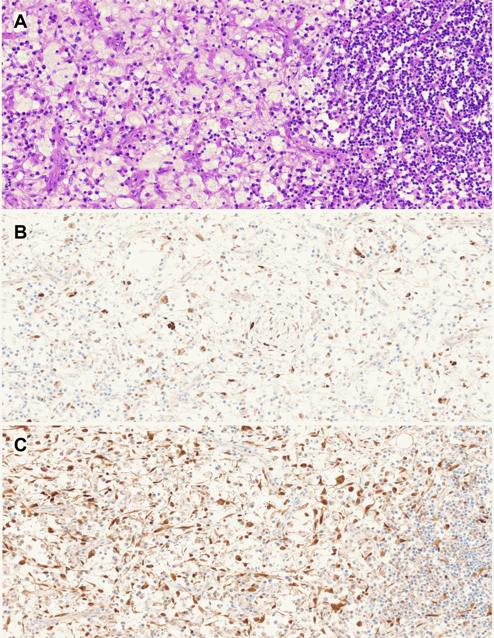
The excised specimen was 8.3×4.2×2.3 cm in size. A microscopic examination at low magnification showed that the lesion was a fibrofatty mass with overlying squamous mucosa. At a higher magnification, there was a noticeable increase in irregular, hyperchromatic nuclei among stromal cells within the fibrous septa separating the adipocytes and dispersed hyperchromatic spindle cells. Immunohistochemistry revealed positivity for MDM2 and CDK4 in the fibrous septa and hyperchromatic spindle cells (
Fig. 3).
 | Fig. 3(A) Fibrous septa between the adipocytes revealed an increased number of stromal cells with slightly irregular, hyperchromatic nuclei scattered fibrous septa containing hyperchromatic spindle cells (H&E, ×400). MDM2 (B) and CDK4 (C) were positive in the fibrous septa and hyperchromatic spindle cells. 
|
The conclusive pathological diagnosis identified the mass as a well-differentiated liposarcoma. Follow-up endoscopy conducted two months post-operation revealed a scar at the intervention site. No signs of recurrence or metastasis were detected on subsequent CT scans, and active surveillance has been scheduled.
Go to :

DISCUSSION
A large esophageal liposarcoma, approximately 10 cm in length and 4 cm in diameter was successfully removed and extracted from the upper esophageal sphincter using the ESD method without complications. The rarity of esophageal liposarcomas, comprising approximately 20% of adult soft tissue sarcomas and more commonly found in the retroperitoneum and limbs, makes this case particularly noteworthy.
2 The slow growth of these tumors in the esophagus can lead to dysphagia and potential esophageal obstruction, often presenting symptoms such as cough, vomiting, and sensations akin to a foreign body. The risk of sudden airway obstruction adds to the clinical urgency, with dysphagia frequently resulting in weight loss caused by delayed diagnosis.
3
Traditionally, a surgical resection has been the primary treatment for esophageal liposarcomas, with an emphasis on tumor removal to alleviate the symptoms and prevent complications.
4
On the other hand, the advent of endoscopic techniques, particularly ESD, has emerged as a promising, less invasive alternative technique, particularly for tumors amenable to endoscopic management owing to their size, location, and morphology. In particular, if the diameter of the lesion is large, the use of a basket instead of a snare to grasp and extract the lesion through the mouth would be effective.
According to the Enzinger and Weiss system, liposarcomas are categorized into five subtypes.
1) Atypical Lipomatous Tumor/Well-Differentiated Liposarcoma, which is characterized by mature adipocytes with slight atypia and low mitotic activity that grows slowly and has low metastatic potential but can recur locally;
2) Dedifferentiated Liposarcoma contains both well-differentiated and high-grade non-lipogenic areas and is more aggressive, with a higher likelihood of metastasis and a poorer prognosis;
3) Myxoid Liposarcoma features a gelatinous matrix and round to oval cells, often with a t(12;16) translocation that has a better prognosis than dedifferentiated liposarcoma but can still metastasize, especially to extrapulmonary sites;
4) Pleomorphic Liposarcoma is the rarest and most aggressive subtype, marked by varied cell shapes and high mitotic activity that frequently metastasizes and has the worst prognosis;
5) Myxoid Pleomorphic Liposarcoma is a rare subtype with myxoid and pleomorphic liposarcoma features, including myxoid areas and pleomorphic lipoblasts that is aggressive, with a high risk of metastasis and poor prognosis.
5
Esophageal liposarcoma was once mistaken for a giant fibrovascular polyp. A giant fibrovascular polyp of the esophagus is a diagnostic term encompassing rare, large, polypoid esophageal masses composed of fibro-adipose tissue. It is considered to represent a reactive non-neoplastic proliferation. On the other hand, a well-differentiated liposarcoma of the esophagus, similar to a giant fibrovascular polyp, has been reported. Endoscopically, both lesions have a cylindrical, elongated structure. Giant fibrovascular polyps are often uniformly thick from the base to the distal part and appear soft. Esophageal liposarcoma, however, is thicker toward the center and somewhat harder. The distinction between well-differentiated liposarcoma and benign lipoma is frequent pathologically, and secondary changes in lipomas may result in an irregular adipocyte size and atrophic adipocytes mixed with inflammatory cells and histiocytes.
6,7 Therefore, giant fibrovascular polyps in the esophagus should be diagnosed with care. MDM2 is a particular marker in spindle cells with atypia and adipocytes. Therefore, it should only be used after excluding the possibility of liposarcoma through MDM2 FISH.
The prognosis and tumor aggressiveness of esophageal liposarcomas vary according to their histological type, location, and resection margin status.
7,8 Well-differentiated liposarcomas have the best prognosis, highlighting the importance of a complete resection. Nevertheless, the potential for new cancer development in the residual esophagus necessitates long-term surveillance, underlining the significance of this case in demonstrating the viability of ESD as a treatment option for large esophageal liposarcomas and expanding the boundaries of endoscopic intervention.
9,10
This report presented a rare endoscopic resection of a large, well-differentiated esophageal liposarcoma without complications. This finding provides evidence of the feasibility of ESD for large esophageal liposarcomas.
Go to :


 PDF
PDF Citation
 Print
Print


 XML Download