Abstract
Background
This study investigates the impact of fluctuating lipid levels on endothelial dysfunction.
Methods
Human aortic and umbilical vein endothelial cells were cultured under varying palmitic acid (PA) concentrations: 0, 50, and 100 μM, and in a variability group alternating between 0 and 100 μM PA every 8 hours for 48 hours. In the lipid variability group, cells were exposed to 100 μM PA during the final 8 hours before analysis. We assessed inflammation using real-time polymerase chain reaction, Western blot, and cytokine enzyme-linked immunosorbent assay (ELISA); reactive oxygen species (ROS) levels with dichlorofluorescin diacetate assay; mitochondrial function through oxygen consumption rates via XF24 flux analyzer; and endothelial cell functionality via wound healing and cell adhesion assays. Cell viability was evaluated using the MTT assay.
Results
Variable PA levels significantly upregulated inflammatory genes and adhesion molecules (Il6, Mcp1, Icam, Vcam, E-selectin, iNos) at both transcriptomic and protein levels in human endothelial cells. Oscillating lipid levels reduced basal respiration, adenosine triphosphate synthesis, and maximal respiration, indicating mitochondrial dysfunction. This lipid variability also elevated ROS levels, contributing to a chronic inflammatory state. Functionally, these changes impaired cell migration and increased monocyte adhesion, and induced endothelial apoptosis, evidenced by reduced cell viability, increased BAX, and decreased BCL2 expression.
Endothelial dysfunction, which is often caused by disruptions in lipid homeostasis, is a major initiator of atherosclerotic diseases. Endothelial cells (ECs), which form the inner lining of vascular surfaces, maintain vascular tone, facilitate blood flow, and regulate permeability [1]. Additionally, they play a vital role in regulating inflammation, hemostasis, thrombosis, fibrinolysis, and angiogenesis [2]. Central to EC functionality is the synthesis of diverse molecules that balance vasoconstriction (e.g., angiotensin [Ang] II and endothelin [ET]-1) and vasodilation (e.g., nitric oxide [NO] and bradykinin), creating a delicate equilibrium essential for circulatory system maintenance. This equilibrium safeguards against disturbances that may otherwise trigger the progression of vascular diseases [3]. Perturbations introduced by stimuli such as lipids, glucose, or blood pressure can induce EC dysfunction, which is characterized by diminished NO production and imbalanced ratios of endothelial vasodilators and vasoconstrictors. These deviations have emerged as precursors of cardiovascular risk and signal the onset of atherosclerosis [4]. Among the many stimuli, the disruption of lipid homeostasis is notable for being a precursor to endothelial dysfunction. Lipids are the main contributors to this cascade. Elevations in low-density lipoprotein cholesterol (LDL-C) levels and the cumulative extent of exposure are well-established cardiovascular risk factors [5-8]. Several studies have suggested an association between hypertriglyceridemia and cardiovascular risks [9-11]. Furthermore, elevated levels of serum-free fatty acids (FAs) have been implicated in an increased risk of cardiovascular diseases [12,13]. Notably, saturated FAs have been implicated in the development of atherosclerosis, a connection supported by mechanisms involving inflammation, oxidative stress, and impaired NO production in ECs [2]. Previous studies by our group and others have demonstrated that lipid variability is associated with the risk of cardiovascular diseases [14-16]; however, the precise mechanisms underlying the increased risk of atherosclerotic diseases resulting from fluctuations in lipid levels remains poorly understood. To address this, we explored the mechanism by which lipid variability leads to endothelial dysfunction using human EC lines and palmitic acids (PAs).
Human aortic endothelial cells (HAECs; Lonza, Walkersville, MD, USA) and human umbilical vein endothelial cells (HUVECs) were used at passage 4–9 for the experiments and cultured in endothelial cell growth medium (EGM)-2 BulletKit (Lonza) supplemented with penicillin-streptomycin. The cells were incubated at 37°C in a humidified atmosphere containing 5% CO2. To analyze the molecular changes occurring under various PA treatment conditions, ECs were either treated with 0, 50, and 100 µM PA, or alternated between 0 and 100 µM PA every 8 hours (variability group) over 48 hours. The ECs in the lipid variability group were exposed to 100 μM of PA in the last 8 hours of culture before every transcriptomic, proteomic, and functional analysis to compare with continuous 100 µM PA group at the same condition.
A 100 mM PA (P5585, Sigma-Aldrich, St. Louis, MO, USA) stock solution was prepared with 0.1 N NaOH at 70°C. The stock solution was conjugated to 10% FA-free bovine serum albumin (BSA; A7030, Sigma-Aldrich) in a heating block for 5 minutes at 60°C for complex formation. This intermediate stock was filter sterilized (0.22 μm) and the required amount was added to the medium for final concentrations.
The cells were treated with TRIzol reagent (Invitrogen, Carlsbad, CA, USA) to extract total RNA. In the reverse transcription reaction, 1 μg of total RNA was used with the PrimeScript 1st strand cDNA synthesis kit (Takara Bio Inc., Tokyo, Japan) following the manufacturer’s instructions. All quantitative polymerase chain reaction (qPCR) reactions were conducted using Power SYBR Green PCR Master Mix (Applied Biosystems, Foster City, CA, USA). Supplemental Table S1 provides the details of the PCR primer sequences.
Western blot analysis was performed as described previously [17]. Total protein lysates were separated by sodium dodecyl sulfate–polyacrylamide gel electrophoresis and transferred onto Immobilon polyvinylidene difluoride membranes (Millipore, Billerica, MA, USA). After blocking, the membranes were incubated with primary antibodies, and subsequently with horseradish peroxidase-conjugated secondary antibodies. The following primary antibodies employed were used: anti-phospho-nuclear factor κB (NF-κB) p65 (p-p65, #3031), anti-NF-κB p65 (p65, #3034), anti-phospho-IkappaB-alpha (p-IκBα, #9241), anti-IκBα (#9242), anti-intercellular adhesion molecule (ICAM, sc-7891), anti-vascular cell adhesion molecule (VCAM, sc8304), anti-E-selectin (sc-14011, Santa Cruz Biotechnology, Santa Cruz, CA, USA), anti-phospho-endothelial nitric oxide synthase (eNos, #9571), anti-B cell CLL/lymphoma 2 (BCL2, #3498), anti-BCL-2-associated X protein (BAX, #2774, Cell Signaling Technology Inc., Danvers, MA, USA), anti-eNos (610299, BD Bioscience, San Jose, CA, USA), anti-inducible nitric oxide synthase (iNOS, 1:1,000, N32030, Transduction Laboratories, Lexington, KY, USA), or anti-β-actin (1:5,000, A5441, Sigma-Aldrich; Merck KGaA, Darmstadt, Germany). Proteins were detected using an enhanced chemiluminescence kit (34577, Thermo Fisher Scientific, Waltham, MA, USA) according to the manufacturer’s instructions. Visualization was performed using the PXi4 Multi-Application Gel Imaging System (Syngene, Cambridge, UK), and densitometry analysis was performed using ImageJ (National Institutes of Health, Bethesda, MD, USA), and GraphPad Prism 8.0 (San Diego, CA, USA). For immunofluorescence staining, cells were fixed in 4% paraformaldehyde in phosphate-buffered saline (PBS), permeabilized with 0.5% Triton X-100. Cells were then incubated with the anti-NF-κB p65 (p65, #8242, Cell Signaling Technology Inc.) overnight at 4°C. Cell nuclei were counterstained with 4´,6-diamidino-2-phenylindole (DAPI). Microscopy analysis was performed using an Olympus IX70 inverted fluorescence microscope (Olympus, Tokyo, Japan).
Concentrations of secretory cytokines (interleukin [IL] 6 and monocyte chemoattractant protein [MCP] 1) were measured via Human Luminex Discovery Assay following the manufacturer’s instructions (#LXSAHM-08, R&D Systems, Minneapolis, MN, USA) using HUVEC supernatants.
To quantify intracellular reactive oxygen species (ROS) levels, HAECs cultured under various conditions of PA exposure were cultured to 70% to 80% of confluency. The cells were then washed with PBS and incubated with 10 μM 2´,7´-dichlorofluorescin diacetate (DCFH-DA, D6883, Sigma-Aldrich) in serum-free medium for 30 minutes to 1 hour at 37°C in a humidified incubator at 5% CO2. After the incubation period, the cells were washed with PBS to remove any extracellular DCFH-DA and subsequently exposed to the experimental conditions. Fluorescence intensity was measured using a fluorescence microplate reader at excitation and emission wavelengths of 488 and 525 nm, respectively. Fluorescence readings were taken at specified time points, and the data were normalized to the control wells to determine the relative ROS levels.
Mitochondrial function in HAECs was measured using the mitochondrial stress test (Seahorse Bioscience, North Billerica, MA, USA) as described previously [17]. Briefly, cells were seeded at a density of 4×10⁴ cells/well in an XF24 flux analyzer plate (Seahorse Biosciences). Mitochondrial inhibitors were added at the following concentrations: 1.0 μM oligomycin, 0.5 mM trifluoromethoxy carbonylcyanide phenylhydrazone, and 0.5 mM rotenone and antimycin A (Seahorse Bioscience).
HAECs were serum-starved for 6 hours under various PA exposure conditions to synchronize and minimize proliferation. A sterile 200-μL pipette tip was used to create a linear scratch (wound) across the cell monolayer. Detached cells and debris were gently washed away with PBS, and fresh EGM was added to the wells. Cells were incubated at 37°C in a 5% CO2 atmosphere. Photomicrographs were captured at 0 and 48 hours postscratching using an inverted microscope to monitor wound closure. The distance between the edges of the scratch was measured and quantified using image analysis software to assess the cell migration rates.
The HAECs were cultured in 12-well plates under various PA exposure conditions. The medium was replaced with serum-free EGM for 6 hours to synchronize the cells. THP1 monocytes (1×10⁶ cells/mL) were pre-stimulated with calcein-acetoxymethyl (AM) (2.5 μg/mL, 17783, Sigma-Aldrich) for 30 minutes. Subsequently, calcein-AM-labeled monocytes were added to the HAEC monolayer and co-incubated for 1 hour at 37°C in a 5% CO2 atmosphere. Non-adhered monocytes were gently washed with PBS, and the adhered cells were quantified using a fluorescence microplate reader (Synergy MX, BioTek, Winooski, VT, USA) with excitation and emission wavelengths set at 495/515 nm. The data were normalized to control wells containing only ECs to calculate relative adhesion rates.
HAECs were seeded onto plates (2×10⁴ cells/well in a 96-well plate) and cultured under various PA exposure conditions for 48 hours. Medium containing BSA was used as a blank. After PA exposure, 3-(4,5-dimethyl-2-thiazolyl)-2,5-diphenyl-2H-tetrazolium bromide (MTT; M2128, Sigma-Aldrich) reagent (20 μL, 5 mg/mL) was added to each well and cells were incubated at 37°C for 4 hours. The absorbance was measured at 570 nm using a fluorescence microplate reader. Cell viability was calculated as follows: cell viability (%)=absorbance of the experimental group/control group×100.
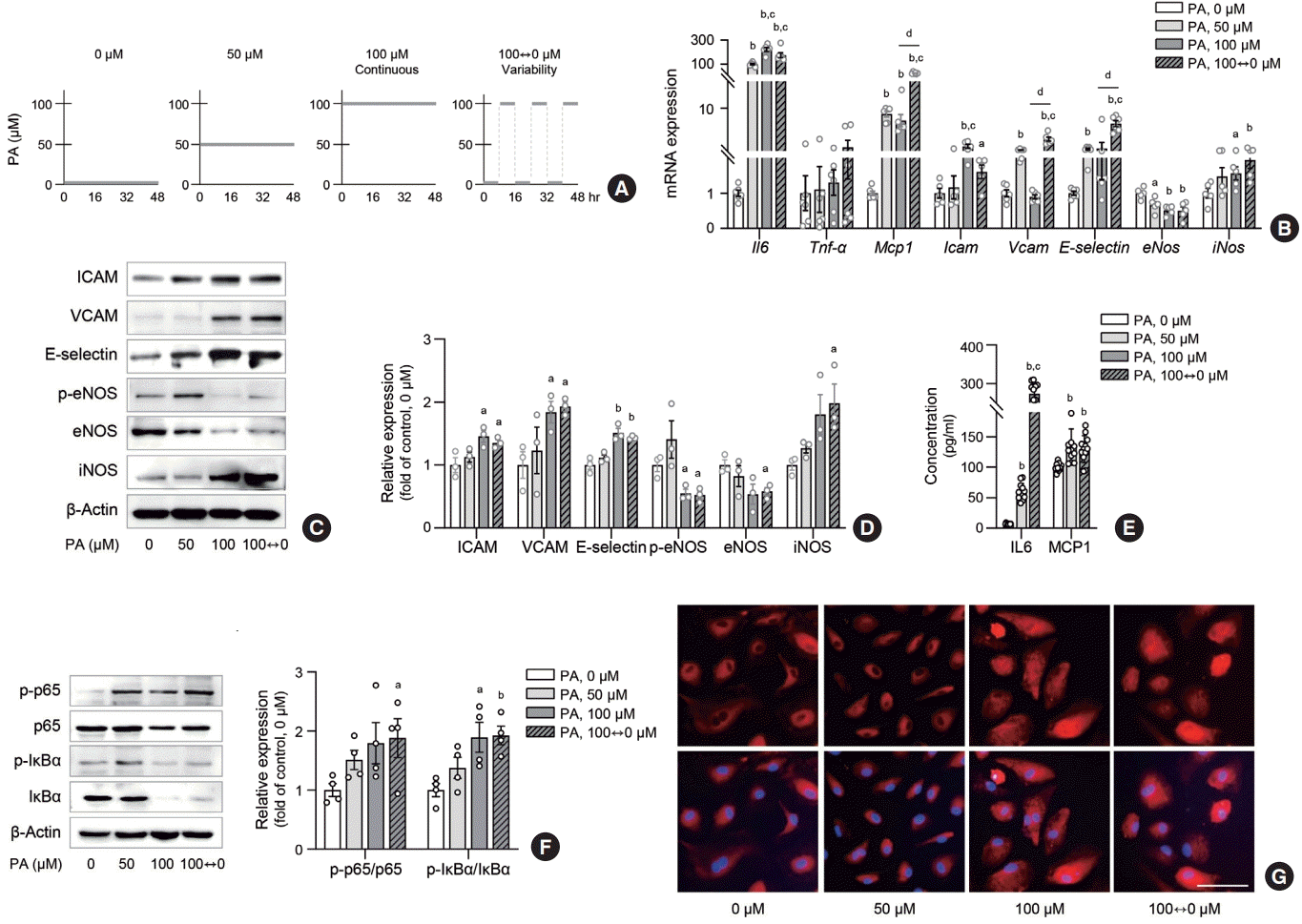
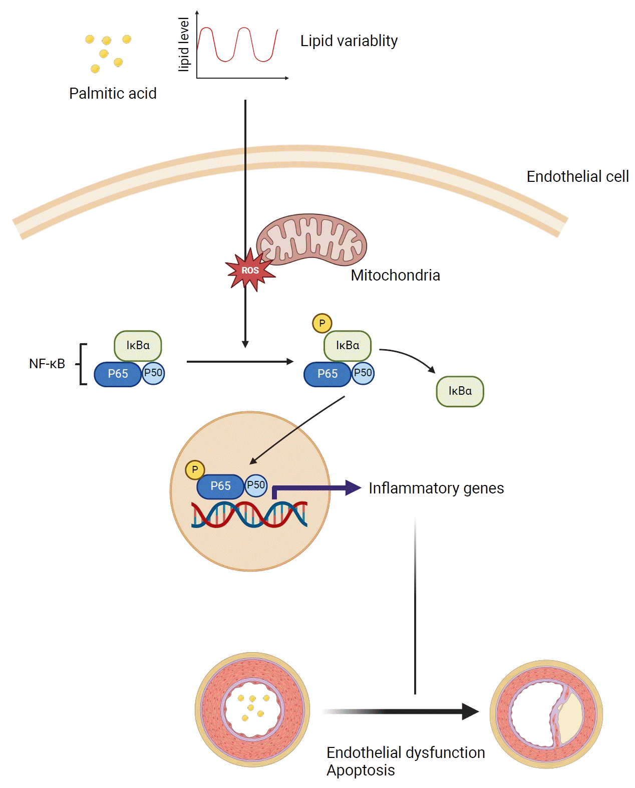
Endothelial inflammation is an early event of endothelial dysfunction. This led us to hypothesize that lipid level fluctuations may increase inflammatory signals in ECs. To test this hypothesis, we exposed HAECs and HUVECs to PA at concentrations of 0, 50, and 100 µM, or alternated between 0 and 100 µM every 8 hours (variability group) over a 48-hour period (Fig. 1A). PA treatment consistently upregulated inflammatory genes, such as Il6, Mcp1, Icam, Vcam, E-selectin, and iNos, while downregulating eNos in HAECs (Fig. 1B). Notably, Mcp1, Vcam, and Eselectin showed even greater upregulation in the variability group compared to the 100 µM PA-treated HAECs. Similar upregulation in inflammatory genes (Il6, Mcp1, Icam, Vcam, E-selectin) were observed in PA-treated HUVECs (Supplemental Fig. S1A). Corresponding to the transcriptomic changes, the protein levels of ICAM, VCAM, E-selectin, and iNOS were significantly increased in the variability group, surpassing the levels observed in the 50 µM PA-treated group and similar to the 100 µM PA-treated group (Fig. 1C, D). Similarly, levels of secreted inflammatory cytokines (IL6, MCP1) were substantially elevated in the variability group compared to the control and 50 µM PA-treated group (Fig. 1E). NF-κB is a transcription factor that mediates inflammatory reactions in various pathophysiological contexts [18]. To further explore the role of NF-κB signaling in the inflammatory response of ECs under different lipid exposure conditions, we conducted Western blot analysis and immunofluorescence staining. We observed an increase in phosphorylation of p65 and IκBα (p-p65/p65 and p-IκBα/IκBα) (Fig. 1F), along with the translocation of NF-κB p65 from the cytoplasm to the nucleus (Fig. 1G) in ECs exposed to lipid variability conditions. These findings suggest that lipid variability activates the NF-κB signaling pathway, leading to human endothelial inflammation.
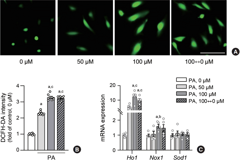
Because oxidative stress commonly underlies inflammation in various pathological contexts, we postulated that it could mediate endothelial inflammation triggered by lipid variability. To confirm whether oxidative stress increased in the lipid variability group, we measured the intracellular levels of ROS in human ECs using the DCFH-DA assay. The fluorescence intensity in the lipid variability group was higher than that of the control or 50 µM PA-treated group, and was comparable to the intensity observed in the 100 µM PA-treated group (Fig. 2A, B). Additionally, the transcriptomic level of heme oxygenase (Ho) 1 was elevated by variable lipid levels to a degree comparable to that of the 100 µM PA-treated group (Fig. 2C, Supplemental Fig. S1B).
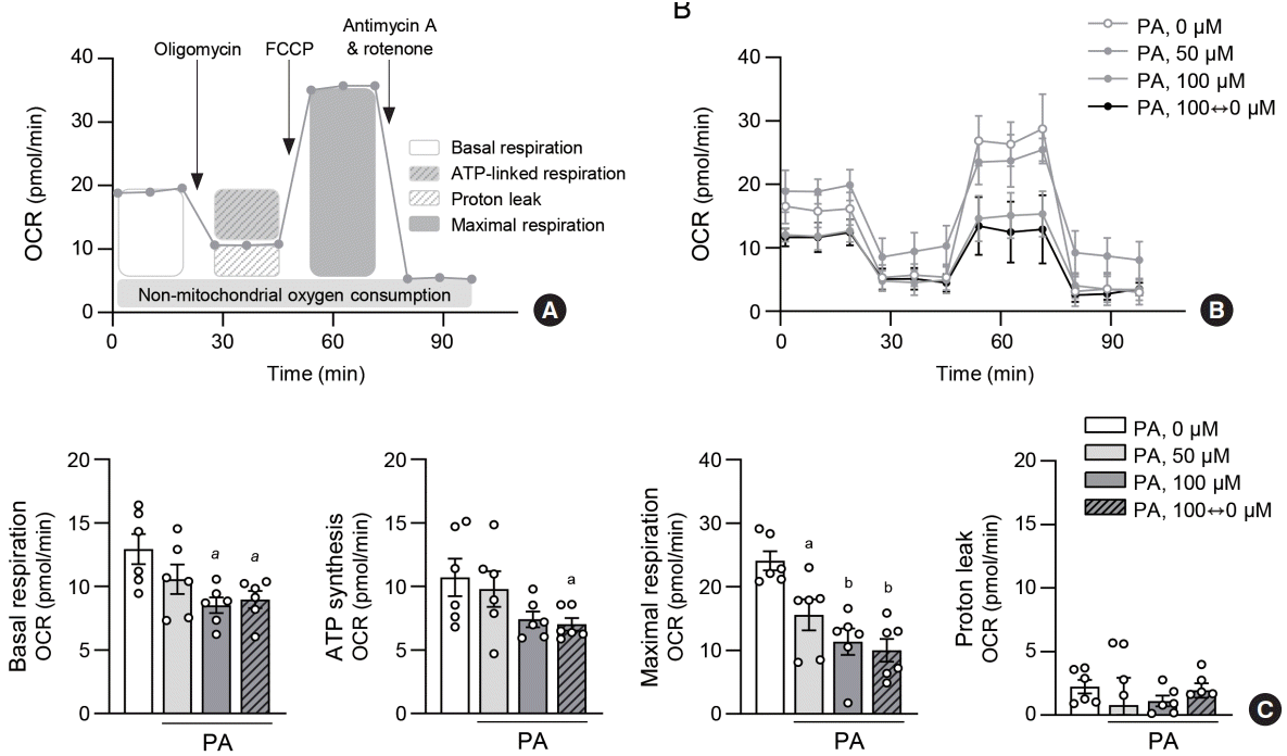
Increased ROS levels are often preceded by mitochondrial dysfunction in pathological conditions. To investigate the effects of lipid variability on mitochondrial metabolism in ECs, we examined the cellular oxygen consumption rate (OCR) of HAECs under various PA treatments (Fig. 3A). Cells incubated with 100 µM PA and alternating 0 and 100 µM PA showed a lower OCR compared to the control and 50 µM PA-treated groups (Fig. 3B). Mitochondrial respiration, including basal and maximal respiration, adenosine triphosphate synthesis, and spare respiratory capacity, was also reduced in the lipid variability group (Fig. 3C). These findings suggest that lipid variability induces mitochondrial dysfunction, leading to ROS accumulation in ECs.
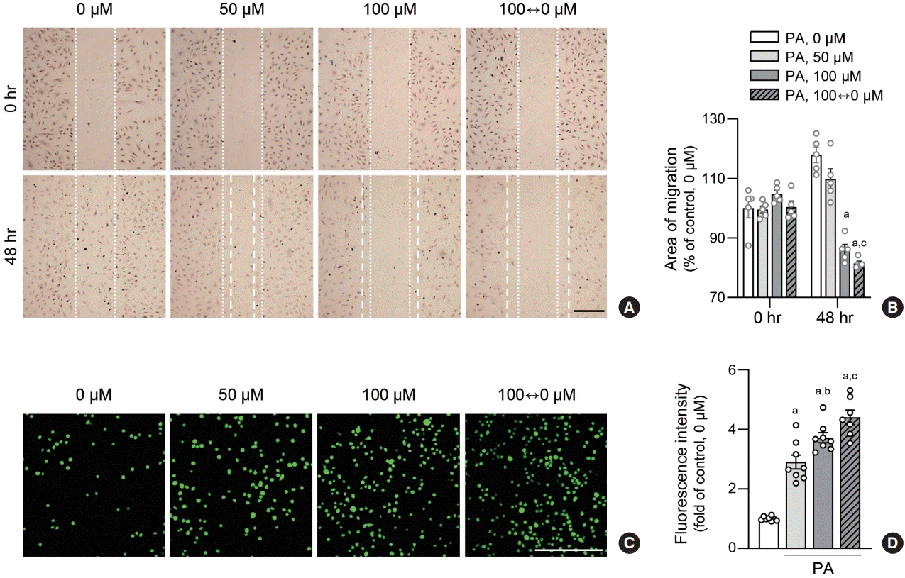
To evaluate whether increased inflammation translates to functional changes in ECs, we conducted wound healing assays on HAECs subjected to varying PA concentrations. Cell migration area was reduced in the lipid variability group compared to the control or 50 µM PA-treated group (Fig. 4A, B). This finding was corroborated by monocyte adhesion assays, where fluorescence-labeled THP1 cells exhibited increased adhesion to HAECs in the lipid variability group, indicating stimulation of monocyte adhesion due to lipid level fluctuations (Fig. 4C, D). These data indicated that lipid variability induces functional abnormalities in human ECs.
We investigated whether lipid variability induces cell death in human ECs. MTT assays demonstrated a decrease in viable cell counts owing to variable lipid levels (Fig. 5A, Supplemental Fig. S1C). The expression of the apoptotic signal BAX was upregulated, whereas that of BCL2 was downregulated in response to lipid variability (Fig. 5B). Overall, lipid variability reduced cell viability by triggering apoptotic signals in human ECs.
In this study, we demonstrated that lipid variability induces endothelial dysfunction by increasing oxidative stress and inflammatory signaling. By treating HAECs and HUVECs with various concentrations of PAs, we consistently demonstrated that variability in lipid levels triggers endothelial dysfunction.
Advancements in lipid-lowering agents have substantially reduced the incidence of cardiovascular diseases [19-23]. However, lipid variability has emerged as an important factor in cardiovascular disease. Despite accumulating clinical evidence [14-16,24], the precise mechanisms by which lipid variability leads to the development of atherosclerotic diseases remain elusive. To the best of our knowledge, this is the first study to delineate the mechanistic link between lipid variability and endothelial dysfunction.
Notably, the degree of alterations in inflammatory genes, endothelial dysfunction, and ROS were consistently amplified in the variability group compared to both the control and the 50 μM PA-treated groups. These changes were comparable to those observed in the 100 μM PA-treated group. Mcp1, Vcam, and E-selectin transcription level was even higher in the variability group compared with the 100 μM PA-treated group. This observation led us to speculate that fluctuations in lipid levels create a distinct milieu that intensifies ROS-driven inflammation and ultimately contributes to endothelial dysfunction.
This study had certain limitations that we were unable to address. Although we demonstrated that lipid variability induces ROS, inflammation, apoptosis, and functional impairment in ECs, we could not directly establish the links between ROS, inflammation, and endothelial dysfunction. The correlation between ROS levels, inflammation, and endothelial dysfunction has been extensively explored in previous studies [25]. In response to stimuli such as endotoxins and cytokines, ECs increase the NF-κB expression, which subsequently upregulates effectors such as VCAM-1, E-selectin, and ICAM [26-28]. This upregulation of effector proteins on the endothelial surface facilitates the recruitment of monocytes and T lymphocytes to the subendothelial region. Activated ECs, smooth muscle cells, monocytes/macrophages, and lymphocytes form a complex local milieu of cytokines, growth factors, and ROS within the arterial wall that perpetuates chronic inflammation and drives the progression of atherosclerotic lesions. In our study, the expression of MCP1, ICAM, VCAM, E-selectin, and eNOS increased in human ECs due to the fluctuation of lipid variability. ICAM, VCAM, E-selectin, and eNOS are part of the pro-inflammatory endothelial gene complex and play a critical role in endothelial dysfunction [25]. ICAM, VCAM, and E-selectin are cell adhesion molecules that participate in angiogenesis [29,30]. We speculated that this altered gene expression contributed to the outcomes observed in the wound healing assay. MCP1 recruits monocytes and macrophages to ECs [31]. The elevated secretion of MCP1 was consistent with observations made in the cell adhesion assay. eNOS catalyzes NO production from L-arginine, and its decreased expression is indicative of endothelial dysfunction. Collectively, our data provide information that establishes a link between lipid variability, inflammation, and endothelial dysfunction.
ROS, which include oxygen ions and peroxides, are highly reactive molecules that are often generated as byproducts of cellular metabolism [32]. Excessive ROS results in oxidative stress, which adversely affects cellular components including proteins, DNA, and lipids. Notably, ROS triggers redox-sensitive signaling pathways that stimulate the production of pro-inflammatory molecules, thus creating an inflammatory environment [25]. This cascade reduces NO levels and increases endothelial apoptosis [33,34]. Our data demonstrated increased ROS levels in ECs under various lipid conditions. We speculate that this could have provided a pro-inflammatory environment, leading to decreased eNOS expression and increased apoptosis in ECs. However, we admit that the ROS-independent mechanism may have contributed to lipid variability-mediated endothelial dysfunction.
In the present study, we focused solely on the effects of fluctuations in PA levels on ECs. Further investigation is necessary to confirm whether fluctuations of other types of lipid (e.g., LDL-C, high-density lipoprotein cholesterol, triglycerides, and apolipoproteins) exert similar effects on ECs. Furthermore, as our study was conducted in vitro, additional in vivo investigations are needed to corroborate the physiological relevance of our findings.
In conclusion, our study demonstrated that lipid variability induces endothelial dysfunction by intensifying inflammation and oxidative stress. We speculate that these data provide valuable translational insights into how fluctuations in lipid levels are associated with an increased risk of cardiovascular diseases. Future in vivo studies investigating the mechanistic links between various lipid types and endothelial dysfunction would improve the robustness of our findings.
ACKNOWLEDGMENTS
Human umbilical vein endothelial cells were kindly provided by Prof. Hyuk-Sang Kwon from The Catholic University of Korea.
This work was supported by the National Research Foundation of Korea (NRF) grant funded by the Korea government (MSIT) (2023R1A2C2004341 to Seung-Hwan Lee, 2021R1C1C1013 016 to Eun Young Lee), Ministry of Education (2022R1I1A1A 01068401 to Joonyub Lee), the Korea Health Industry Development Institute (KHIDI), funded by the Ministry of Health & Welfare, Republic of Korea (HI20C1234 to Joonyub Lee), and 2022 Research fund of Seoul St. Mary’s Hospital, The Catholic University of Korea (to Seung-Hwan Lee).
REFERENCES
1. Eelen G, de Zeeuw P, Treps L, Harjes U, Wong BW, Carmeliet P. Endothelial cell metabolism. Physiol Rev. 2018; 98:3–58.
2. Ghosh A, Gao L, Thakur A, Siu PM, Lai CW. Role of free fatty acids in endothelial dysfunction. J Biomed Sci. 2017; 24:50.
3. Pober JS, Sessa WC. Evolving functions of endothelial cells in inflammation. Nat Rev Immunol. 2007; 7:803–15.
4. Gimbrone MA Jr, Garcia-Cardena G. Vascular endothelium, hemodynamics, and the pathobiology of atherosclerosis. Cardiovasc Pathol. 2013; 22:9–15.
5. Mortensen MB, Dzaye O, Botker HE, Jensen JM, Maeng M, Bentzon JF, et al. Low-density lipoprotein cholesterol is predominantly associated with atherosclerotic cardiovascular disease events in patients with evidence of coronary atherosclerosis: the Western Denmark Heart Registry. Circulation. 2023; 147:1053–63.
6. Kannel WB, Dawber TR, Kagan A, Revotskie N, Stokes J 3rd. Factors of risk in the development of coronary heart disease: six year follow-up experience. The Framingham Study. Ann Intern Med. 1961; 55:33–50.
7. Prospective Studies Collaboration, Lewington S, Whitlock G, Clarke R, Sherliker P, Emberson J, et al. Blood cholesterol and vascular mortality by age, sex, and blood pressure: a meta-analysis of individual data from 61 prospective studies with 55,000 vascular deaths. Lancet. 2007; 370:1829–39.
8. Zhang Y, Pletcher MJ, Vittinghoff E, Clemons AM, Jacobs DR Jr, Allen NB, et al. Association between cumulative low-density lipoprotein cholesterol exposure during young adulthood and middle age and risk of cardiovascular events. JAMA Cardiol. 2021; 6:1406–13.
9. Nordestgaard BG. Triglyceride-rich lipoproteins and atherosclerotic cardiovascular disease: new insights from epidemiology, genetics, and biology. Circ Res. 2016; 118:547–63.
11. Hokanson JE, Austin MA. Plasma triglyceride level is a risk factor for cardiovascular disease independent of high-density lipoprotein cholesterol level: a meta-analysis of population-based prospective studies. J Cardiovasc Risk. 1996; 3:213–9.
12. Chen X, Liu L, Palacios G, Gao J, Zhang N, Li G, et al. Plasma metabolomics reveals biomarkers of the atherosclerosis. J Sep Sci. 2010; 33:2776–83.
13. Feinleib M. Seven countries: a multivariate analysis of death and coronary heart disease. JAMA. 1981; 245:511–2.
14. Kim MK, Han K, Kim HS, Park YM, Kwon HS, Yoon KH, et al. Cholesterol variability and the risk of mortality, myocardial infarction, and stroke: a nationwide population-based study. Eur Heart J. 2017; 38:3560–6.
15. Bangalore S, Breazna A, DeMicco DA, Wun CC, Messerli FH; TNT Steering Committee and Investigators. Visit-tovisit low-density lipoprotein cholesterol variability and risk of cardiovascular outcomes: insights from the TNT trial. J Am Coll Cardiol. 2015; 65:1539–48.
16. Boey E, Gay GM, Poh KK, Yeo TC, Tan HC, Lee CH. Visitto-visit variability in LDL- and HDL-cholesterol is associated with adverse events after ST-segment elevation myocardial infarction: a 5-year follow-up study. Atherosclerosis. 2016; 244:86–92.
17. Rhee M, Kim JW, Lee MW, Yoon KH, Lee SH. Preadipocyte factor 1 regulates adipose tissue browning via TNF-αconverting enzyme-mediated cleavage. Metabolism. 2019; 101:153977.
18. Liu T, Zhang L, Joo D, Sun SC. NF-κB signaling in inflammation. Signal Transduct Target Ther. 2017; 2:17023.
19. Cholesterol Treatment Trialists’ (CTT) Collaboration, Baigent C, Blackwell L, Emberson J, Holland LE, Reith C, et al. Efficacy and safety of more intensive lowering of LDL cholesterol: a meta-analysis of data from 170,000 participants in 26 randomised trials. Lancet. 2010; 376:1670–81.
20. Sabatine MS, Giugliano RP, Keech AC, Honarpour N, Wiviott SD, Murphy SA, et al. Evolocumab and clinical outcomes in patients with cardiovascular disease. N Engl J Med. 2017; 376:1713–22.
21. Rubins HB, Robins SJ, Collins D, Fye CL, Anderson JW, Elam MB, et al. Gemfibrozil for the secondary prevention of coronary heart disease in men with low levels of high-density lipoprotein cholesterol. Veterans Affairs High-Density Lipoprotein Cholesterol Intervention Trial Study Group. N Engl J Med. 1999; 341:410–8.
22. Amini M, Zayeri F, Salehi M. Trend analysis of cardiovascular disease mortality, incidence, and mortality-to-incidence ratio: results from global burden of disease study 2017. BMC Public Health. 2021; 21:401.
23. Kim K, Ginsberg HN, Choi SH. New, novel lipid-lowering agents for reducing cardiovascular risk: beyond statins. Diabetes Metab J. 2022; 46:517–32.
24. Park J, Kang M, Ahn J, Kim MY, Choi MS, Lee YB, et al. Mean and variability of lipid measurements and risk for development of subclinical left ventricular diastolic dysfunction. Diabetes Metab J. 2022; 46:286–96.
25. Gimbrone MA Jr, Garcia-Cardena G. Endothelial cell dysfunction and the pathobiology of atherosclerosis. Circ Res. 2016; 118:620–36.
26. Bevilacqua MP, Pober JS, Mendrick DL, Cotran RS, Gimbrone MA Jr. Identification of an inducible endothelial-leukocyte adhesion molecule. Proc Natl Acad Sci U S A. 1987; 84:9238–42.
27. Gahmberg CG, Tolvanen M, Kotovuori P. Leukocyte adhesion: structure and function of human leukocyte beta2-integrins and their cellular ligands. Eur J Biochem. 1997; 245:215–32.
31. Maus U, Henning S, Wenschuh H, Mayer K, Seeger W, Lohmeyer J. Role of endothelial MCP-1 in monocyte adhesion to inflamed human endothelium under physiological flow. Am J Physiol Heart Circ Physiol. 2002; 283:H2584–91.
32. Taniyama Y, Griendling KK. Reactive oxygen species in the vasculature: molecular and cellular mechanisms. Hypertension. 2003; 42:1075–81.