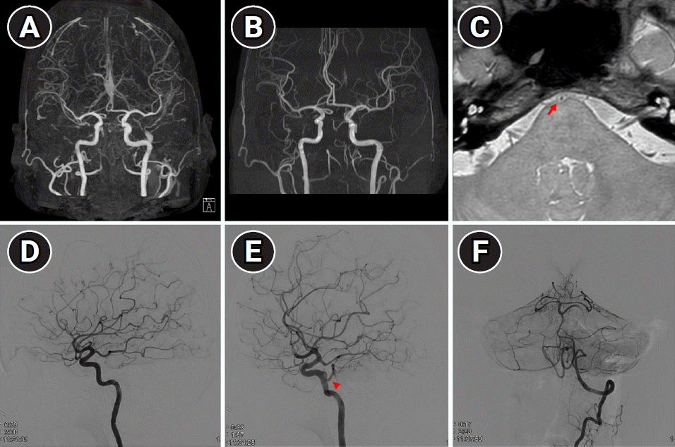
A 55-year-old woman presented with acute memory impairment, initially suspected as transient global amnesia. To rule out alternative causes, she underwent diffusion-weighted imaging (DWI) with time-of-flight magnetic resonance angiography (TOF-MRA) and computed tomography angiography (CTA). While the DWI showed no abnormalities, the basilar artery was not visualized in either CTA or TOF-MRA (Fig. 1A and B). Due to the non-visualized basilar artery, she was admitted for further investigation.
High-resolution magnetic resonance imaging (Fig. 1C) and conventional angiography (Fig. 1D-F) were subsequently performed. These examinations revealed a hypoplastic basilar artery with bilateral fetal posterior cerebral arteries (PCAs) and a left persistent primitive trigeminal artery (PPTA). Since basilar artery patency was confirmed, the patient was discharged without any interventions.
The incidence of PPTA is very low, with one Japanese cohort finding a mere 0.68% occurrence [1]. Among these cases, approximately 28% were presented with severe basilar artery hypoplasia. In our case, the presence of bilateral fetal PCAs further accentuated the hypoplastic basilar artery, creating an occlusion-like appearance. Notably, the proton density-weighted sequence in high-resolution magnetic resonance imaging helped differentiate occlusion from hypoplasia. Given the invasive nature of cerebral angiography, utilizing high-resolution magnetic resonance imaging in similar clinical situations could be a prudent alternative.
Notes
Fig. 1.
Multiple images of brain magnetic resonance imaging and conventional angiography. Computed tomography angiography (A) and time-of-flight magnetic resonance imaging (B) showed a non-visualized basilar artery. High-resolution magnetic resonance imaging (C) revealed hypoplastic basilar artery at the level of pons (red arrow). (D-F) Conventional angiography images demonstrate fetal-type posterior cerebral arteries and left persistent primitive trigeminal artery (red arrowhead).


 PDF
PDF Citation
 Print
Print


 XML Download