Abstract
In the age of globalization of infectious diseases, qualified personnel is needed for the diagnosis of parasitic diseases in the laboratory. This review aimed to introduce the methods for stool examination and identification of helminth eggs for the diagnosis of helminthic infections in laboratory and field surveys. The formalin-ether sedimentation technique (FEST) and the Kato-Katz egg counting technique (KKECT) are mainly described as representative stool examinations. The FEST is somewhat complicated and troublesome, but it is useful for differentiating small trematode eggs from opisthorchiid and heterophyid flukes. KKECT is useful in field surveys of large populations in areas endemic for soil-transmitted helminthiases. Helminth eggs are divided into four groups based on the presence or absence of the operculum and embryo. The eggs of Clonorchis sinensis and heterophyid flukes including Metagonimus spp. are relatively small and contain an operculum and an embryo (miracidium). Meanwhile, eggs of diphyllobothriid tapeworms, echinostomatid flukes, Paragonimus westermani, Fasciola hepatica, and Fasciolopsis buski are relatively larger, operculated, and contain germ cells and yolks instead of the embryo. The eggs of cyclophyllidian tapeworms, Taenia spp. and Hymenolepis spp., and blood flukes, Schistosoma spp., are embryonated but do not have an operculum. Nematode eggs have no operculum and embryo, but those of hookworms and pinworms sometimes have developed larvae inside. This review provides valuable insights into the methods of stool examination and helminth egg identification for the diagnosis of helminthic infections in the laboratory and field surveys.
Helminths are commonly referred to as worms, worm parasites, or intestinal worms. Many worm parasite are members of this animal group. Taxonomically, there are five phyla related to human parasites in the helminth group: Nemathelminthes, Platyhelminthes, Nematomorpha, Acanthocephala, and Anellnida. However, members of 3 classes, Nematoda, Trematoda, and Cestoidea in the phyla Nemathelminthes and Platyhelminthes, are considered medically important groups. Members of Nematoda mainly have a life cycle including both sexes, male and female worms, and produce sexually mature eggs from adult females after mating; however, Trematoda and Cestoidea are hermaphroditic, and their eggs are commonly discharged from fully mature adults [1–3]. Parasites, especially helminths, have the instinct for species preservation and continuously discharge their eggs from their habitats into the environment to maintain their life cycle. Therefore, helminth eggs detected from various samples are a crucial clue for some circumstances, such as helminthic infections in humans and animals, contamination in the soil of playgrounds, and endemic evidence in ancient times in archeoparasitology [4–6].
In the Republic of Korea (Korea), soil-transmitted helminthiases (STH), including ascariasis, trichuriasis, and hookworm infections, were widely prevalent until the 1970s and were the main public health threat to be solved at that time. However, their prevalence has decreased remarkably because of systematic nationwide control programs [7]. Helminthic diseases are no longer a health problem. However, the prevalence of foodborne helminthiases (FBH), including clonorchiasis and metagonimiasis, is still endemic in the Korean riverside areas. The eradication project for FBH has been continuously performed by the Division of Vectors and Parasitic Diseases of the Korea Disease Control and Prevention Agency (DCPA) [8–12]. In this eradication project, the exact identification of small trematode eggs (STE) is necessary to differentiate between liver flukes (Clonorchis sinensis) and intestinal heterophyid flukes including Metagonimus spp. in fecal specimens. The exact identification of STE is also necessary in the individual stool examination performed in the diagnosis department in hospitals, the Department of Parasitology and Tropical Medicine in Medical Colleges, the Diagnosis Laboratory in the Korea Association of Health Promotion (KAHP), and the Division of Vectors and Parasitic Diseases of the DCPA [12–14]. Therefore, in the context of helminthic infections in Korea, the formalin-ether sedimentation technique (FEST) should mainly be adopted for stool examinations by well-trained laboratory personnel.
In contrast, non-governmental organizations (NGO), including the KAHP, have been used to control parasites, especially helminths, in endemic countries of Southeast Asia and Africa. Although the target parasites to be controlled in the projects differed by survey subject and region, most of the surveys were performed using the Kato-Katz egg counting technique (KKECT). This method has been very useful in helminth control projects, especially soil-transmitted helminths such as Ascaris lumbricoides, Trichuris trichiura, and hookworms, in endemic countries of Southeast Asia and Africa [15–19].
Laboratory diagnosis of helminthic diseases is performed in two steps, detection and identification, during stool examination by trained laboratory personnel. Detection methods and skills are important for the accurate diagnosis of helminthic infections in stool examinations. The ability to recognize parasites, especially helminth eggs, should be trained and is ultimately needed to differentiate them from non-parasite artifacts for the exact identification of helminth eggs through microscopy [20,21]. In contrast, Palmieri et al. [22] emphasized the importance of parasitology education, especially the training to identify and diagnose parasitic infections, as an emerging need. These authors also mentioned the increasing need for well-trained and qualified healthcare professionals in the laboratory to diagnose parasitic diseases due to the globalization of infectious diseases. Therefore, in this review, we focused on two topics: methods for stool examination and the identification of helminth eggs.
Stool examinations are usually performed to diagnose parasitic infections (helminths and protozoa). Examination of the infection status of helminths is largely divided into two methods, qualitative and quantitative. A qualitative method examines prevalence (infection rate) and is divided into two methods: smear (direct smear and cellophane thick smear) and concentration methods (sedimentation and flotation techniques). The cellophane thick smear (CTS), also known as Kato's method, has been popularly adopted in field surveys of large populations [15–19]. The concentration method, especially the FEST, is applied to small-scale surveys or individual examinations because their procedures are complicated and timeconsuming. Quantitative methods check the intensity of infection (worm burden) and are only used to estimate the number of worms infecting a certain person or population. As representative quantitative techniques, Stoll’s dilution egg counting and KKECT are recommended; however, the former method is complicated and troublesome. Therefore, in this review, we focus on describing the procedures for the FEST and KKECT.
Direct smears are commonly used to detect the trophozoites of Entaemeba histolytica and Giardia lamblia from the diarrheic stools of acute-phase patients. This method is not useful for examining helminthic infections because the egg detection rate is very low, except for diphyllobothiid tapeworms.
The cellophane thick smear (Kato’s method) is performed using a cellophane (No. 600) strip pre-soaked in Kato’s solution instead of a cover slip. Kato’s solution is a mixture of glycerin (500 mL), distilled water (DW, 500 mL), and 3% malachite green (recently, methylene blue has been highly recommended) solution (5 mL). This method is discussed in detail in the quantitative technique.
The Faust’s zinc sulfate flotation technique is commonly used to detect cysts and/or oocysts of intestinal protozoa, such as E. histolytica, G. lamblia, and Cryptosporidium parvum, in the stool of patients in the chronic phase. It is not suitable for the diagnosis of helminthic infections because the specific gravity of helminth eggs is relatively high.
FEST is somewhat complicated and troublesome; however, it is better than the Kato's method for the exact identification of helminth eggs, particularly the STE of opisthorchiid and heterophyid flukes.
FEST procedure (Supplementary Fig. 1 in the online-only Data Supplement)
1. Sample a thumb head-size amount of feces into a beaker with DW.
2. Stir with a wooden applicator to form a stool suspension.
3. Filter the stool suspension through two layers of wet gauze.
4. Centrifuge the conical tip tube with the filtered stool suspension at 500 × g for 3 min.
5. Discard the supernatant in a tube.
6. Add 10% formalin (10 mL) and ether (3 mL) to the tube.
7. Shake the tube vigorously for 15 s after placing a rubber stopper or cap on it.
8. Centrifuge the tube at 500 × g for 3 min.
9. Gently remove the plug of debris and discard the supernatant.
10. Drop the sediment onto the slide glass.
11. Cover the glass slip (cover slip) and examine it under a light microscope.
In stool examinations using FEST in FBH-endemic riverside areas of Korea (2011–2023), the prevalence of clonorchiasis was the highest (4.8%), followed by heterophyid fluke infection (1.5%), trichuriasis (0.2%), and Gymnophalloides seoi infection (0.06%) (Table 1) [12,13]. Results of stool examinations with FEST in health checkups in the KAHP revealed a similar pattern, with a higher prevalence of FBH, such as clonorchiasis (1.5%) and heterophyid fluke infections (0.34%), including metagonimiasis, compared to STH, that is, ascariasis and trichuriasis (0.13%) (Table 2) [14]. These findings suggest that FEST is the gold standard for the diagnosis of helminthic infections, including clonorchiasis and metagonimiasis, in Korea.
1. Fill a 0.1 N NaOH or KOH solution into the 56 mL scale of a Stoll’s flask (Supplementary Fig. 2 in the online-only Data Supplement).
2. Add the fecal sample in the Stoll’s flask to a volume of 60 mL (1 g of feces is dissolved in 15 mL of fecal solution).
3. Sample the fecal solution (0.15 or 0.075 mL) with a Stoll pipette after shaking the flask, and drop it onto the slide glass when the feces are completely dissolved in the solution.
4. Cover the glass slip, examine the fecal specimen under a light microscope, and count the number of eggs.
5. EPG (No. eggs per gram of feces), n (correction factor for the condition of the stool) × No. of eggs × 100 (200 for the 0.075 mL sample)
6. EPD (No. eggs per day): EPG × whole-day stool (200 g in adults)
7. EPDPF (EPDPW), No. of eggs per day per female (worm)
Ascaris lumbricoides: 200,000, Trichuris trichiura: 5,000, hookworms: 10,000, Clonorchis sinensis: 3,000.
8. No. of worms: EPD/EPDPF (EPDPW)
Materials (Supplementary Fig. 3 in the online-only Data Supplement)
1. Plastic template: 50 mg (hole: 9 mm diameter × 1 mm thick). 41.7 mg (hole: 6 mm in diameter × 1.5 mm thick), 20 mg (hole: 6.5 mm in diameter × 0.5 mm thick).
2. Plastic spatula.
3. 60–105 nylon mesh.
4. Slide glass.
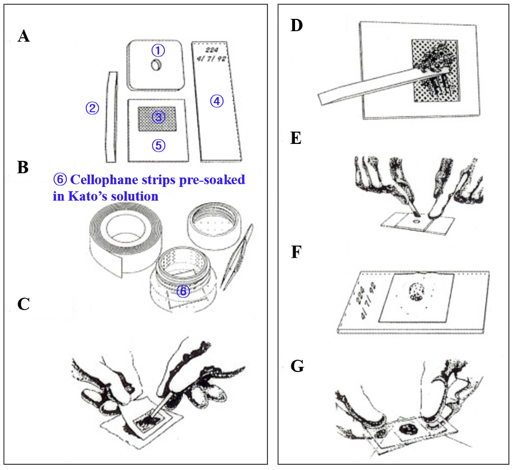
5, Newspaper (Fig. 1A).
6. Cellophane: 40–50 µm thick (25 × 30 mm), flat-bottom jar with lid, and forceps (Fig. 1B).
1. Place a small amount of fecal sample on a newspaper and press the nylon mesh on top so that some of the feces are sieved through the mesh and accumulate on top (Fig. 1C).
2. Scrape the flat-sided spatula across the upper surface of the mesh to collect the sieved feces (Fig. 1D).
3. Place the template at the center of a glass slide and add feces from the spatula to completely fill the hole (Fig. 1E).
4. Remove the template carefully so that the mound of feces remained on the slide.
5. Cover the fecal mound with the presoaked cellophane strip (Fig. 1F).
6. Invert the slide and firmly press the fecal sample against cellophane strips to evenly spread the feces (Fig. 1G).
7. Maintain CTS specimens at room temperature for more than 1 h for clearance (Supplementary Fig. 3 in the online-only Data Supplement).
8. Examine the fecal specimen under a light microscope and count the number of eggs.
9. EPG: No. of eggs × 20 (50 mg template), No. eggs × 24 (41.7 mg template).
10. EPD = EPG × whole-day stool amount (200 g in adults)
11. No. of the infected worms: EPD/EPDPF (EPDPW)
Control and elimination programs for helminthiases have been performed using the Kato-Katz thick smear technique by NGOs including KAHP, the Korea International Cooperation Agency, and the Korea Foundation for International Healthcare in endemic countries (Myamar, Cambodia, and Lao PDR) in Southeast Asia [15–18]. The individual prevalence of helminthiasis differed between the survey regions and subjects. However, in these programs, STHs, such as ascariasis (17.7%), trichuriasis (14.0%), and hookworm infections (14.9%), were the main target diseases, and FBHs, that is opisthorchiid and heterophyid fluke infections (12.1%), were additional health problems (Table 3). Eom et al. [17] conducted a Korea-Laos collaborative project for the control of foodborne trematode infections in Lao PDR (2007–2011). They examined 6,178 fecal samples using a Kato-Katz thick smear and Stoll’s egg counting technique to determine the infection status of helminths. The prevalence of STE was the highest (55.6%), followed by hookworm infection (27.8%), trichuriasis (6.5%), and ascariasis (3.7%).
In contrast, McCarthy et al. [23] reviewed the diagnostic technologies relevant to the control of helminth infections, either available or under development. They also discussed the critical gaps to be identified and the opportunities to improve diagnostic technologies for control and elimination programs as a research agenda for helminthic diseases in humans. Some research groups have adopted new devices and kits to efficiently detect helminth eggs. Boonyong et al. [24] compared the detection performance of a fully automatic digital feces analyzer (Orienter Model FA280) with that of formalin-ethyl acetate concentration technique. Lefaure et al. [25] evaluated the detection efficacy of the Parasep® device, which was adapted to the Faust’s flotation technique, with conventional techniques. Sarirah et al. [26] evaluated the detection efficacy of the MiniFLOTAC and compared it with that of KKECT, a quantitative technique recommended by the World Health Organization (WHO). Khanna et al. [27] also evaluated the detection rate of Mini Parasep® solvent-free method in the rapid diagnosis of intestinal helminthiases through comparing it with that of formalin-ethyl acetate sedimentation technique (FEAST, has been recently recommended instead of FEST by WHO) [28]. Recently, the Korea DCPA developed the ParaEgg kit and adopted it for stool examinations in the FBHendemic riverside areas of Korea (personal communication). The detection efficiency of new devices or kits is promising compared to that of conventional techniques in stool examinations; however, some limitations, such as sensitivity, specificity, practical difficulty, cost, and infrastructure demand, need to be solved preemptively for their widespread use.
The cellotape anal swab is used for the diagnosis of pinworm, Enterobius vermicularis, and sometimes beef tapeworm, Taenia saginata, infections.
The identification of helminth eggs is somewhat difficult, even for trained laboratory personnel. However, the helminth eggs are similar to the avian eggs. They commonly have hard chitinous shells, germ cells, yolks, and sometimes embryos (Supplementary Fig. 4 in the online-only Data Supplement), but they have different compositions and unique morphologies according to the helminth species. Each species of helminth eggs is differentiated according to their unique morphology and size. In this review, we discuss the morphologies and differential keys for the identification of helminth eggs from human fecal samples.
The eggs of the opisthorchiid fluke (C. sinensis) and heterophyid flukes including Metagonimus spp. are relatively small and include an operculum and an embryo (miracidium).
Clonorchis sinensis: Ovoid and sesame seed-shaped with a miracidium inside, 27–35 × 15–20 µm in size, have a dominantly developed (conspicuous) operculum and shoulder rim, a shell surface with muskmelonlike wrinkles, and maximum width in the posterior 1/3 level (Fig. 2A, 2 B).
Heterophyids: Ovoid in shape with a miracidium inside, commonly having an inconspicuous operculum and shoulder rim, smooth shell surface without muskmelon wrinkle pattern, and maximum width in the median (Fig. 2C–2F). More than 11 species, including Metagonimus spp., Heterophyes nocens, Heterophyopsis continua, Stellantchasmus falcatus, Pygidiopsis summa, Centrocestus armatus, Stictodora fuscata, Stictodora lari, and Acanthotrema felis, have been reported in Korea. They are divided into 3 groups according to their lengths: < 25 µm, 26–30 µm, and > 31µm.
In the CTS specimens, the eggs of opisthorchiid flukes are indistinguishable from those of heterophyid f lukes including Metagonimus spp. (Supplementary Fig. 5 in the online-only Data Supplement). However, they can be easily differentiated based on the dominantly developed operculum and muskmelon surface appearance in FEST fecal specimens. Unlike opisthorchiid flukes, the eggs of heterophyid flukes had a smooth shell surface.
Stictodora fuscata: Ovoid with a miracidium inside, larger (34–38 × 20–23 µm) than other heterophyid f luke eggs, with thick shells, which are sometimes dark brown in color (Fig. 2F).
Plagiorchis muris: Elliptical with a miracidium inside, 32–38 × 20–24 µm in size, thick shell, conspicuous operculum, and sometimes an abopercular knob (Fig. 2G).
Eurytrema pancreaticum: Oval with a miracidium inside, 48–53 × 25–33 µm in size, thick shell, inconspicuous operculum, dark brown in color (Fig. 2H).
Gymnophalloides seoi: Very small with a miracidium inside, 20–25 × 11–15 µm in size, very thin shell, and large operculum (Fig. 2I).
The eggs of Diphyllobothrium spp., echinostomatid flukes, Neodiplostomum seoulense, Paragonimus westermani, Fasciola hepatica, and Fasciolopsis buski are relatively larger in size and have an operculum, but do not have an embryo. They consist of germ cells and yolk inside.
Echinostomatids: The echinostomes infecting humans belong to 4 species, Echinostoma cinetorchis (99–116 × 66–76 µm), Isthmiophora hortensis (127–139 × 71–81 µm), Echinochasmus japonicus (76–87 × 52–63 µm), and Acanthoparyphium tyosenense (78–93 × 63–73 µm), have been reported in Korea. Echinostomatid eggs differentiate from other fluke eggs in this group by the presence of abopercular wrinkles, and are roughly divided into 2 groups based on their average length: < 100 µm and > 101 µm. They are commonly oval in shape, with a thin shell, a small operculum, and abopercular wrinkles (Fig. 3A–3D).
Paragonimus westermani: Elliptical and asymmetrical in shape, 80–100 × 45–65 µm in size, have a wide operculum, shoulder rim, and abopercular thickening (Fig. 3E).
Neodiplostomum seoulense: Elliptical and asymmetrical in shape, 86–99 × 55–63 µm in size, have thin shell, and an inapparent operculum (Fig. 3F).
Fasciola hepatica: Very big, 145 × 80 µm in average size, with thin shell, and inconspicuous smaller operculum (Fig. 3G).
Fasciolopsis buski: Very big, 147 × 75 µm in average size, with thin shell, and inconspicuous smaller operculum.
Diphyllobothrium spp.: Ovoid and symmetrical in shape, 58–76 × 40–51 µm in size, with a thick shell, and smaller operculum, and sometimes with an abopercular knob (Fig. 3H).
The eggs of cyclophyllidian tapeworms, such as Taenia spp. and Hymenolepis spp., and blood flukes, Schistosoma spp., are embryonated, but they do not have an operculum.
Taenia spp.: Round, 30–45 µm in diameter size, with thick and striated embryophore, and have a hexacanth embryo. Three species, Taenia asiatica, T. saginata, and T. solium, are not microscopically differentiable at the species level (Fig. 4A).
Hymenolepis nana: Round, 30–47 µm in diameter size, with transparent shell, and have a hexacanth embryo with polar filaments (Fig. 4B, 4C).
Hymenolepis diminuta: Round, 70–80 µm in diameter size, with thick shell, and have a hexacanth embryo with polar thickenings (Fig. 4D).
Schistosoma japonicum: Ovoid, 70–100 × 50–65 µm in size, with thin shell, a small lateral spine and a miracidium inside.
Schistosoma mansoni: Long, elliptical, 114–175 × 45–68 µm in size, with thin shell, a stout lateral spine, and a miracidium inside.
Schistosoma haematobium: Long, elliptical, 112–170 × 40–70 µm in size, with thin shell, a terminal spine, and a miracidium inside.
The nematode eggs have no operculum and embryo, but those of hookworms and pinworms are sometimes observed in embryonated stages.
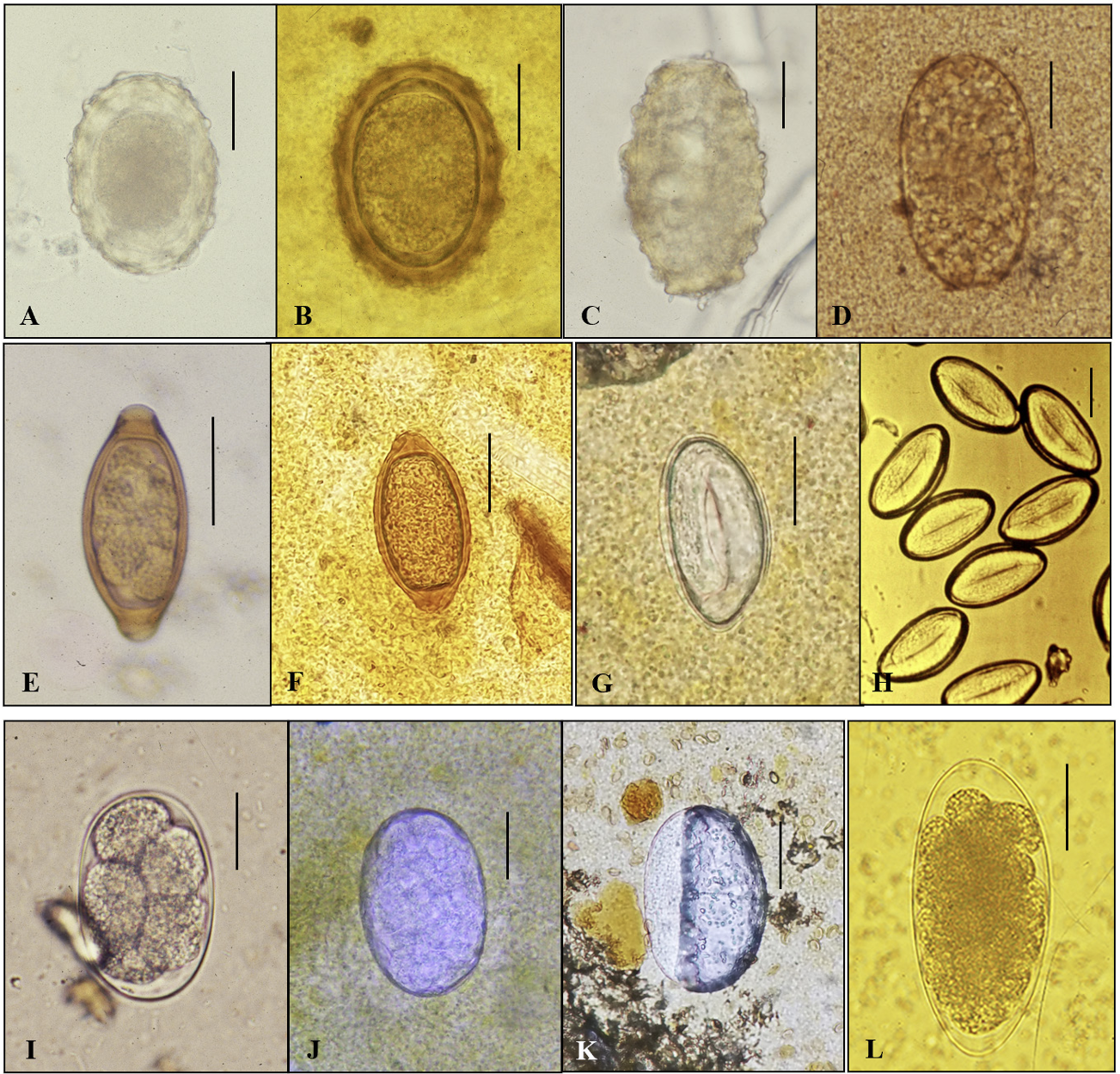
Ascaris lumbricoides (F): Subspherical or round, 50–75 × 35–50 µm in size, with 3 layered shells: outer mammilated protein coat, middle chitinous, and inner lipoid layers. In fresh stool, they retain a single blastomere (Fig. 5A, 5 B). They sometimes develop in CTS specimens as 2-, 4-, 8-, 16-, 32-cells, and morular stages in a warm environment and rarely have a decorticated (no protein coat) appearance. Fertilized eggs of A. lumbricoides are clearly differentiated from toxocarid (Toxocara spp.) eggs from cats and dogs by their morphological shape, being nearly round with a thinner shell, but nearly identical to those of Ascaris suum from pigs.
Ascaris lumbricoides (U): Long, elliptical, 80–90 × 40–45 µm in size, thin shell with poorly developed protein coat and granules inside (Fig. 5C, 5D). A variety of shapes are frequently observed in the CTS specimens (Supplementary Fig. 6 in the online-only Data Supplement). Characteristic broken shell fragments with a protein coat and internal granules are helpful in identifying unfertilized A. lumbricoides eggs.
Trichuris trichiura: Barrel shape, 50–55 × 22–23 µm in size, with thick shell and polar mucoid plugs on both sides and a single blastomere in fresh stool specimens (Fig. 5E, 5F).
Recently, two types (standard- and large-sized) of whipworm (T. trichiura) eggs from school children in Yangon, Myanmar were molecularly confirmed to be the same species. The size of standard-eggs was 52–58 × 21–28 μm and that of large- eggs was 65–76 × 30–35 μm in the study by Ryoo et al. [29]. The large eggs were slightly smaller than the known size of Trichuris vulpis (dog whipworm). Some parasitologists strongly suspect T. vulpis eggs in CTS specimens from prevalent countries, such as Lao PDR, Cambodia, and Myanmar.
Enterobius vermicularis: Persimmon seed shape, 50–60 × 20–30 µm in size, with double-layered shell and larva. Pinworm eggs are detected in the cellotape anal swab specimens but are sometimes found in the fecal specimens of CTS (Fig. 5G, 5H).
Hookworms: Ovoid, 55–75 × 35–45 µm in size, have hyaline thin shells with blunting of both ends, and 4–8 cell stage in the fresh fecal specimen (Fig. 5I). They rapidly develop and are embryonated in warm environments, and sometimes show an air-bubble-like appearance (Fig. 5J, 5 K). In this case, they are difficult to recognize. To solve this problem, the microscope should be finely adjusted. The definite and continuous demarcation of egg shells may be helpful in the diagnosis of hookworm eggs. As humaninfecting hookworms, 3 species, i.e., Ancylostoma duodenale, A. cylanicum, and Necator americanus, have been reported.
Trichostrongylus orientalis: Long, elliptical, 75–95 × 45–50 µm in size, with a thin hyaline shell and a morular stage inside (Fig. 5L). These nematode eggs are easily differentiated from hookworm eggs by their large size, one pointed end (both blunted ends in hookworms), and the internal morular stage.
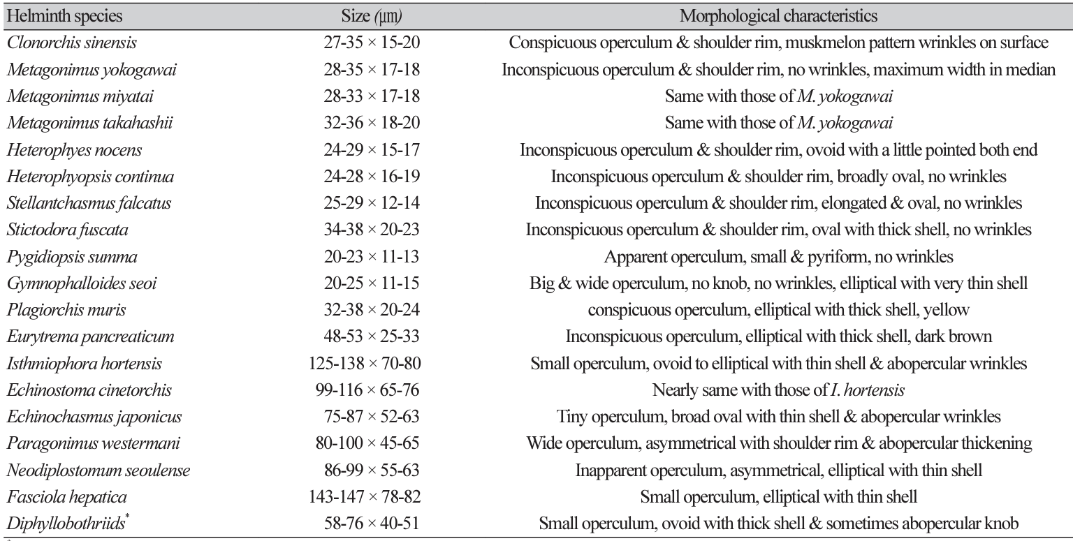
The morphological characteristics of helminth eggs are summarized in Tables 4 and 5.
Helminth eggs with an operculum ............................................................................................................................. 1
Helminth eggs without an operculum .................................................................................................................... 10
1. Operculated eggs with a developed embryo (miracidium) ............................................................................... 2
Operculated eggs with a germ cell and yolks ...................................................................................................... 7
2. Egg shell rough with muskmelon appearance .................................................................... Clonorchis sinensis
Egg shell smooth, no muskmelon appearance .................................................................................................... 3
3. Very small (< 25 µm in length), big operculum, thin, and transparent shell ............. Gymnophalloides seoi
Small with thick shell ............................................................................................................................................... 4
4. Very small (< 23 µm in length), with a prominent operculum ......................................... Pygidiopsis summa
Small (>25 µm in length) ........................................................................................................................................ 5
5. Small (25–35 µm in length) ............................................................................................................. Heterophyids*
Small (> 35 µm in length) with thick shell ........................................................................................................... 6
6. Small (35–38 µm in length) with thick shell .......................................................................... Stictodora fuscata
Medium (45–50 µm in length) with thick dark shells .............................................. Eurytrema pancreaticum
*Metagonimus spp., Heterophyes nocens, Heterophyiopsis continua, Stellantchasmus falcatus, Centrocestus armatus, Stictodora lari, and Acanthotrema felis.
7. Unembryonated eggs with abopercular wrinkles .................................................................. Echinostomatids#
Unembryonated eggs without abopercular wrinkles ......................................................................................... 8
8. Very big (> 140 µm in length) in size, thin shell, small or inconspicuous operculum ............................................................................................. Fasciola spp. and Fasciolopsis buski
Relatively big (< 100 µm in length) in size .......................................................................................................... 9
9. Symmetrical in shape, < 80 µm in length, with thick shell and small operculum .................................................................................................................... Diphyllobothrium spp.
Asymmetrical in shape, 80–100 µm in length, wide operculum and abopercular thickening ............................................................................................................................. Paragonimus spp.
#Echinostoma spp., Isthmiophora hortensis, and Echinochasmus japonicus.
10. Non-operculated eggs with an developed embryo ......................................................................................... 11
Non-operculated eggs with an undeveloped embryo .................................................................................... 14
11. Round or elliptical in shape, with embryopore (egg shell) and a hexacanth embryo ............................... 12
Very large or large, thin shell; lateral or terminal spine in the shell surface and miracidium ................... 13
12. Round, with thick and striated embryopore, a hexacanth embryo ................................................ Taenia spp.
Round or elliptical, transparent shell, a hexacanth embryo with polar filaments at both opposite sites ........................................................................................................................... Hymenolepis nana
Round or elliptical, thick-shelled, a hexacanth embryo with polar thickenings at both opposite sites .................................................................................................................... Hymenolepis diminuta
13. Ovoid in shape, with a small lateral spine and miracidium ..................................... Schistosoma japonicum
Long, elliptical in shape, with a stout lateral spine and miracidium .......................... Schistosoma mansoni
Long and elliptical, with terminal spine and miracidium ................................... Schistosoma haematobium
14. Egg shell with rough surface (mammilated protein coat) ............................................................................. 15
Egg shell with smooth surface ........................................................................................................................... 16
15. Round or elliptical, thick shell with cospicuous mammilated coat ......................... Ascaris lumbricoides (F)
Elliptical or elongated, with thin shells and granules inside ..................................... Ascaris lumbricoides (U)
16. Hyaline thin shell ................................................................................................................................................. 17
Thick shell ............................................................................................................................................................. 18
17. Ovoid, medium (< 75 µm in length) in size, with 4–8 cell stages inside .................................. Hookworm@
Long elliptical, large (> 75 µm in length), with a morular stage inside .............. Trichostrongylus orientalis
18. Barrel shape, with polar mucoid plugs ................................................................................. Trichuris trichiura
Persimmon seed (stone) shaped with a larva ............................................................. Enterobius vermicularis
@Ancylostoma duodenale, A. cylanicum, and Necator americanus.
The prevalence of soil-transmitted helminthiases, including ascariasis, trichuriasis, and hookworm infections, has markedly decreased owing to systematic nationwide control programs in Korea. The prevalence of foodborne helminthiases, including clonorchiasis and metagonimiasis, is continuously maintained in riverside areas. The infection status of helminths urged us to adopt FEST to differentiate STE, whether C. sinensis eggs or not, in stool specimens. Although some modified concentration techniques with new devices or kits have been introduced and adopted in stool examinations, conventional basic FEST is described in this review to assist laboratory personnel. Recently, Soares et al. [30] reviewed detection techniques for parasites in human stool and discussed with comparative results the advantages, limitations, and perspectives of each detection method for professionals and specialists in this field. However, this review focuses on the basic detection techniques, FEST and KKECT, as representative (qualitative and quantitative) stool examinations.
It is ultimately necessary to differentiate helminth eggs from non-parasitic artifacts for the exact diagnosis of helminthiases by microscopic stool examinations. In this review, helminth eggs were largely divided into 4 groups (Groups I–IV) based on the presence or absence of the operculum and embryo to facilitate microscopic identification by laboratory personnel. According to the basic framework for the identification of helminth eggs, members of Groups I and II have an operculum, and those of Groups I and III have a larva inside. Members in Groups III and IV have no operculum, and in Groups II and IV have germ cells and yolks instead of larvae inside (Table 6). Recently, digital identification and quantification of helminth eggs has been adopted as a new approach (automatic detection), which uses various image processing algorithms [31–35]. These studies suggest that the identification of helminth eggs using artificial intelligence-based technique can be popularly adopted in stool examinations in the near future. However, in this review, we describe only the morphologies and differential keys for the exact identification of helminth eggs from human fecal samples. In conclusion, this review provides valuable insights into the methods of stool examination and helminth egg identification for the diagnosis of helminthic infections through laboratory and field surveys.
Supplementary materials
Supplementary materials The online-only Data Supplement is available with this article at https://doi.org/10.5145/ACM.2024.27.2.3.
Conflict of interest
The author has no conflicts of interest concerning the work reported in this review.
REFERENCES
1. Seo BS. Clinical parasitology. 2nd ed. Seoul; Ilchokak, 1978: 360-70.
.
2. Beaver PC, Jung R, et al. eds. Clinical pathophysiology. 9th ed. Philadelphia; Lea & Febiger, 1984: 733-48.
.
3. Chai JY, Hong ST, et al. eds. Seo and Lee’s clinical parasitology. Seoul; Seoul National University Press, 2011: 22-7.
.
4. Mohd-Zain SN, Rahman R, Lewis JW. Stray animals and human defecation are sources of soil-transmitted helminth eggs in playgrounds in Peninsular Malaysia. J Helminthol 2015;89:740-7.
.
5. Campos MC, Beltrán M, Fuentes N, Moreno G. Helminth eggs as parasitic indicators of fecal contamination in agricultural irrigation water, biosolids, soils, and pastures. Biomedica 2018;38:42-53.
.
6. Chai JY, Seo M, Shin DH. Paleoparasitological research on ancient helminth eggs and larvae in the Republic of Korea. Parasites Hosts Dis 2023;61:345-87
.
7. Korea Centers for Disease Control and Prevention. A national survey of the prevalence of intestinal parasitic infections in Korea in 2012. The 8th Report. Osong: Korea National Institutes of Health; 2013.
.
8. Seo BS, Lee SH, Cho SY, Chai JY, Hong ST, Han IS, et al. An epidemiological study on clonorchiasis and metagonimiasis in riverside areas of Korea. Korean J Parasitol 1981;19:13750.
.
9. Cho SH, Lee KY, Lee BC, Cho PY, Cheun HI, Hong ST, et al. Prevalence of clonorchiasis in the southern endemic areas of Korea in 2006. Korean J Parasitol 2008; 46:133-7.
.
10. Jeong YI, Shin HE, Lee SE, Cheun HI, Ju JW, Kim JY, et al. Prevalence of Clonorchis sinensis infection among residents of five major rivers in the Republic of Korea. Korean J Parasitol 2016;54:215-9.
.
11. Yoo WG, Sohn WM, Na BK. Current status of Clonorchis sinensis and clonorchiasis in Korea: epidemiological perspectives integrating data from humans and intermediate hosts. Parasitology 2022;149:1296-305.
.
12. Lee MR, Shin HE, Back SO, Lee YJ, Lee HI, Ju JW. Status of helminthic infections in residents of river basins in the Republic of Korea over 10 Years (2011-2020). Korean J Parasitol 2022;60:187-93.
.
13. Korean Association for Health Promotion (KAHP). KAHP annual report. Seoul: Korean Association for Health Promotion; 2021:49, 2022:34, 2023:38.
.
14. Ju JW, Lee MR, Shin HE, Kim YJ, Lee SE, Kim HJ, et al. Current status of the elimination project for intestinal parasitic diseases in areas endemic for food-borne trematodiasis. Week Health Dis 2017;11:426-32.
.
15. Chai JY, Sohn WM, Hong SJ, Jung BK, Hong SJ, Cho S, et al. Effect of mass drug administration with a single dose of Albendazole on Ascaris lumbricoides and Trichuris trichiura infections among schoolchildren in the Yangon Region, Myanmar. Korean J Parasitol 2020;58:195-200.
.
16. Yong TS, Chai JY, Sohn WM, Eom KS, Jeoung HG, Hoang EH, et al. Prevalence of intestinal helminths among Cambodian inhabitants (2006-2011). Korean J Parasitol 2014;52:661-6.
.
17. Eom KS, Yong TS, Sohn WM, Chai JY, Min DY, Rim HJ, et al. Prevalence of helminthic infections among inhabitants of Lao PDR. Korean J Parasitol 2014;52:51-6.
.
18. Rim HJ, Chai JY, Min DY, Cho SY, Eom KS, Hong SJ, et al. Prevalence of intestinal parasite infection on a national scale among primary school children in Laos. Parasitol Res 2003;91:267-72.
.
19. Kim JY, Sim SB, Cung EJ, Rim HJ, Chai JY, Min DY, et al. Effectiveness of mass drug administration for neglected tropical diseases in schoolchildren in Zanzibar, Tanzania. Korean J Parasitol 2020;58:109-19.
.
20. Garcia LS. Clinical microbiology procedure handbook. American Society for Microbiology Press; 2010: 2265.
.
21. Garcia LS, Arrowood M, Kokoskin E, Paltridge GP, Pillai DR, Procop GW, et al. Practical guidance for clinical microbiology laboratories: diagnosis of parasites in the gastrointestinal tract. Clin Microbiol Rev 2017;15;31:e00025-17.
.
22. Palmieri JR, Elswaifi SF, Fried KK. Emerging need for parasitology education: training to identify and diagnose parasitic infections. Am J Trop Med Hyg 2011; 84:845–6.
.
23. McCarthy JS, Lustigman S, Yang GJ, Barakat RM, García HH, Sripa B, et al. Research agenda for helminth diseases in humans: diagnostics for control and elimination programs. PLoS Negl Trop Dis 2012;6:e1601.
.
24. Boonyong S, Hunnangkul S, Vijit S, Wattano S, Tantayapirak P, Loymek S, et al. Highthroughput detection of parasites and ova in stool using a fully automatic digital feces analyzer, Orienter model fa280. Parasites Vectors 2024;17:13.
.
25. Lefaure B, Kebbabi C, Monnin N, Machouart M, Debourgogne A. Evaluation of a flotation adapted Parasep® for stool ova and parasite examination. J Parasitol 2019;105:480-3.
.
26. Sarirah M, Wijayanti MA, Murhandarwati EEH. Comparison of mini-flotac and Kato-Katz methods for detecting soil-transmitted helminth eggs in 10% formalin-preserved stools stored for > 12 months. Trop Biomed 2019;36:677–86.
.
27. Khanna V, Sagar S Khanna R, Chawla K. A comparative study of formalin-ethyl acetate sedimentation technique and Mini Parasep® solvent-free method in the rapid diagnosis of intestinal parasites. Trop Parasitol 2018;8:29-32.
.
28. World Health Organization. Bench aids for the diagnosis of intestinal parasites. 2nd ed. Geneva: WHO; 2019.
.
29. Ryoo S, Jung BK, Hong S, Shin H, Song H, Kim HS, et al. Standard- and large-sized eggs of Trichuris trichiura in the feces of schoolchildren in the Yangon region, Myanmar: morphological and molecular analyses. Parasites Hosts Dis 2023;61:317-24.
.
30. Soares FA, Benitez AN, dos Santos BM, Loiola SHN, Rosa SL, Nagata WB, et al. Historical review of parasite recovery techniques for detection in human stools. Rev Soc Bras Med Trop 2020;53:e20190535.
.
31. Inácio SV, Ferreira Gomes J, Xavier Falcão A, Nagase Suzuki CT, Bertequini Nagata W, Nery Loiola SH, et al. Automated diagnosis of canine gastrointestinal parasites using image analysis. Pathogens 2020;9:139.
.
32. Jim´enez B, Maya C, Vel´asquez G, Barrios JA, Perez M, Román A, et al. Helminth egg automatic detector (HEAD): improvements in development for digital identification and quantification of helminth eggs and their application online. Exp Parasitol 2020;217:107959.
.
33. Alva A, Cangalaya C, Quiliano M, Krebs C, Gilman RH, Sheen P, et al. Mathematical algorithm for the automatic recognition of intestinal parasites. PLoS One 2017;12:e0175646.
.
34. Jimenez B, Maya C, Velasquez G, Torner F, Arambula F, Barrios JA, et al. Identification and quantification of pathogenic helminth eggs using a digital image system. Exp Parasitol 2016;166:164-72
.
35. Yang YS, Park DK, Kim HC Choi MH, Chai JY. Automatic identification of human helminth eggs in microscopic fecal specimens using digital image processing and an artificial neural network. IEEE Trans Biomed Eng 2001;48:718–30.
.
Table 1
Results of stool examinations with the formalin-ether sedimentation technique in FBH endemic riverside areas of Korea [12,13]
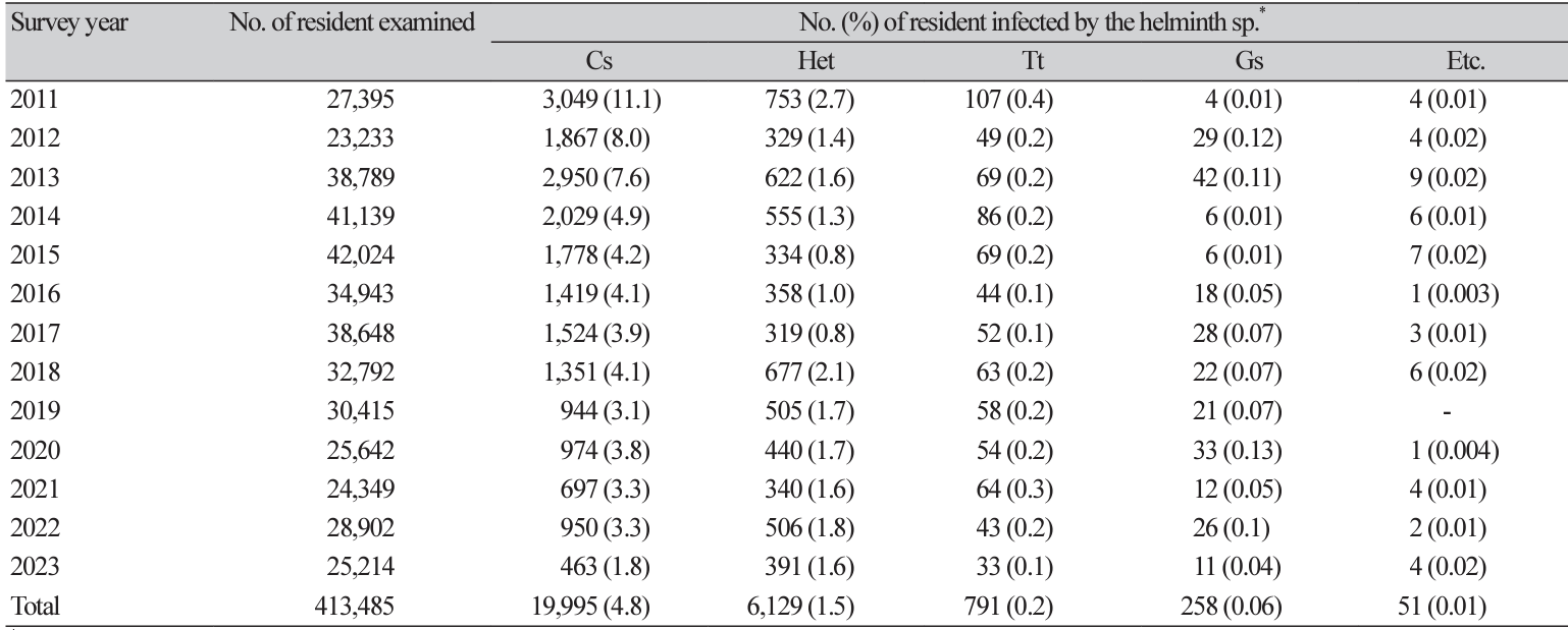
Table 2
Results of stool examinations with the formalin-ether sedimentation technique in health check-ups in Korea Association of Health Promotion [14]

Table 3
Results of stool examinations with the Kato-katz thick smear technique in Southeast Asian countries*

*By projects of Korea International Cooperation Agency (KOICA), Korea Association of Health Promotion (KAHP) and Korea Foundation for International Healthcare (KOFIH) in Myamar, Cambodia and Lao PDR; †Ascaris lumbricoides; ‡Trichuris trichiura ; §hookworm; ||small trematode eggs including opisthorchiid and heterophyid flukes. [15] Chai et al. (2020): Schoolchildren in Yangon Region, Myanmar; [16] Yong et al. (2014): Inhabitants of Cambodia (2006-2011); [17] Eom et al. (2014): Inhabitants of Lao PDR (2007-2011); [18] Rim et al. (2004): Primary schoolchildren of Lao PDR (2000-2002).
Fig. 2.
The embryonated small-sized eggs with an operculum (Group I). A. Clonorchis sinensis; B. SEM-view of C. sinensis egg; C. Metagonimus yokogawai; D. Heterophyes nocens; E. Pygidiopsis summa; F. Stictodora fuscata; G. Plagiorchis muris; H. Eurytrema pancreaticum; I. Gymnophalloides seoi. Scale bar = 10 ㎛.
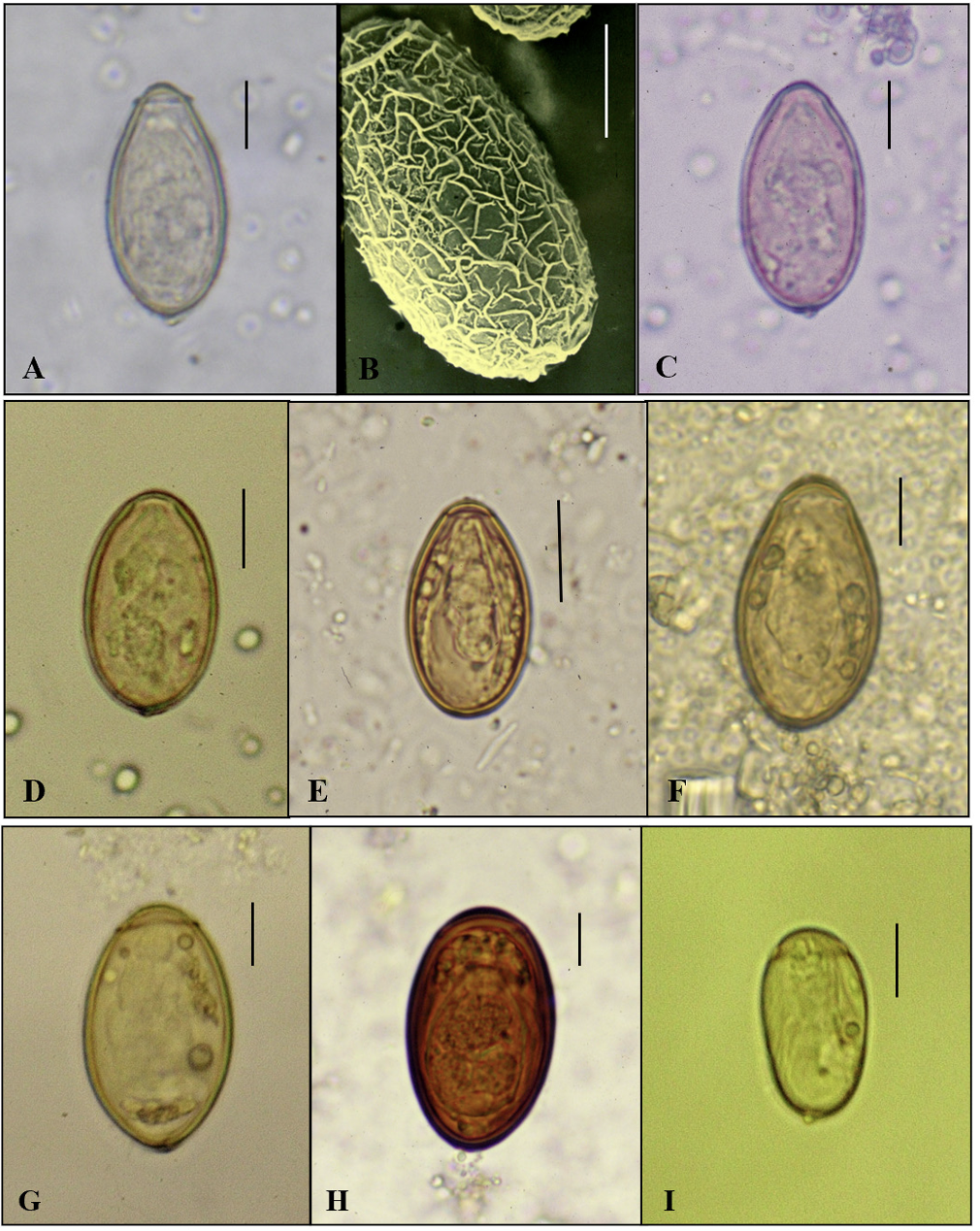
Fig. 3.
The unembryonated large-sized eggs with an operculum (Group II). A. Echinostomatid egg in CTS specimen; B. Isthmiophora hortensis; C. Echinostoma cinetorchis; D. Echinochasmus japonicus; E. Paragonimus westermani; F. Neodiplostomum seoulense; G. Fasciola hepatica; H. Dibothriocephalus nihonkaiensis. Scale bar = 50 ㎛. CTS, cellophane thick smear.
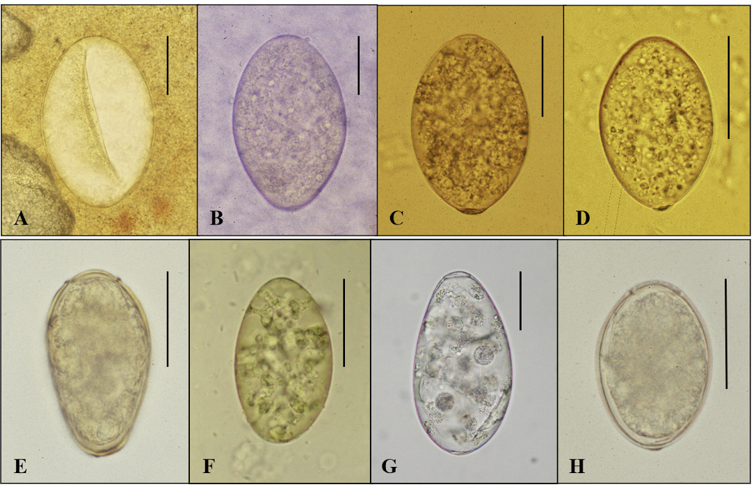
Fig. 4.
The embryonated (cestode) eggs without an operculum (Group III). A. Taenia sp. in CTS specimen; B. Hymenolepis nana; C. H. nana in CTS specimen; D. Hymenolepis diminuta. Scale bar = 15 ㎛. CTS, cellophane thick smear.

Fig. 5.
The nematode eggs (Group IV). A. Ascaris lumbricoides (F); B. A. lumbricoides (F) in CTS specimen; C. A. lumbricoides (U); D. A. lumbricoides (U) in CTS specimen; E. Trichuris trichiura; F. T. trichiura in CTS specimen; G. Enterobius vermicularis in CTS specimen; H. E. vermicularis in a specimen of cellotape anal swab; I. Hookworm; J. Hookworm (morular stage) in CTS specimen; K. Air-bubble like hookworm egg of which a mature larva already escaped; L. Trichostorongylus orientalis. Scale bar = 25 ㎛. CTS, cellophane thick smear.


 PDF
PDF Citation
 Print
Print


 XML Download