Abstract
This report presents a unique case of checkrein deformities in both halluces following isolated intramuscular sarcoidosis, a rare occurrence given the infrequent musculoskeletal involvement in sarcoidosis. Typically resulting from flexor hallucis longus tendon entrapment by scar tissue post-trauma, the checkrein deformity reported in this paper presented with unusual metatarsophalangeal joint flexion and interphalangeal joint extension during ankle dorsiflexion. A 49-year-old woman with a history of intramuscular sarcoidosis presented with a great toe deformity and discomfort while wearing shoes, leading to a diagnosis of dynamic deformity, possibly attributed to tendon tethering by sarcoidosis. Surgical treatments, including abductor hallucis muscle intratendinous tenotomy, flexor hallucis longus Z-plasty lengthening, Weil osteotomy, and Kirschner wire fixation, significantly improved the functional scores and patient discomfort. This report underscores the importance of recognizing dynamic deformities and the potential for rare diseases, such as sarcoidosis, to cause such conditions, highlighting the need for careful diagnosis and tailored surgical intervention for atypical checkrein deformities.
A 49-year-old woman visited the outpatient clinic with a chief complaint of great toe deformity and discomfort. She reported that the deformation of her toes has been progressing for approximately two years. She expressed discomfort due to the dorsal side of the MTP joint protruding, which causes pain from friction when wearing shoes. In addition, due to the deformity, she feels that she frequently trips while walking. She denied any recent history of trauma or lower limb surgery. Her medical history revealed that she had been diagnosed with extensive intramuscular nodular lesions at both lower extremities, indicating sarcoidosis. She had previously received steroid therapy for her diagnosis.
Physical examination revealed that, in a standing position, the dorsal side of the MTP joint was protruded and both the halluces were slightly abducted (Fig. 1A). The alignment of the hindfoot was neutral. During passive ankle dorsiflexion, the first MTP joint was flexed while the IP joint was extended (Fig. 1B). The MTP joint was tightened, limiting the extension motion of the hallux at the MTP joint level. The range of motion in the ankle joint was normal, and the power of ankle dorsiflexion/plantar flexion was also normal. In addition, no abnormalities were observed in the pulse.
Flexion of the MTP joint was observed on weight-bearing lateral plain radiographs, and no signs of arthritis were observed in the other MTP and IP joints (Fig. 2). Dynamic ultrasonography revealed no significant findings in the flexor tendon. Magnetic resonance imaging (MRI) revealed only mild tenosynovitis in the anterior compartment of the lower leg.
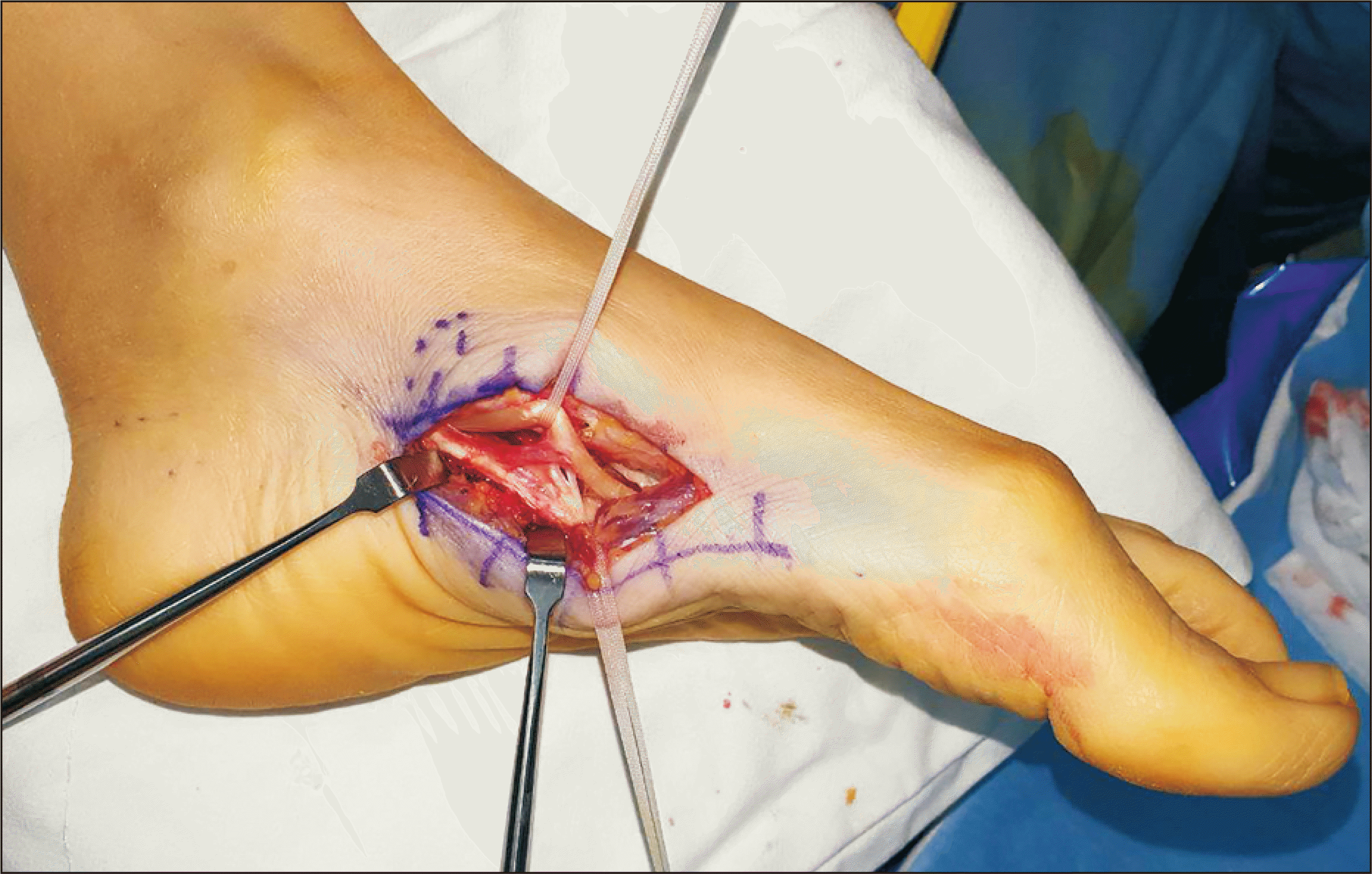
The patient complained of a deformity and associated discomfort over a long period and was advised to undergo surgery. The patient agreed with this recommendation and underwent a surgical intervention. We planned a staged operation to avoid discomfort during walking that could result from a simultaneous operation on both sides. Tightness in the abductor hallucis muscle was identified, and an intratendinous tenotomy was performed on the medial side of the midfoot. After identifying the master knot of Henry (Fig. 3), flexor hallucis longus Z-plasty lengthening was performed at midfoot level. However, we determined that correction solely through soft tissue procedures was insufficient. To achieve MTP joint congruency and adjust the working distance of the flexor muscles, we believed that subsequent bony procedure was necessary. Through a dorsal approach, a Weil osteotomy of the first metatarsal bone was performed to reduce the length of the first ray. Then, for the hyperextension deformity of the first IP joint, Kirschner wire fixation was performed to achieve a neutral position. The same procedure was performed on the contralateral side six months after the initial surgery.
A short leg cast was maintained for one month after surgery, with partial weightbearing, primarily on the heel, was allowed. One month postoperatively, the cast was replaced with a short leg removable thermoplastic splint, and the temporarily fixed Kirschner wire was removed. Two months postoperatively, the patient was advised to wear a postoperative shoe brace and allowed to bear full weight. Four months after surgery, the patient walked well while wearing shoes. At the final follow-up (left side: 14 months and right side: 8 months postoperatively), the Foot and Ankle Outcome Score (FAOS) improved to 96.4 points (FAOS symptoms: 89; FAOS pain: 100; FAOS activities of daily living: 93; FAOS sports and recreation: 100; and FAOS quality of life: 100), which represents a significant improvement from the preoperative scores of 69 points (FAOS symptoms: 72; FAOS pain: 44; FAOS activities of daily living: 86; FAOS sports and recreation: 88; and FAOS quality of life: 55) (Fig. 4, 5). The patient reported no discomfort while walking or running and mentioned that she no longer trips while walking. In addition, there was no transfer metatarsalgia under the second metatarsal head.
This case holds significance as the first report demonstrating a checkrein deformity following isolated intramuscular sarcoidosis. Although the exact reason for lower leg involvement remains unclear, it is believed that an environment conducive to reducing the working distance of flexor muscles was created, leading to the manifestation of a dynamic deformity. In addition, both halluces were abducted preoperatively. This can be attributed to the abductor hallucis muscle attached to the base of the proximal phalanx,8) leading to a tightened MTP joint and flexion of the halluces.
In this case, the patient presented with pain and discomfort in the MTP joint; however, in reality, a dynamic deformity was identified. If the physical examination had been conducted only in a sitting position, the flexion of the MTP joint might not have been significantly apparent, potentially leading to failure in detecting the cause of discomfort. Furthermore, imaging studies, including ultrasonography and MRI, did not reveal any significant findings elucidating the lesion. Moreover, the absence of a specific history of trauma, including fractures or surgical interventions, could have left the etiology unclear. Therefore, assessing dynamic deformities become essential by performing ankle joint maneuvers and examining patients in a standing position. Furthermore, this case underscores the complexity of checkrein deformities and highlights the need for a thorough investigation of the underlying causes, which can extend beyond the common etiologies of trauma-induced scar formation. In addition, due to the rarity of checkrein deformities, a standard treatment approach was unclear. We focused on addressing the patient’s primary concerns, specifically the inability to smoothly perform toe-off owing to flexion of the MTP joint and the sensation of tripping over. Surgical intervention facilitated the release of the tightened MTP joint, thereby adjusting soft tissue tension and enabled correction of the deformity through the shortening effect of the Weil osteotomy.
In conclusion, this case report’s unique presentation and surgical management of checkrein deformities in the context of intramuscular sarcoidosis provided valuable insights into the diagnosis and treatment of rare musculoskeletal manifestations.
Notes
REFERENCES
1. Kyung MG, Cho YJ, Lee DY. 2024; Management of checkrein deformity. Clin Orthop Surg. 16:1–6. doi: 10.4055/cios23229. DOI: 10.4055/cios23229. PMID: 38304213. PMCID: PMC10825257.

2. Boszczyk A, Zakrzewski P, Pomianowski S. 2015; Hallux checkrein deformity resulting from the scarring of long flexor muscle belly - case report. Ortop Traumatol Rehabil. 17:71–6. doi: 10.5604/15093492.1143539. DOI: 10.5604/15093492.1143539. PMID: 25759157.

3. Meyer N, Sutter R, Schirp U, Gutzeit A. 2017; Extensive intramuscular manifestation of sarcoidosis with initially missed diagnosis and delayed therapy: a case report. J Med Case Rep. 11:246. doi: 10.1186/s13256-017-1403-3. DOI: 10.1186/s13256-017-1403-3. PMID: 28835264. PMCID: PMC5569518.

4. Wessendorf TE, Bonella F, Costabel U. 2015; Diagnosis of sarcoidosis. Clin Rev Allergy Immunol. 49:54–62. doi: 10.1007/s12016-015-8475-x. DOI: 10.1007/s12016-015-8475-x. PMID: 25779004.

5. Otake S. 1994; Sarcoidosis involving skeletal muscle: imaging findings and relative value of imaging procedures. AJR Am J Roentgenol. 162:369–75. doi: 10.2214/ajr.162.2.8310929. DOI: 10.2214/ajr.162.2.8310929. PMID: 8310929.

6. Dhomps A, Foret T, Streichenberger N, Skanjeti A, Tordo J. 2019; Isolated muscular sarcoidosis revealed by hypercalcemia and 18F-FDG PET/CT. Clin Nucl Med. 44:824–5. doi: 10.1097/RLU.0000000000002678. DOI: 10.1097/RLU.0000000000002678. PMID: 31274562.

7. Karthik V, Roshan R, Jabbar PK, Nair A. 2023; Isolated muscular sarcoidosis presenting as hypercalcaemic renal failure. BMJ Case Rep. 16:e257439. doi: 10.1136/bcr-2023-257439. DOI: 10.1136/bcr-2023-257439. PMID: 37848272.

8. Brenner E. 1999; Insertion of the abductor hallucis muscle in feet with and without hallux valgus. Anat Rec. 254:429–34. doi: 10.1002/(SICI)1097-0185(19990301)254:3<429::AID-AR14>3.0.CO;2-5. DOI: 10.1002/(SICI)1097-0185(19990301)254:3<429::AID-AR14>3.0.CO;2-5.


 PDF
PDF Citation
 Print
Print


 XML Download