Journal List > Anat Cell Biol > v.57(2) > 1516087502

Ramalho Rodrigues, Neto, Andrade Fernandes, Barreto, Barros Maciel do Amaral, Karoline de Araújo Deca, Freire de Albuquerque Figueiredo, Dantas de Lucena, Bezerra da Silva, Sales, and Oliveira: Morphological analysis of the jugular foramen in dry human skulls in northeastern Brazil

Abstract

The jugular foramen (JF) is located between the temporal and occipital bones. The JF is a primary pathway for venous outflow from the skull and passage of nerves. Variations are common in this region and may have clinical and surgical implications. To analyze the sexual dimorphism and JF morphology in skulls from Northeastern Brazil. 128 human skulls from the Anatomy Laboratory of the Federal University of Paraíba, 64 male and 64 female, were selected and the JFs analyzed for bone septation and the presence of a dome. Data analysis considered P<0.05 as significant. On at least one side, complete septation was observed in 26 skulls (20.3%), incomplete septation in 93 skulls (72.6%) and 61 skulls (47.6%) did not present septation. In 114 skulls (89%), 47.6% female and 41.4% male, have a unilateral presence of the dome and 71 (55.4%) have it bilaterally. Posterolateral compartment diameters and JF area had higher values on the right side in the total sample and separated by sex (P<0.05). Most morphometric variables of the anteromedial compartment were higher in male than in female (P<0.05), fact that was not observed in the posterolateral compartment (P>0.05). This study showed a higher prevalence of complete septation in males compared to females. Morphometric analysis presented a peculiar morphology of the JF in this study. These results suggests that the surgical approach to diseases that affect the JF may be peculiar to the studied population, confirming the importance of morphological analysis of the skull base.

Introduction

The jugular foramen (JF) is a bony foramen situated in the posterior cranial fossa, positioned between the temporal and occipital bones at the posterior extremity of the petro-occipital fissure, anterolaterally of the foramen magnum. This foramen is related to important structures such as the inferior carotideal opening anteriorly, internal acoustic meatus anterosuperiorly, the medial face of the styloid process covering laterally and the hypoglossal channel medially [1, 2].
The JF is an important structure at this region since it is a primary pathway for venous outflow from the skull and is a passageway for nerve structures of the peripheral nervous system through the base of the skull [3]. The following structures pass through the JF: the distal sigmoid sinus (SS), that continues as jugular bulb and the internal jugular vein (IJV), the inferior petrosal sinus, the meningeal branches of pharyngeal and ascending occipital arteries, besides the cranial nerves glossopharyngeal (CN IX), vagus (CN X) and accessory (CN XI) [2, 4-6].
There is currently a lack of agreement regarding the subdivision of compartments within the JF [4-8]. But historically, this foramen has divided into the 2 compartments, the anteromedial pars nervosa and posterolateral pars venosa, divided by the intrajugular process [3, 4, 7, 9-12]. The pars nervosa contains the glossopharyngeal nerve (IX) and the inferior petrosal sinus, while the pars venosa contains the posterior meningeal branch of the ascending pharyngeal artery, the jugular bulb from the SS, meningeal branches of the occipital artery and the vagus (X) and accessory nerves (XI) [3-5, 10, 13, 14]. However, for some authors, it seems that the JF has three compartments: the anterior portion hosts the inferior petrosal sinus, the middle portion shelters the three cranial nerves CN IX, CN X, and CN XI and the posterior portion has the IJV and the meningeal branches of pharyngeal and ascending occipital arteries [4, 5, 8, 10].
The literature also reports the presence of a domed (PD) bone at the superior part of the jugular bulb [1, 5, 15-17]. When the SS is positioned posteriorly within the mastoid, the dome of the bulb is typically low. Conversely, in cases where the SS is located further forward, bending sharply upward into the jugular bulb, the opposite effect may occur. A dome that is positioned medially and anteriorly can pose challenges during surgical access to the middle ear. Therefore, the anatomical position of the dome of the jugular bulb holds significant importance in the preoperative evaluation before any ear and skull base surgery [10, 15-17].
Previous studies reported variations of the JF architecture, including differences in shape, as well as anteroposterior (APD) and mediolateral diameter of the foramen [8, 17]. Additionally, variations related to gender, antimere dominance and race were also documented [1, 5, 10, 18]. This emphasizes the significance of comprehending the anatomical variations, and morphometry of the JF in order to deepen the anatomical understanding of this particular structure. This region is affected by a significant number of pathological processes including glomic tumors, schwannomas, intracranial meningioma, metastatic lesions, nerve palsies as the Vernet’s syndrome and infiltrative inflammatory processes from surrounding structures highlighting the middle ear [1, 10, 18, 19].
Thus, given the insufficient understanding of the morphology of the JF, the aim of this study was to analyze morphometrically the JF, its sexual dimorphism and variations in dry skulls in Northeastern Brazil to allow a more accurate and safer practice by professionals working with this body region.

Materials and Methods

This study analyzed 128 dry skulls of Northeastern Brazil (64 females and 64 males), belonging to the Human Anatomy Laboratory of the Universidade Federal da Paraíba (UFPB). Only undamaged dried skulls of adults were analyzed. Sexing of skulls was documented through anthropometric standards suggested by Vanrell [20] by two independent and well-trained technicians. The laboratory does not have a record of the ethnicity of skulls.
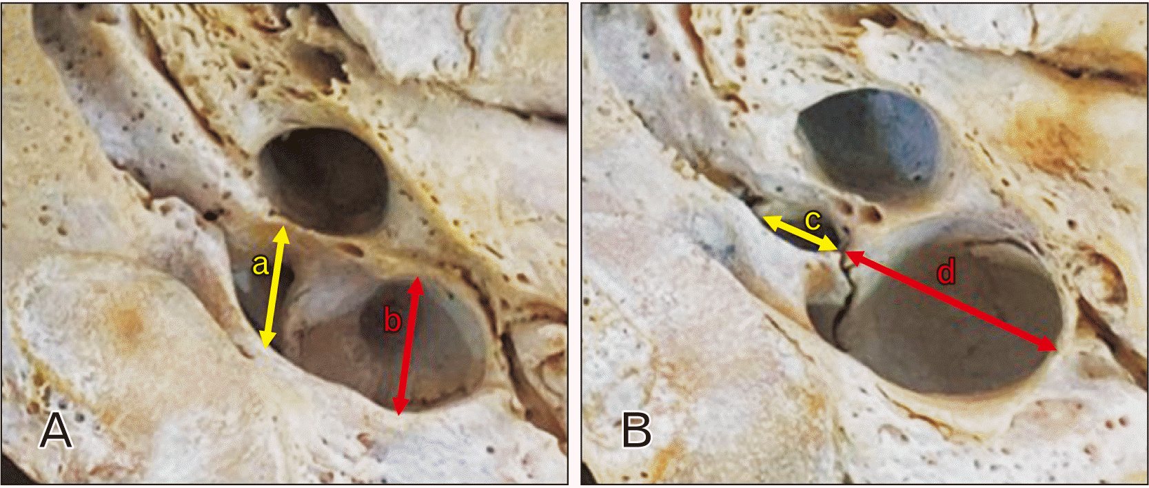
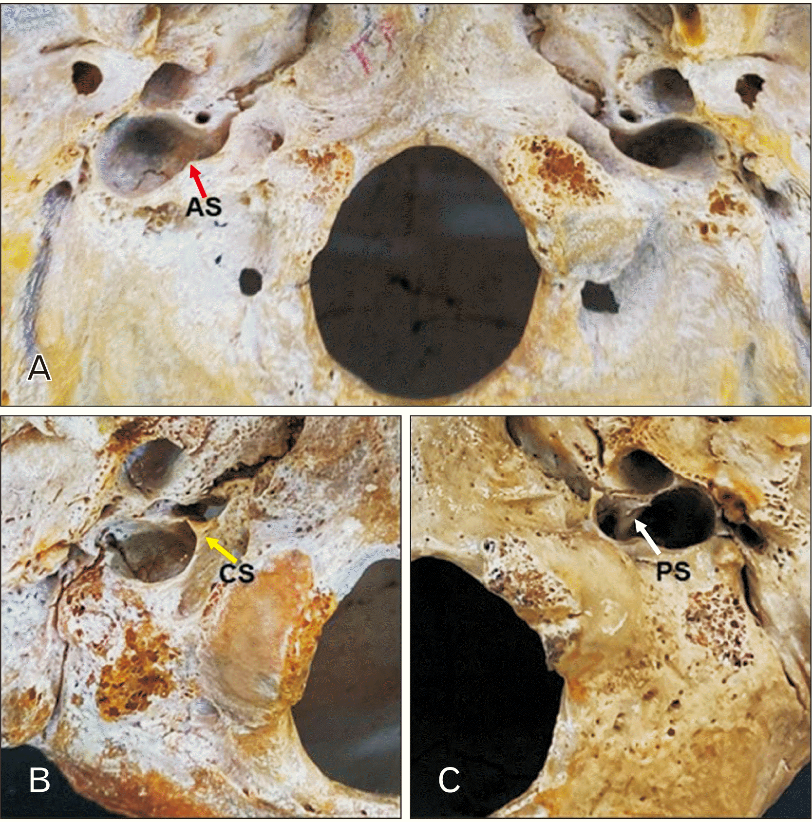
For morphometric analysis, a digital caliper Eccofer® with capacity of 150 mm and accuracy up to 0.02 mm was used. The APD and lateromedial diameters (LMD) of anteromedial and posterolateral compartments of JF in both sides were measured, as shown in Fig. 1. In septate or partially septate JF (Fig. 2), the total lateromedial diameter (TLMD) was the sum of the LMD distance of the anteromedial compartment, and the LMD distance of the posterolateral compartment.
The area was calculated using the formula [21]:
A=π×APD×LMD4
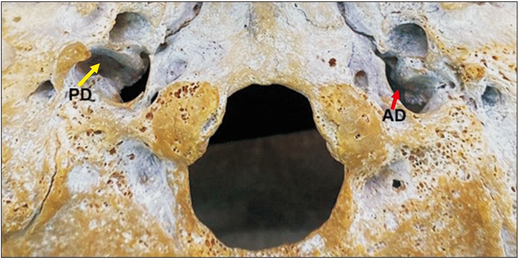
The incidence of septation was cataloged by the presence of intrajugular process in complete (CS), partial (PS) or absent septation (Fig. 2). The occurrence of the domed bone roof was categorized by the presence or absence (AD) of it, as shown in Fig. 3.
The data analysis was performed using Jamovi version 2.3.24. Comparison was done between genders and between right and left antimers. The QQ plot curve and the W Shapiro–Wilk test were used to determine the normality of the samples according to each group analyzed. Data from the QQ plot suggestive of normality and data from the W Shapiro–Wilk test that presented P>0.05 were considered parametric and their measurements were recorded as mean±SD. Data with QQ plot suggestive of non-normality and W Shapiro–Wilk test presenting P<0.05 were considered non-parametric and their measurements recorded as median±interquartile range. Comparison between groups for paired or unpaired samples was done by t-test and Mann–Whitney U-Test, respectively. Values of P<0.05 were considered as significant.
The present study was selected in accordance to the Brazilian Federal Law 8.501 (November 30, 1992). An institutional and ethical approval (CAAE number registration: 58097822.0.0000.5188 - Medical Sciences Center of the Federal University of Paraíba, Brazil) was obtained.

Results

One hundred and twenty-eight dried skulls were included in the analysis, 64 males (50.0%) and 64 females (50.0%), corresponding to 256 JF.
The incidence of JF septation in this study was summarized in Table 1. CS in the JF, on at least one side, was observed in 26 skulls (20.3%), 5 females and 21 males (3.9% and 16.4%, respectively). Unilateral CS on the right side was observed in 12 skulls (9.3%), 3 females and 9 males (2.3% and 7%, respectively). Unilateral CS on the left side was observed in 8 skulls (6.3%), 1 female and 7 males (0.8% and 5.5%, respectively). One female and 5 males skulls had CS bilaterally (0.8% and 3.9%, respectively).
Ninety-three skulls had PS on at least one side, corresponding to 72.6% of the total sample, 51 females skulls and 42 males skulls (39.8% and 32.8%, respectively). The partial presence of unilateral septation on the right side was observed in 21 skulls (16.4%), 10 females skulls and 11 males skulls (7.8% and 8.6%, respectively). The presence of unilateral PS on the left side was observed in 24 skulls, 13 females and 11 males (10.1% and 8.6%, respectively). Twenty-eight females and 20 males skulls showed bilateral incomplete septation (21.8% and 15.6%, respectively).
The absence of septation in the JF, on at least one side, was observed in 61 skulls, corresponding to 47.6% of the total sample, 33 females skulls and 28 males skulls (25.7% and 21.9%, respectively). The absence of unilateral septation on the right side was observed in 19 skulls, 12 females and 7 males (9.4% and 5.5%, respectively). The absence of unilateral septation on the left side was observed in 20 skulls, 11 females and 9 males (8.6% and 7%, respectively). Ten females and 12 males skulls did not present septation bilaterally in the same skull (7.8% and 9.3%, respectively).
The PD in the JF (Table 2), on at least one side, was observed in 114 skulls, corresponding to 89% of the total sample, with 61 females skulls and 53 males skulls (47.6% and 41.4%, respectively). The PD unilaterally on the right side was observed in 30 skulls (23.4%), 15 females and 15 males (11.7% and 11.7%, respectively). The PD unilaterally on the left side was observed in 13 skulls (10.2%), 7 females and 6 males (5.5% and 4.7%, respectively). Thirty-nine females and 32 males skulls presented the dome bilaterally (30.4% and 25%, respectively).
The AD in the JF, on at least one side, was observed in 57 skulls, corresponding to 44.5% of the total sample, 25 females skulls and 32 males skulls (19.5% and 25%, respectively). The AD unilaterally on the right side was observed in 13 skulls (10.2%), 7 females and 6 males (5.5% and 4.7%, respectively). The absence of a single dome laterally on the left side was observed in 30 skulls (23.4%), 15 females and 15 males (11.7% and 11.7%, respectively). Three females and 11 males skulls showed absence of dome bilaterally (2.3% and 8.5%, respectively).
Regarding the morphometric analysis (Table 3), the APD and LMD of the posterolateral compartment, and the area of the JF had higher values on the right side comparing to the left side, not only in the total sample but also in the sample separated by sex (P<0.05). The APD of the anteromedial compartment of the JF was also larger on the right (P=0.011), but only when the total sample (both male and female skulls) was considered. The LMD of the anteromedial compartment of the JF didn’t have any differences between the right and left sides (P>0.05). The TLMD of the JF presented higher values on the right side comparing to the left side at the total sample (P=0.001) and at the female sample (P=0.005), but not at the male (P=0.054).
A gender analysis of the morphometric data was also performed in this study (Table 4). On the right side, no differences were observed in all measured parameters between male and female genders (P>0.05), except the APD of the anteromedial compartment of the JF, which was larger in males than in females (P=0.004). On the left side, only the APD and LMD of the anteromedial compartment of the JF along with the TLMD were larger in males than in females (P<0.05). The same results were observed when comparing these measurements in the total sample (P<0.05).

Discussion

Due to its anatomical variety and intrinsic relationships with neurovascular structures, the shape and size of the JF are of great physiological and radiological importance, being a fundamental part of the study of the skull base [10].

The septation

The JF may be subdivided into two important compartments: one venous and one neural compartment. Thus, it is of great value to understand the relationship between these structures and the frequency of compartmentalization by fibrous or bony septum, in order to perform a more precise microsurgical approach [22]. Studying the anatomical organization of the JF in relation to existing lesions may provide a method to categorize and better understand the growth patterns of JF tumors, thus allowing correct removal and preservation of the neurovascular structures in the region [4].
In the present study, the presence of CS was identified in 20.3% of the skulls, identified both unilaterally (15.6%) and bilaterally (4.7%). Moreover, males had a higher prevalence of CS comparing to females. Similar prevalence of bilateral septation was also found in previous studies by Sturrock [23] and Pereira et al. [1], in which 0%–4% of the skulls analyzed presented the variation. This compartmentalization pattern can compress the structures that pass through the JF, and can compress several cranial nerves, characterizing the JF syndrome (Vernet’s syndrome) [10]. Therefore, the data collected in the present study seem to show a trend towards a higher risk in male compared to female (16.4% vs. 3.9%, respectively).
Tubbs et al. [3] in his study has classified meningeal morphologies of the JF in four types of dural septations. This included type I, where a dural septation was seen between CN IX anteriorly and CNs X and XI posteriorly. Type II was defined as a JF with no meningeal septation. Type III was defined as a foramen with a septation between CNs IX and X anteriorly and CN XI posteriorly. Type IV with multiple septations within the JF that divided the various rootlets of the CNs. Ossification of these septations can make surgical manipulation more dangerous because there would be less give when moving the CNs exiting the JF. He concludes that the various types of foramina as identified might alter surgical approaches [3].

The dome

The presence of the JF dome was also evaluated, which is usually located under the mastoid segment of the facial nerve (60%), or in the mastoid and tympanic cavities in 22% and 17% of the bones, respectively. Since the position of the dome is multifactorial and with several variations, its evaluation was not analyzed in the study. The evaluation of the presence of the dome is of great importance in the preoperative evaluation of ear and skull base surgeries [17].
The dome was present bilaterally in 71 skulls (55.5%), only on the right side in 30 skulls (23.4%) and only on the left side in 11 skulls (10.2%). There were no important differences between the genders studied, but clearly there is a higher prevalence of the dome on the right side. The data were similar to those presented by two Northern Indian studies [10, 22], in which the dome was present bilaterally in 62%–66% of the sample. Interestingly, a Southern Indian population presented bilateral dome in 20% of the sample [18]. Therefore, it is noted that within the same country there are morphological differences in the incidence of domes. This would apply to Brazil, especially as it is a continental country like India.
That variability reinforces an accurate knowledge and radiological verification of possible variations, which are essential to avoid problems on surgical positioning and to decide the approach individually. Several cases of jugular glomus tumors invade the middle ear cavity by penetrating this wall [10, 24, 25]. The AD indicates that the jugular bulb is absent or poorly developed, thus not forming the floor of the middle ear cavity. In these cases, there is a lower risk of these tumors invading the middle ear [10].

The total lateromedial diameter

The dimensions and configuration of the JF are linked to the size of the IJV and the presence or absence of a superior jugular bulb. The difference on the right and left sizes of the IJV is noticed in the human embryo at 8 weeks post conception, due to the variation of the development of the right and left brachiocephalic veins [10, 22, 26, 27]. It has been noted that the right foramen is frequently larger than the left [6, 22, 28], fact that was confirmed in this study (P=0.001). This may occur due to the superior sagittal sinus that could drain into the right transverse sinus [2, 10, 22].
The TLMD measurement in the present study was closer to the Ethiopian population [8] and larger than other populations, such as Southern [1] and Midwestern [5] Brazilians, and Indian studies [10, 22]. Otherwise, it was smaller than the Southern Indians [18]. The dominance between genders was not compared in most of these studies [1, 5, 10]. In the current study, only the female sample had a larger right JF comparing to the left side (P=0.005). Thus, the chances of compression of neurovascular structures passing through JF may be more common on the left side and also approach to the tumors in this area may be more difficult.
The results of our study contrast with other studies that claim that the right JF is larger through the exocranial facet in males and could be a risk factor for compression of the neurovascular meshwork in the presence of a JF tumor [10, 22, 27]. This variability between studies, even among Brazilian populations from different regions can be explained by racial and individual factors, by virtue of the historical miscegenation of Latino America.
Additionally, present study reported mean TLMD to be 17.0 mm and 16.3 mm and APD to be 9.05 mm and 8.0 mm on the right and left side, respectively, suggesting an oval shape of the JF with its long axis along the TLMD. This finding in our study was also similar to other studies done in the Southern [1] and Midwestern [5] regions of Brazil.

The LMD and APD of posterolateral and anteromedial compartments

Most studies analyzed morphometrically only the TLMD and the APD of the posterolateral compartment and did not consider the LMD and the APD of the posterolateral and anteromedial compartments singly [1, 5, 8, 10, 18, 22].
In this study, the LMD and the APD of the posterolateral compartment in males and females had higher values on the right side compared to the left side (P<0.05). The same was observed in another Brazilian study [1], except in the female skulls, where they did not see differences between the right and left sides for the APD of the posterolateral compartment. The current study had an average APD of the posterolateral compartment smaller than several studies in the literature [1, 5, 8, 10, 22]. Regarding the sexual dimorphism, the LMD and the APD of the anteromedial compartment were larger in males on both sides, compared to females (P<0.05). Interestingly, no differences between genders were observed for these measurements in the posterolateral compartment of both sides (P>0.05). It was not possible to compare the LMD and APD of the anteromedial compartment due to the absence of these measurements singly in other research papers.
The findings of the posterolateral compartments measurements, regardless gender, supports the theory that the IJV passes through the posterolateral compartment [4] and can usually present variations between sides. This occurs due to the frequent drainage of the superior sagittal sinus into the right transverse sinus, increasing the pressure and size of the IJV that will pass into the posterolateral compartment on the right. Note that the anteromedial compartment remains unchanged, with the exception of the APD of the anteromedial compartment, which had higher values on the right side compared to the left when the sample was not separated according to gender. However, when male and female skulls were isolated, no difference was observed between the right and left sides. which can be explained by the certain invariability of the presentation of the cranial nerves that pass through this compartment, maintaining their similar morphology between the right and left sides.
The differences between the anteromedial compartment of males and females on both sides raise a question regarding the possible factors influencing the observed data. Maybe, just the size of male nerves is inherently larger than female nerves, since even to categorize skulls into male and female it is necessary to observe larger bone accidents in male compared to female, such as the glabella, mastoid process and styloid process. Further investigation and research would be necessary to gain a comprehensive understanding of these differences and their underlying causes.

The area

This study presented a larger JF area on the right side compared to the left side when the total sample was analyzed (P<0.001), or when male and female skulls were isolated (P=0.006 and P<0.001, respectively). However, no differences of the area on the right and left sides (P=0.276 and P=0.252, respectively) between genders were observed. The sexual dimorphism in this Brazilian study presented a smaller area on both sides compared to previous studies [1, 5, 8, 10, 22], with the left JF having the lowest values.
This suggests that the chances of compression of neurovascular structures passing through JF may be more common on the left side, which is smaller [10]. There is a considerable number of diseases inherent to this region, as glomic tumors, schwannomas, intracranial meningioma, metastatic lesions, nerve palsies as the Vernet’s syndrome and infiltrative inflammatory processes [1, 10, 18]. Surgical interventions could be more difficult to perform in our population, mainly the left JF, since it has a smaller area comparing to others populations, including Brazilians from other regions. These results reinforce the careful evaluation of the JF area by the surgeons, especially in bolder approaches, such as lateral suboccipital approach, retrosigmoid and transjugular craniotomy.
Hence, when contrasting with various studies, it becomes evident that there is a significant diversity in JF morphology and morphometry, due to genetic, environmental, ethnic, racial factors, measurement methods and sample sizes. It’s noteworthy that this variability is observed both across different countries and within the boundaries of the same country. This study had limitations as the difficulties to measure the diameters on the extracranial side and no other previous study to compare the measures of the anteromedial compartment. So, further researches are needed to compare these variations and certify those differences, since an accurate knowledge of the intricate anatomy of the JF is crucial for clinical and surgical interventions.
In conclusion, this study showed a higher prevalence of CS in males compared to females, which may make it difficult to approach tumors in this gender. The dome was present in more than 55% of the studied skulls, was more frequent on the right side and does not seem to have important differences when gender was considered. It has been observed that the right foramen is often larger than the left, especially in the posterolateral compartment. When genders were analyzed, the anteromedial compartment of the JF was larger in male than in female. The same was not observed for the posterolateral compartment. These results seem to demonstrate that the surgical approach to JF diseases may represent a greater challenge in this population, given the peculiar morphological characteristics found in this study, thus increasing the importance of anatomical knowledge of this region of the skull base.

Notes

Author Contributions

Conceptualization: ASBO, THAS, IBS. Data acquisition: RRR, DFNN, JVAF, LOB, VBMA, DKAD, VLFAF. Data analysis or interpretation: ASBO, JDL, THAS, IBS. Drafting of the manuscript: ASBO, JDL, IBS. Critical revision of the manuscript: ASBO, JDL, IBS. Approval of the final version of the manuscript: all authors.

Conflicts of Interest

No potential conflict of interest relevant to this article was reported.

References

1. Pereira GAM, Lopes PTC, Santos AMPV, Krebs WD. 2010; Morphometric aspects of the jugular foramen in dry skulls of adult individuals in Southern Brazil. J Morphol Sci. 27:3–5.
2. Standring S. Gray's anatomy: the anatomical basis of clinical practice. 40th ed. Churchill Livingstone;2008. p. 423–34.
3. Tubbs RS, Griessenauer CJ, Bilal M, Raborn J, Loukas M, Cohen-Gadol AA. 2015; Dural septation on the inner surface of the jugular foramen: an anatomical study. J Neurol Surg B Skull Base. 76:214–7. DOI: 10.1055/s-0034-1543973. PMID: 26225304. PMCID: PMC4433395.
crossref
4. Bond JD, Zhang M. 2020; Compartmental subdivisions of the jugular foramen: a review of the current models. World Neurosurg. 136:49–57. DOI: 10.1016/j.wneu.2019.12.178. PMID: 31926358.
crossref
5. Caldeira JVC, Godas AG de L, Carvalho GB de A, Silva KRT da, Machado AR da SR, Almeida PF de, Silva AV da. 2020; Measurement of the jugular foramen in dry skulls from the midwest region of Brazil. Braz J Health Rev. 3:14614–28. Portuguese. DOI: 10.34119/bjhrv3n5-256.
6. Thintharua P, Chentanez V. 2023; Morphological analysis and morphometry of the occipital condyle and its relationship to the foramen magnum, jugular foramen, and hypoglossal canal: implications for craniovertebral junction surgery. Anat Cell Biol. 56:61–8. DOI: 10.5115/acb.22.105. PMID: 36635090. PMCID: PMC9989787.
crossref
7. Shruthi BN, Pavan PH, Shaik SH. 2015; Study on morphological variations in structure of the jugular foramen. Int J Anat Res. 3:1533–35. DOI: 10.16965/ijar.2015.268.
crossref
8. Jegnie MJ, Asmare Y, Muche A. 2020; Anthropometric measurements of jugular foramen in Ethiopian dried adult skulls. Ethiop J Health Biomed Sci. 10:55–63.
9. Shapiro R. 1972; Compartmentation of the jugular foramen. J Neurosurg. 36:340–3. DOI: 10.3171/jns.1972.36.3.0340. PMID: 5059973.
crossref
10. Das SS, Saluja S, Vasudeva N. 2016; Complete morphometric analysis of jugular foramen and its clinical implications. J Craniovertebr Junction Spine. 7:257–64. DOI: 10.4103/0974-8237.193268. PMID: 27891036. PMCID: PMC5111328.
crossref
11. Akdag UB, Ogut E, Barut C. 2020; Intraforaminal dural septations of the jugular foramen: a cadaveric study. World Neurosurg. 141:e718–27. DOI: 10.1016/j.wneu.2020.05.271. PMID: 32522647.
crossref
12. Fang Y, Olewnik Ł, Iwanaga J, Loukas M, Dumont AS, Tubbs RS. 2022; Variations and classification of bony septations of the jugular foramen: an anatomic and histologic study with application to imaging and surgery of the skull base. World Neurosurg. 163:e464–70. DOI: 10.1016/j.wneu.2022.04.010. PMID: 35398325.
crossref
13. Dichiro G, Fisher RL, Nelson KB. 1964; The jugular foramen. J Neurosurg. 21:447–60. DOI: 10.3171/jns.1964.21.6.0447. PMID: 14171303.
crossref
14. Keles B, Semaan MT, Fayad JN. 2009; The medial wall of the jugular foramen: a temporal bone anatomic study. Otolaryngol Head Neck Surg. 141:401–7. DOI: 10.1016/j.otohns.2009.05.030. PMID: 19716021.
15. Graham MD. 1977; The jugular bulb: its anatomic and clinical considerations in contemporary otology. Laryngoscope. 87:105–25. DOI: 10.1288/00005537-197701000-00013. PMID: 187882.
crossref
16. Graham MD. 1975; The jugular bulb. Its anatomic and clinical considerations in contemporary otology. Arch Otolaryngol. 101:560–4. DOI: 10.1001/archotol.1975.00780380038010. PMID: 1164239.
crossref
17. Cömert E, Kiliç C, Cömert A. 2018; Jugular bulb anatomy for lateral skull base approaches. J Craniofac Surg. 29:1969–72. DOI: 10.1097/SCS.0000000000004637. PMID: 29944552.
crossref
18. Shruthi BN, Pavan PH, Shaik HS, Henjarappa KS. 2015; Morphometric study of jugular foramen. Int J Intg Med Sci. 2:164–6. DOI: 10.18535/ijmsci/v2i8.01. PMID: 25336833. PMCID: PMC4201011.
19. Ramina R, Maniglia JJ, Fernandes YB, Paschoal JR, Pfeilsticker LN, Neto MC, Borges G. 2004; Jugular foramen tumors: diagnosis and treatment. Neurosurg Focus. 17:E5. DOI: 10.3171/foc.2004.17.2.5. PMID: 15329020.
crossref
20. Vanrell JP. Odontologia legal & antropologia forense. Guanabara Koogan;2009.
21. Ahmed MM, Jeelani M, Tarnum A. 2015; Anthropometry: a comparative study of right and left sided foramen ovale, jugular foramen and carotid canal. Int J Sci Stud. 3:88–94.
22. Gupta C, Kurian P, Seva KN, Kalthur SG, D'Souza AS. 2014; A morphological and morphometric study of jugular foramen in dry skulls with its clinical implications. J Craniovertebr Junction Spine. 5:118–21. DOI: 10.4103/0974-8237.142305. PMID: 25336833. PMCID: PMC4201011.
crossref
23. Sturrock RR. 1988; Variations in the structure of the jugular foramen of the human skull. J Anat. 160:227–30. PMID: 3253258. PMCID: PMC1262066.
24. Chong VF, Fan YF. 1996; Jugular foramen involvement in nasopharyngeal carcinoma. J Laryngol Otol. 110:987–90. DOI: 10.1017/S0022215100135534. PMID: 8977870.
crossref
25. Wysocki J, Chmielik LP, Gacek W. 1999; Variability of magnitude of the human jugular foramen in relation to conditions of the venous outflow after ligation of the internal jugular vein. Otolaryngol Pol. 53:173–7. Polish.
26. Padget DH. Contributions to embryology. Vol. 36, The development of the cranial venous system in man: from the viewpoint of comparative anatomy. Carnegie Institution of Washington Publication;1957. p. 79–140.
27. Vlajković S, Vasović L, Daković-Bjelaković M, Stanković S, Popović J, Cukuranović R. 2010; Human bony jugular foramen: some additional morphological and morphometric features. Med Sci Monit. 16:BR140–6. PMID: 20424543.
28. Vijisha P, Bilodi AK, Anandaraj L. 2013; Morphometric study of jugular foramen in Tamil Nadu region. Natl J Clin Anat. 2:71–4. DOI: 10.4103/2277-4025.297875.
crossref

Fig. 1
Linear measurements of the jugular foramen (JF). (A) a: Anteroposterior diameter (APD) of the anteromedial compartment of the JF; b: APD of the posterolateral compartment of the JF. (B) c: Lateromedial diameter (LMD) of the anteromedial compartment of the JF; d: LMD of the posterolateral compartment of the JF. Total LMD=c+d.
acb-57-2-213-f1.tif
Fig. 2
Types of septation of the jugular foramen. (A) Absent septation (AS). (B) Complete septation (CS). (C) Partial septation (PS).
acb-57-2-213-f2.tif
Fig. 3
Incidence of bony dome roof of the jugular foramen. Present dome (PD) or absent dome (AD).
acb-57-2-213-f3.tif
Table 1
Incidence of jugular foramen septation in both sexes
Sex Complete Partial Absent
Only right Only left Bilaterally Only right Only left Bilaterally Only right Only left Bilaterally
Male (n=64) 9 (7.0) 7 (5.5) 5 (3.9) 11 (8.6) 11 (8.6) 20 (15.6) 7 (5.5) 9 (7.0) 12 (9.4)
Female (n=64) 3 (2.3) 1 (0.8) 1 (0.8) 10 (7.8) 13 (10.1) 28 (21.9) 12 (9.4) 11 (8.6) 10 (7.8)

Data is presented as number (%). Number is total number of skulls analyzed in the study; percentage is percentage distribution in relation to the total sample.

Table 2
Prevalence of jugular foramen dome in both sexes
Sex Present Absent
Only right Only left Bilaterally Only right Only left Bilaterally
Male (n=64) 15 (11.7) 6 (4.7) 32 (25.0) 6 (4.7) 15 (11.7) 11 (8.6)
Female (n=64) 15 (11.7) 7 (5.5) 39 (30.5) 7 (5.5) 15 (11.7) 3 (2.3)

Data is presented as number (%). Number is total number of skulls analyzed in the study; percentage is percentage distribution in relation to the total sample.

Table 3
Mean or median distance±SD or IQR values of the morphometric analysis of the jugular foramen antimers
Measurements Total sample (n=256) Male (n=128) Female (n=128)
Right Left P-value Right Left P-value Right Left P-value
APD (posterolateral compartment) (mm) 9.05±2.62 8.00±2.40 <0.001 9.10±2.63 7.80±2.15 0.007 9.19±1.77a) 8.16±2.10a) 0.003
LMD (posterolateral compartment) (mm) 11.90±3.03 11.00±3.20 <0.001 12.00±3.07 11.30±3.63 0.003 11.50±1.91a) 10.40±2.06a) 0.001
APD (anteromedial compartment) (mm) 5.30±1.73 5.00±1.55 0.011 5.69±1.24a) 5.34±1.13a) 0.058 5.10±1.65 4.60±1.42 0.123
LMD (anteromedial compartment) (mm) 6.67±1.65a) 6.87±1.44a) 0.186 6.86±1.70a) 7.14±1.44a) 0.191 6.48±1.58a) 6.59±1.40a) 0.586
TLMD (mm) 17.00±4.05 16.30±3.92 0.001 17.80±3.10a) 17.00±2.70a) 0.054 16.90±2.37a) 16.00±2.55a) 0.005
Area (mm) 121.00±46.60 106.00±45.10 <0.001 125.00±56.60 110.00±44.30 0.006 119.00±38.90 103.00±42.30 <0.001

Number is total number of jugular foramina analyzed in the study. APD, anteroposterior diameter; LMD, latero-medial diameter; TLMD, total latero-medial diameter (a sum of LMD posterolateral part and LMD anteromedial part). a)Values described as mean±SD. The other linear measurements were described as median±interquartile range (IQR).

Table 4
Mean or median distance±SD or IQR values of the sexual dimorphism analysis of the jugular foramen
Antimers Sex APD (posterolateral compartment) (mm) LMD (posterolateral compartment) (mm) APD
(anteromedial compartment) (mm)
LMD (anteromedial compartment) (mm) TLMD (mm) Area (mm)
Right (n=128) Male (n=64) 9.10±2.63 12.00±3.07 5.69±1.24a) 6.86±1.70a) 17.80±3.10a) 125.00±56.60
Female (n=64) 8.90±2.55 11.80±2.63 5.07±1.14a) 6.48±1.58a) 16.90±2.37a) 119.00±38.90
P-value 0.977 0.202 0.004 0.188 0.082 0.276
Left (n=128) Male (n=64) 7.80±2.15 11.00±2.46a) 5.45±1.43 7.14±1.44a) 17.00±2.70a) 110.00±44.30
Female (n=64) 8.10±2.63 10.40±2.06a) 4.60±1.42 6.59±1.40a) 16.00±2.55a) 103.00±42.30
P-value 0.941 0.152 0.002 0.031 0.029 0.252
Total (n=256) Male (n=128) 8.60±2.40 11.50±2.29a) 5.52±1.20a) 7.01±1.58a) 17.40±2.92a) 114.00±51.30
Female (n=128) 8.55±2.60 11.00±2.05a) 4.91±1.14a) 6.54±1.49a) 16.50±2.49a) 111.00±38.90
P-value 0.962 0.084 <0.001 0.014 0.005 0.139

Number is total number of jugular foramina analyzed in the study. APD, anteroposterior diameter; LMD, latero-medial diameter; TLMD, total latero-medial diameter (a sum of LMD posterolateral part and LMD anteromedial part). a)Values described as mean±SD. The other linear measurements were described as median±interquartile range (IQR).

TOOLS
Similar articles