

 PDF
PDF Citation
 Print
Print


Introduction
Pediatric orthopaedic knee-related conditions can occur in various age groups. Although rare in neonates and infants, these problems can stem from several origins such as congenital, developmental, infection, syndromic or skeletal dysplasia [1]. Conditions such as congenital dislocation or synostosis of the knee occur more frequently in infants. These conditions have limited information on treatment options and outcomes, thus, surgical interventions are the most common route of treatment [2, 3]. Regional nerve blocks for pediatric knee surgery are emerging as safe and effective techniques for postoperative pain [4]. Therefore, pain management and analgesic strategies to facilitate patient rehabilitation and reduced opioid consumption are significant [5].
Several regional techniques are employed for postoperative knee pain management. The femoral triangle, femoral nerve and adductor canal block anaesthetize the anterior medial portion of the knee [6, 7]. The femoral and sciatic nerve block, which may result in motor weakness of the lower extremity [5, 8], and the tibial nerve block, which has the potential to block the common peroneal nerve [9, 10]. More recently, an ultrasound-guided nerve block that targets the posterior aspect of the knee, with motor-sparing effects has been described. The interspace between the popliteal artery and the capsule of the posterior knee (IPACK) block is a novel block, in which anesthesiologists use ultrasound guidance to anaesthetize the articular branches of the tibial, common fibular (peroneal) and obturator nerves in the popliteal fossa to offer pain relief to the posterior aspect of the knee while preserving leg/foot sensorimotor function [10].
The location of the IPACK injection was originally characterised by one fingerbreadth above the base of the patella by Dr. Sanjay Sinha [11]. However, Thobhani et al. [12] and Kampitak et al. [5] conducted the block further distally at the level of the femoral condyles [13]. Additionally, a modified approach has been described, which involves injecting at the level of the articular branches of the tibial nerve, also known as genicular nerves due to the neighbouring genicular arteries [14]. A more recent technique, described as the lateral approach has also been adopted. This requires the patient to lie in a supine position, while the needle is inserted into the lateral thigh at the popliteal crease [15]. With the development of newer techniques, there lies a need to compare and contrast approaches to guide clinicians in choosing the best procedure for efficient coverage.
To our knowledge, there are only a handful of papers reporting on the spread of the IPACK block in an adult population [5, 6, 10]. Whereas, only two papers report on its clinical use in a paediatric [16] and adolescent [17] sample. Therefore, this anatomical cadaveric study aims to evaluate the injectate spread following the IPACK block within a South African paediatric sample, to further add to the limited literature. While the secondary aims are, to determine the mechanism of action, to compare the injectate distribution associated with proximal versus distal insertion sites and to quantity the nerves and articular branches affected by the block.
Go to : 
Material and Methods
Three unembalmed paediatric neonatal cadavers (2 females, 1 male), subject to cryopreservation, were obtained through the university donor collection programme. Cadavers with an intact distal thigh, knee, and proximal lower leg without prior damage were used for this study. Ethical approval was obtained from the Sefako Makgatho Health Sciences Research Ethics Committee (SMUREC/M/25/2022), Garankuwa, Pretoria, South Africa, including the Head of Department from Anatomy to conduct this study. This study was conducted in line with the principles of the Declaration of Helsinki. Additionally, all cadaveric material was handled in accordance with the South African National Health Act, 61 of 2003. Using a portable ultrasound machine (Vinno A5, colour Doppler), with a 4.5–6 MHz curvilinear array probe, the IPACK block was replicated bilaterally in each cadaver. Methylene blue dye was then injected into the IPACK using the proximal and distal techniques. The methylene blue mixture consisted of 10 ml of iodinated contrast material, diluted in 85 ml of normal saline. Based on the existing rationale for lower extremity nerve blocks in paediatric patients, 0.3 ml/kg (weight range of 2–10 kg) [18] was used.
Proximal injections were performed with the cadaver in a supine position. With the hip abducted, externally rotated and knee flexed, the probe was placed transversely over the anteromedial thigh one fingerbreadth superior to the base of the patella. A 22 gauge (70 mm) echogenic needle was then inserted posterior to the femoral shaft in an anteromedial to posterolateral direction, using the in-plane approach. The needle tip was directed towards the interspace between the distal femur and the popliteal artery (vessels) (Fig. 1A, B). Methylene blue dye was injected into the interspace and unremittingly during needle withdrawal. Distal injections were performed with the cadaver in a prone position. The probe was placed in a transverse orientation, superior to the popliteal crease at the superior level of the femoral condyles (where the femoral condyles converge with the femoral shaft). A 22 gauge (70 mm) echogenic needle was then inserted from medial to lateral using the in-plane approach. The needle tip was directed towards the intercondylar fossa between the posterior capsule of the knee and the popliteal artery (vessels) (Fig. 1C, D). Again, methylene blue dye was injected into the interspace and unremittingly during needle withdrawal. Thirty minutes following the dye injections, cadavers were dissected to track the spread of the injectate within the interspace, exposing the articular branches involved.
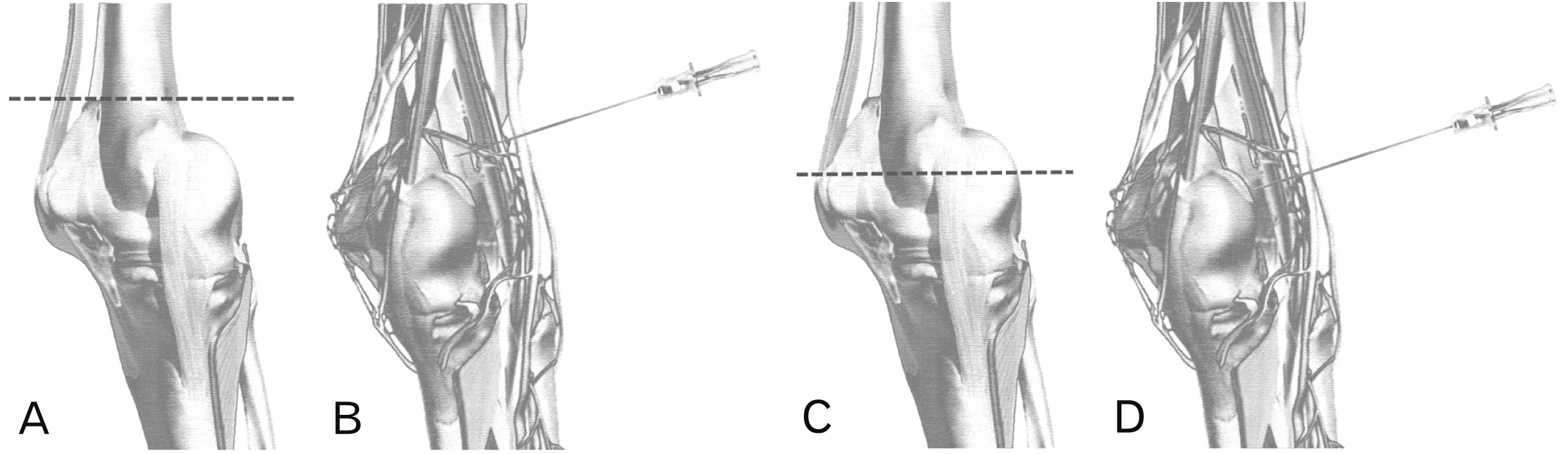 | Fig. 1(A) Schematic drawing of the lateral view of the knee joint displaying the level of the proximal technique (black dotted line). (B) Posteromedial view of the knee displaying the needle insertion for the proximal technique. (C) Schematic drawing of the lateral view of the knee joint displaying the level of the distal technique (black dotted line). (D) Posteromedial view of the knee displaying the needle insertion for the distal technique.
|
For the dissection procedure, cadavers were placed in a prone position. A vertical skin incision was made along the midline of the posterior leg from the gluteal fold to the lower leg (superior calf). Followed by a horizontal skin incision at the level of the popliteal crease. The skin was then reflected laterally to expose the posterior compartment of the thigh and leg. The superficial fascia and subcutaneous fat (fatty lobules) were removed using blunt dissections. The boundaries of the popliteal fossa, namely: superolateral (biceps femoris muscle), superomedial (semimembranosus and semitendinosus muscles), and inferior boundaries (lateral and medial head of the gastrocnemius and the plantaris muscles) were delineated and retracted laterally. The following superficial nerves could then be identified: the sciatic nerve dividing into its two branches, the tibial and common fibular (peroneal) nerves. Subsequently, the deep fascia and fat were dissected without altering the original positions and integrity of the surrounding nerves. Deep to that, the obturator nerve, popliteal artery and vein were identified. The accompanying articular branches of the posterior knee; the superior branches of the tibial nerve, the inferior branches of the tibial nerve, the posterior branch of the common fibular (peroneal) nerve, the posterior branch of the obturator nerve and the branches of the genicular nerves were documented. Further deeper dissection, in anterolateral and anteromedial directions, was done to expose the bony landmarks that correlated with the spread of the injectate dye.
Go to :
Results
This study set out to investigate the spread of dye following the performance of the IPACK block bilaterally in fresh unembalmed cadavers (n=6). Once the dye was introduced, the cadavers were dissected to track the spread of dye within the interspace. Table 1 below summarizes the spread of dye following proximal and distal injections.
Table 1
Summary of the spread of dye and structures stained following proximal and distal interspace between the popliteal artery and the capsule of the posterior knee injections
![]()
The results displayed effective spread in all but one case (therefore n=5). For the proximal injection, no surface (superficial) staining was seen. Upon deeper dissection, staining was noted around the capsule of the knee and its associated nerves (Fig. 2). Cephalad-caudal dye spread spanned from the level just inferior to the adductor hiatus to the superior border of the femoral condyles, however, limited spread was noted towards the medial compartment of the knee. Distal injections displayed no surface (superficial) staining. At a deeper level, staining was noted on the posteromedial border of the semimembranosus (and semitendinosus) muscles, the capsule of the knee and its associated nerves (Fig. 3). Cephalad-caudal dye spread was traced from the adductor hiatus to the tibial tuberosity. Additionally, spread towards the medial and lateral compartments of the knee was noted. Lastly, one case was noted as a failed block as no staining was seen within the fossa. Further dissection revealed superior staining at the supracondylar area. This could be a result of needle displacement prior to the administration of the injectate.
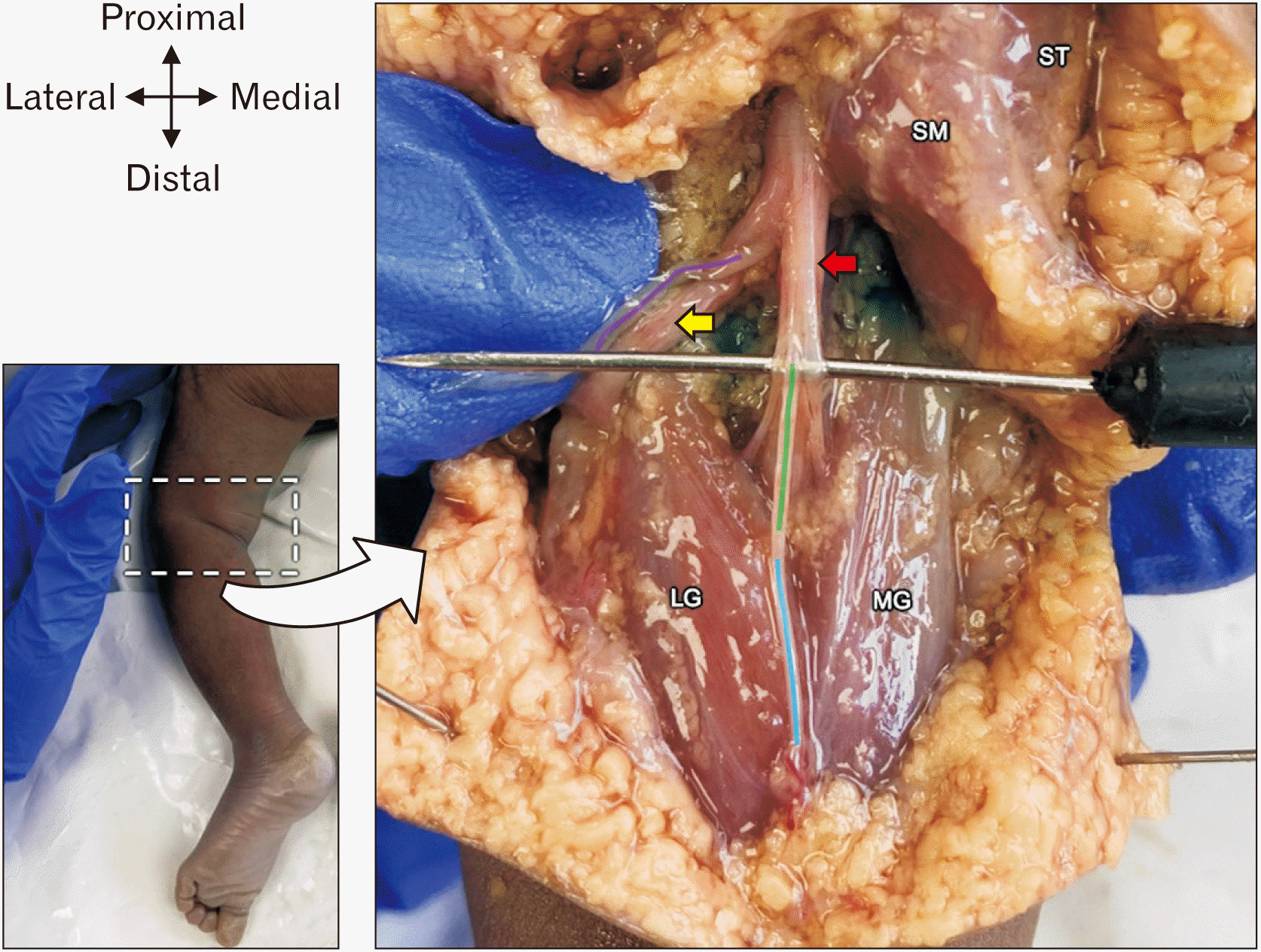 | Fig. 2Dissection following a proximal interspace between the popliteal artery and the capsule of the posterior knee injection. Purple line, branch of the sural nerve from the common fibular (peroneal) nerve (cut and reflected); green line, branch of the sural nerve from the tibial nerve; blue line, sural nerve; red arrow, tibial nerve; yellow arrow, common fibular (peroneal) nerve; LG, lateral head of gastrocnemius muscle; MG, medial head of gastrocnemius muscle; SM, semimembranous muscle; ST, semitendinosus muscle.
|
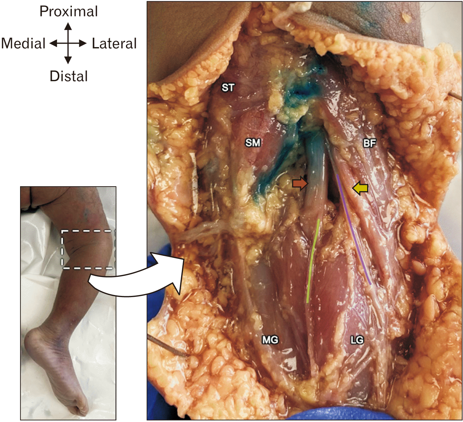 | Fig. 3Dissection following a distal interspace between the popliteal artery and the capsule of the posterior knee injection. Purple line, branch of the sural nerve from the common fibular (peroneal) nerve (cut); green line, branch of the sural nerve from the tibial nerve; red arrow, tibial nerve; yellow arrow, common fibular (peroneal); LG, lateral head of gastrocnemius muscle; MG, medial head of gastrocnemius muscle; BF, biceps femoris muscle; SM, semimembranous muscle; ST, semitendinosus muscle.
|
Furthermore, proximal injections resulted in no staining of the nerve trunks—tibial and common fibular (peroneal) nerves, with some staining of the articular branches if they were present. While the distal injections resulted in staining of all four articular branches—superior and inferior tibial branches, the posterior branch of common fibular (peroneal) and the posterior branch of the obturator nerve—as well as the tibial and common fibular (peroneal) nerves (Fig. 4). Additional staining of the superior lateral genicular nerve and artery, as well as the superior medial genicular nerve and the artery, was noted in four out of the five cases (Fig. 5). Overall, the superior and inferior articular branches of the tibial nerve were stained in all cases (5/5), while the posterior branch of the common fibular (peroneal) and posterior branch of the obturator nerves were stained in four out of the five cases and three out of the five cases, respectively. Staining of the genicular vessels was noted more frequently following distal injections.
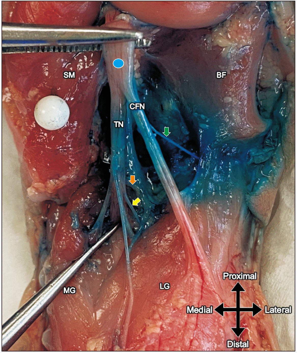 | Fig. 4Staining of methylene blue dye following a distal interspace between the popliteal artery and the capsule of the posterior knee injection. Green arrow, posterior branch of the common fibular (peroneal) nerve; orange arrow, articular branch of the superior branch of the tibial nerve; yellow arrow, articular branch of the inferior branch of the tibial nerve; blue dot, sciatic nerve; LG, lateral head of gastrocnemius muscle; MG, medial head of gastrocnemius muscle; BF, biceps femoris muscle (cut); SM, semimembranous muscle (cut); TN, tibial nerve; CFN, common fibular (peroneal) nerve.
|
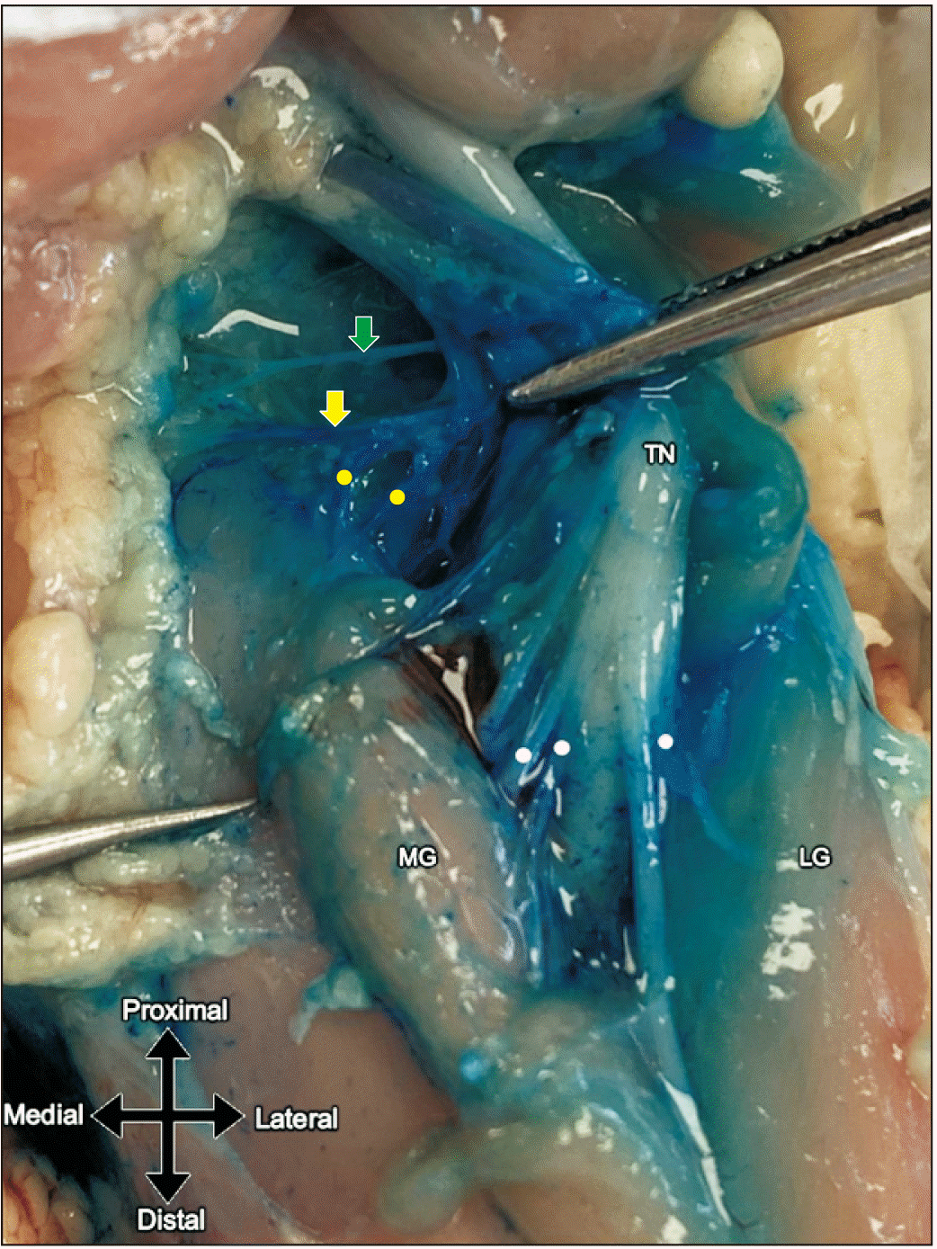 | Fig. 5Staining of methylene blue dye following a distal interspace between the popliteal artery and the capsule of the posterior knee injection. Green arrow, superior medial genicular nerve; yellow arrow, superior medial genicular artery; yellow dots, articular branches from the superior medial genicular artery; white dots, muscular branches from the tibial nerve; TN, tibial nerve; LG, lateral head of gastrocnemius muscle; MG, medial head of gastrocnemius muscle.
|
Overall, both techniques produced posterior staining and interestingly, anterior staining to a certain extent. Additionally, the proximal technique preserved staining of the main trunks, while the distal technique did not.
Go to :
Discussion
Recently there has been an expansion of regional anesthesia for pain management in a pediatric population. Pediatric orthopaedic knee surgery requires evidence-based studies demonstrating the safety and efficacy of peripheral nerve blocks. These blocks are advantageous as they are efficient and offer site-specific analgesia and a decreased need for opioids [4]. The IPACK block, a novel interspace block that is centralised in the popliteal fossa, targets the popliteal plexus responsible for innervating the posterior capsule of the knee joint. This block is hypothesised to be an alternative to the existing traditional blocks used for total knee arthroplasty and anterior cruciate ligament repair [5]. This cadaveric study aims to replicate the spread of the IPACK block in a neonatal sample while comparing proximal versus distal spread.
The results displayed adequate coverage within the popliteal fossa when using 0.3 ml/kg of injectate. Most of the articular branches were stained irrespective of the technique used. The inadvertent anterior spread in all cases could be explained by the needle trajectory during the insertion and withdrawal, as well as the continuous injectate administration upon needle withdrawal for both the proximal and distal techniques. The wide distribution of spread of the injectate when simulating the IPACK block, reveals a potential for pain relief of the anterior aspect of the knee depending on the nerve involved.
To date, there has only been a handful of studies reporting on the spread of the IPACK block in an adult population [6, 10], while no studies report on a paediatric population.
From the cadaveric studies (a total of three), authors either used frozen, embalmed or lightly embalmed adult cadavers as opposed to the fresh unembalmed paediatric cadavers used in the current study (Table 2). Not only the age (adult versus neonate), and degree of embalmment, but also discrepancies in the material (latex versus dye), concentrate volume and techniques led to diverse findings.
Table 2
Summary of the approach and nerves stained following an interspace between the popliteal artery and the capsule of the posterior knee block by previous authors
| Author (yr) | Approach | Sample size | Sample type | Injectate | Nerves affected by the spread |
|---|---|---|---|---|---|
| Niesen et al. (2019) [10] | Posterior aspect of the femur at the transitional level of the femoral condyles to the shaft & medial epicondyle and lateral epicondyle. | 10 | Fresh frozen adult cadavers | 20 ml latex |
· Tibial nerve (30%) · Common fibular nerve (20%) · Medial genicular nerves (90%) · Posterior branch of obturator nerve (0%) |
| Kampitak et al. (2019) [5] | Lateral border of the popliteal artery & space between the popliteal artery and femoral condyle. | 10 | Embalmed adult cadavers | 8 ml methylene blue dye |
· Tibial nerve · Articular branches of tibial nerve · Popliteal plexus |
| Tran et al. (2019) [6] | Proximal injection: one finger breath superior to the base of the patella & distal injection: one finger breath superior to the femoral condyles. | 14 | Lightly embalmed cadavers | 10 ml methylene blue dye (per injection) |
· Tibial nerve for both techniques (43%) · Common fibular nerve in proximal technique (57%) · Common fibular nerve in distal technique (71%) · Genicular branch of the posterior division of the obturator nerve (100%) · Posterior branch of the common fibular for both techniques (80%) · Superior branch of tibial nerve in distal technique (85%) · Inferior branch of tibial nerve in distal technique (42%) |
![]()
Niesen et al. [10], conducted a study on 10 fresh frozen adult cadavers to evaluate the IPACK spread using latex. The block was performed unilaterally at the posterior aspect of the femur at the transitional level of the femoral condyles to the shaft, proximal to the popliteal crease. Using the in-plane approach, 10 ml of coloured latex was deposited at the posterior aspect of the lateral femur, while another 10 ml was deposited at the posterior aspect of the medial femur. Subsequent to latex polymerisation, dissections were performed. Similar to the current study, injectate spread was observed in the popliteal fossa in a medial to lateral and proximal to distal direction.
Niesen et al. [10], further demonstrated spread in three specimens to the tibial nerve, of which two displayed possible contact with the common fibular (peroneal) nerve, unlike the current study which only displayed spread to these nerves with distal injections. Another specimen in their study demonstrated spread to the common fibular (peroneal) nerve only. Additionally, all but one specimen, as in this study, exhibited spread to the medial genicular artery. The authors concluded that consistent spread to the medial genicular artery suggests blockade of the articular branches from the tibial nerve that accompanies the artery resulting in posterior knee analgesia [10]. Obstruction of these nerves may contribute to the sensory blockade of the posterior knee, however, at the risk of possible unintended motor blockade [19]. Discrepancies between the spread noted by Niesen et al. [10] and the current study could result from the injectate being administered at two separate areas, rather than multiple aliquots during needle withdrawal which were done in this study.
Kampitak et al. [5], examined the distribution of methylene blue dye in 10 embalmed adult cadavers. The block was replicated unilaterally, at the level of the femoral condyles in the popliteal fossa. The dye (8 ml) was introduced at two points: firstly, at the lateral border of the popliteal artery and secondly at the space between the popliteal artery and femoral condyle. Upon dissection, methylene blue dye spread to the articular branches of the tibial nerve and the popliteal plexus within the fossa. Further staining of the tibial nerve was observed. Additionally, staining of the common fibular (peroneal) nerves was not noted in any of the samples. The posterior branch of the obturator nerve was not found in their study [5]. Contrary to the current study, spread revealed bilateral staining of the posterior branch of the obturator nerve in three cases following both the proximal and distal techniques. This could be a result of the single injectate administration, suggesting greater continuous spread when compared to multiple injectate points.
A similar study was conducted by Tran et al. [6], to evaluate the injectate spread in 14 lightly embalmed adult cadavers. Proximal injection techniques were performed on seven of the cadavers, while distal injection techniques were performed on the remaining seven cadavers. For the procedure, 10 ml of methylene blue dye was introduced one fingerbreadth superior to the base of the patella (proximal injection) or superior to the femoral condyle (distal injection) using the in-plane approach. Cephalad to caudad spread between the adductor hiatus and level of the superior border of the femoral condyles was noted in the proximal injections, while the distal injections displayed cephalad to caudad spread between the adductor hiatus and the knee joint line. Additionally, proximal injections demonstrated extensive medial spread to the knee joint which extended anteriorly towards the suprapatellar bursa. However, distal injection spread was limited to the adductor tubercle and femoral epicondyle, medially. Lateral spread to the lateral femoral condyle was also noted. Overall, proximal injections resulted in greater coverage of the anteromedial aspect of the joint capsule, while distal injections resulted in greater coverage of the posterior and anterolateral aspects of the joint capsule [6]. When compared to the current study, lateral spread to the anterior knee capsule, with limited medial spread was noted for both techniques, but more so in the distal technique. The distal injection revealed a limited medial spread in only one case. This can be explained by the medial-to-lateral withdrawal of the needle, which could have influenced the anterolateral spread.
Tran et al. [6], further reported on the frequency of nerves stained. Results revealed the tibial nerve was stained in 43% of the cases for both techniques, whereas the common fibular (peroneal) nerve was stained in 57% and 71% of cases for the proximal and distal techniques, respectively. With regards to the articular branches, the genicular branch of the posterior division of the obturator nerve was stained in both techniques (100%). The posterior branch of the common fibular (peroneal) (80%), superior (85%) and inferior (42%) branches of the tibial nerve were more frequently stained in the distal rather than proximal technique. Additionally, the distal technique revealed a higher frequency of staining in the anterior branch of the common fibular (peroneal), superior lateral genicular and saphenous nerves. Staining of the medial branch of the nerve to vastus intermedi and superior medial genicular nerves was noted superiorly following the proximal techniques [6]. The authors concluded that the distal injection may provide better relief for posterior knee pain. Furthermore, the anteromedial and anterolateral spread following both techniques suggests that the IPACK block may play a role in alleviating anterior knee pain. Similar to Jinadu et al. [7], the current study displayed staining of the tibial, common fibular (peroneal) and articular branches (all four branches to the posterior capsule) more frequently following the distal technique. However, the proximal technique evaded staining the main trunks of the sciatic nerve (Fig. 1), potentially avoiding complications such as motor weakness or foot drop. Greater nerve coverage following distal injections is most likely due to the close proximity of the injectate needle to the popliteal plexus.
The results from the current pediatric cadaveric study is further substantiated by the clinical studies presented in Table 3, as successful coverage of the posterior aspect of the knee following the IPACK block was noted in all cases.
Table 3
Summary of the approach and volume used following successful interspace between the popliteal artery and the capsule of the posterior knee blocks in pediatric and adolescent patients
| Author (yr) | Approach | Sample number | Patient age in years | Volume of anaesthesia |
|---|---|---|---|---|
| Harb et al. (2020) [16] | Immediately superior to the femoral condyles, 2 cm from the lateral border of the popliteal artery. | 3 | 13 | 12 ml 0.5% ropivacaine |
| 14 | 10 ml 0.25% bupivacane | |||
| 14 | 20 ml 0.5% ropivacaine | |||
| Nguyen et al. (2020) [17] | At the level of the femoral condyles, between the lateral border of the popliteal artery and the femoral condyle. | 3 | 13 | 10.2 ml 0.2% ropivacaine |
| 14 | 12.6 ml 0.2% ropivacaine | |||
| 16 | 22.6 ml 0.2% ropivacaine |
![]()
Overall, we postulate that the mechanism of action of the various distributions of the spread of the injectate is attributed to the insertion of the injectate directly posterior to the capsule, together with the injection pressure. This technique allows for spread to travel further posteriorly and mediolaterally along the vascular structures without affecting superficial structures. Additionally, our observations further support the study by Niesen et al. [10], which concluded that spread along the genicular vessels is a possible mechanism of action for targetting the articular branches innervating the posterior capsule of the knee joint.
Although other authors propose that an increase in volume may result in an increased spread [20], further research exploring optimal concentration, dose and volume to provide effective coverage in a paediatric population is required. While this study focused on a neonatal sample, there are no foreseeable reasons why this block will not be appropriate for various paediatric age groups if the correct dosage and volume are applied.
Limitations
A foremost limitation of the study was the sample size, due to the sensitivity of the procurement procedure for paediatric cadavers. Furthermore, the limited age range of the cadavers obtained restricted our results to a neonatal sample, rather than various paediatric age groups. An additional limitation was the use of cadaveric material to replicate the spread in a living model. This may be an inaccurate representation due to the lack of in vivo factors such as tissue resistance, muscle tone, or body temperature which may limit or even enhance the spread of injectate [10, 21]. Other limitations include the speed of injectate, specifically, the variability in the volume and the mixture of dye may not replicate the exact spread of local anaesthesia in vivo [6].
In conclusion, the results from this study augment the few cadaveric reports evaluating the spread of the IPACK block. Furthermore, to our knowledge, this is the only study to date that investigates the IPACK spread in a neonatal sample. Both the proximal and distal techniques provide a similar spread of methylene blue dye in the popliteal fossa, staining the articular branches that are responsible for providing sensory innervation to the posterior capsule of the knee joint. Results from this study reveal staining of some articular branches while preserving the main nerve trunks following the proximal technique. However, greater spread to the articular branches was noted following the distal technique. We believe that the IPACK block can be seen as a more holistic and viable alternative to many lower limb blocks for various age groups (using the correct dosage) in the paediatric population, as it allows for a wider spread in the posterior and medial-lateral compartments of the knee.
Go to :
 XML Download