Abstract
In the present study, anatomical assessment of zygomaticofacial foramina (ZFFs) and zygomatic canals communicating with ZFFs were performed using cadaver micro-computed tomography images. It was suggested that all ZFFs were located above the jugale (Ju)-zygomaxillare (Zm) line, which is the reference line connecting the Ju and Zm, and most were located in the zygomatic body area (ZBA). The anteroposterior position of the ZFF in the ZBA was within a middle to posterior region and was most often located slightly posteriorly in males and closer to the middle of the region in females. The mean distance from the Ju-Zm line to the ZFF in the ZBA was 12.36 mm (standard deviation [SD] 1.52 mm) in males and 11.48 mm (SD 1.61 mm) in females. In zygomatic canals communicating with ZFFs, most zygomatic canals were type I canals, communicating from the zygomaticoorbital foramen and harboring the zygomaticofacial nerve, and the others were type II canals, communicating from the zygomaticotemporal foramen and located near the posterior margin of the frontal process. These results provide useful anatomical information for preventing nerve injury during surgical procedures for zygomatic implant treatment.
Zygomatic implant treatment, introduced by Brånemark, is effective for the rehabilitation of severely resorbed maxillae. The zygomatic implant is placed in the second premolar region such that it traverses the maxillary sinus and is embedded within the body of the zygomatic bone [1-7]. During the surgical procedure, the infraorbital nerve could be damaged by excessive subperiosteal dissection, inadvertent transection, or inadvertent compression by the tip of the retractor causing postoperative paresthesia, anesthesia, or dysesthesia [7-10]. It is also believed that damage to the zygomaticofacial nerve (ZFN) may be caused during the procedure [10-12].
The zygomatic nerve originates in the pterygopalatine fossa, enters the orbit through the inferior orbital fissure, and is divided into two branches that form the ZFN and zygomaticotemporal nerve (ZTN). These branches enter the zygomatic canal (within the zygomatic bone) through the zygomaticoorbital foramen (ZOF) and continue to the temporal and facial regions through the zygomaticotemporal foramen (ZTF) and zygomaticofacial foramen (ZFF), respectively (Fig. 1). The skin on the cheek region is innervated predominantly by the ZFN emerging from the ZFF [13-15]. Therefore, the location of the ZFF, where the ZFN emerges, is considered to be an important area to pay attention to when reflecting a mucoperiosteal flap around the cheek region. Previous studies have mainly reported numbers and locations of zygomatic foramina using dry skulls [16-25], with some reporting anatomical findings of the ZFN exiting the zygomatic foramen [26]. Recently, there has also been a report that involved observation of the zygomatic canal using micro-computed tomography (CT) analysis of seven cadavers [27]. Although these anatomical reports have provided knowledge that enables the prevention of iatrogenic injuries during surgical procedures [18-22], there are few reports of anatomical studies focusing on zygomatic implant treatment.
To obtain useful anatomical information for zygomatic implant treatment, a reference line suitable for measurement is needed. In anatomical studies, it has been reported by Kato et al. [28] that the jugale (Ju)-zygomaxillare (Zm) line can be used as a landmark for the zygomatic implant placement site. Furthermore, the Ju-Zm line may be considered a guideline for the subperiosteal detachment of the zygomatic bone, because the lateral surface of the zygomatic bone is peeled from the zygomatic alveolar crest toward the notch [2-7].
In the present study, the location and diameter of ZFFs and the courses of zygomatic canals communicating with ZFFs were investigated to provide insights that will further aid in the prevention of iatrogenic damage to the nerve during surgical procedures for zygomatic implant treatment.
The research was performed with the approval of the Ethics Committee of the Nippon Dental University School of Life Dentistry at Tokyo (approval no. NDU-T2020-19).
The study sample comprised cadavers of Japanese adults used for anatomy education. All cadavers were kept in the anatomical storage room of The Nippon Dental University School of Life Dentistry at Tokyo. A total of 104 sides of zygomatic bone specimens fixed in 10% formalin from 52 cadavers (26 males and 26 females; average age, 86.4 years; age range, 61–101 years) were examined. These were randomly selected without consideration for the presence or absence of teeth.
The sample size calculation was conducted using G-Power version 3.1.9.7 (Heinrich Heine Universität Düsseldorf Allgemeine Psychologie und Arbeitspsychologie) software for Student’s t-test with 80% power and 5% significance. The minimum sample size to detect significant differences between groups was calculated to be 52 persons (26 per group).
Cadavers with intact zygomatic bones were subjected to micro-CT analysis (ScanXmate-D100SS270; Comscantecno). Samples were placed with the Camper plane parallel to the sample stage. A total of 1,471 slice images (slice thickness of 50 µm; pixel size of 50 µm in 1,856×1,856 matrices) were obtained as Tagged Image File Format (TIFF) or Digital Imaging and Communications in Medicine (DICOM) files for each sample with a tube voltage of 100 kV, tube current of 200 μA, and a 0.8-mm Cu filter. The DICOM files were converted to Standard Triangulated Language (STL) files using Molcer software version 1.8.0.0 (White Rabbit).
To enable three-dimensional (3D) analysis of the imaging data, images were imported as TIFF files to TRI/3D-BON software (Ratoc System Engineering) and as STL files to GOM Inspect 2017 software (GOM). TRI/3D-BON software was primarily used to visualize the zygomatic canals, and GOM Inspect software was used to determine ZFF position.
The canals and foramina of the zygomatic bone were identified on micro-CT images (Fig. 2). The canal connecting the orbital, temporal, or lateral surfaces of the zygomatic bone was defined as the zygomatic canal, and the foramina connecting to the canal from those respective surfaces were defined as the ZOF (Fig. 2A), ZTF (Fig. 2B), and ZFF (Fig. 2C). The identified canals and foramina were analyzed anatomically and measured.
The zygomatic foramina (ZOF, ZTF, and ZFF) identified on micro-CT images were counted, with the number of each recorded. A slice image at the ZFF level was selected (Fig. 2C), and the ZFF diameter was measured using Adobe Photoshop 2022 software (Adobe Inc.), as shown in Fig. 2D. The ZFF location was determined based on a reference line connecting the Ju and Zm (Ju-Zm line), which has relevance to zygomatic implant treatment (Fig. 3A–C). The Ju-Zm line is an anthropological reference, where Ju is the most concave point at which the temporal and frontal processes of the zygomatic bone meet, and Zm is the most inferior point of the zygomaticomaxillary suture. The lateral surface of the zygomatic bone was divided into three regions, the frontal process area (FPA), zygomatic body area (ZBA), and temporal process area (TPA) (Fig. 3A). To determine the anteroposterior location of the ZFF in the ZBA, the zygomatic bone was anteroposteriorly divided into five equal parts (A, anterior area; AM, anterior middle area; M, middle area; PM, posterior middle area; P, posterior area) based on the Ju-Zm line, facilitating the assessment of the anteroposterior location of the ZFF (Fig. 3B). Based on the location of the ZFF in the ZBA, the distance between the ZFF and Ju-Zm line was measured (Fig. 3C).
A 3D representation of the zygomatic canals is shown in Fig. 4A. The courses of zygomatic canals vary. The present study focused on the zygomatic canal communicating with the ZFF (Fig. 4B), with the course of the canal classified as either type I or II, depending on whether the canal communicated with the ZOF (Fig. 4B, C). Type I canals were further classified as type Ia or Ib, depending on whether they communicated with the ZTF.
Table 1 shows the numbers of zygomatic foramina (ZOF, ZTF, and ZFF) identified on micro-CT images. The ZFF, the focus of the present study, was either absent or ranged in number from one to four foramina per zygomatic bone. One and two ZFFs were most commonly identified in males and females, respectively; females had significantly more ZFFs than males.
Diameters of the ZFFs were measured on micro-CT slice images (Fig. 2C, D). Mean ZFF diameters were 1.09 mm (standard deviation [SD] 0.31 mm) in males and 1.03 mm (SD 0.32 mm) in females (Fig. 5). No significant sex-related difference in ZFF diameter was observed.
To determine the overall location of the ZFF on the lateral surface of the zygomatic bone, the zygomatic bone was divided into three areas (FPA, ZBA, and TPA) (Fig. 3A). The proportion of ZFFs located in the ZBA was 79.3% in males and 77.5% in females, whereas that of the ZFF in the FPA was 20.7% in males and 22.5% in females. The ZFF was not observed in the TPA in either males or females (Fig. 6). Furthermore, there was no significant difference in ZFF numbers in any area between males and females.
The anteroposterior location (A, AM, M, PM, and P) (Fig. 3B) of the ZFF in the ZBA was investigated. No ZFF was observed in area A in either males or females. Moreover, ZFFs were observed in area AM in 1.4% of males and 7.5% of females, area M in 26.1% of males and 39.8% of females, area PM in 52.2% of males and 30.1% of females, and area P in 20.3% of males and 22.6% of females (Fig. 7). A significant difference in the anteroposterior location between males and females was seen.
Focusing on zygomatic canals communicating with ZFFs (Fig. 4A, B), their courses are shown in Figs. 4C, 9. Type I was 93.2% (type Ia, 49.3%; type Ib, 43.8%) in males and 85.7% (type Ia, 59.0%; type Ib, 26.7%) in females (Table 2). Type II canals were identified in 6.8% of males and 14.3% of females.
In addition, ZFF sites (Fig. 3A) in type I canals and type II canals were investigated. ZFFs with type I canals were located in the ZBA in 84.1% of males and 88.6% of females and in the FPA in 15.9% of males and 11.4% of females (Table 2). Conversely, in both males and females, all ZFFs with type II canals were located in the FPA, and none were located in the ZBA. Furthermore, all type II ZFFs were located near the posterior margin of the frontal process of the zygomatic bone, and the ZFF was immediately behind the ZTF.
Paresthesia, anesthesia, and dysesthesia in the cheek region have been reported as postoperative complications in zygomatic implant treatment [7-12]. The reason for these complications could result from intra-operative subperiosteal dissection [8, 10-12] or implant placement [10]. The aim of the present study was to provide anatomical information that will help prevent iatrogenic damage to the nerve during surgical procedures for zygomatic implant treatment. The locations and diameters of ZFFs were investigated anatomically, and the courses of zygomatic canals communicating with ZFFs were examined in cadavers by micro-CT analysis. Because the ZFN and the ZTN, which are branches of the zygomatic nerve, are located within the zygomatic bone, it is difficult to determine their courses. Most previous studies investigated numbers and locations of zygomatic foramina in dry skulls by macroscopic observation [16-25]. In the present study, the canal connecting the orbital, temporal, or lateral surfaces of the zygomatic bone was defined as the zygomatic canal on micro-CT images, and the openings of the foramina of the zygomatic canal were defined as ZOF, ZTF, and ZFF respectively. Therefore, the assessment performed was considered to be highly objective and reproducible. However, the present results showed an overall trend toward a higher number of zygomatic foramina compared to previous reports [23, 24], because smaller foramina can be detected with high accuracy.
In the present study, the Ju-Zm line, which was reported on previously [28], was used as an anatomical landmark for zygomatic implant placement. Moreover, it was also considered that the Ju-Zm line can be used as a guideline for subperiosteal detachment of the zygomatic bone, because the lateral surface of the zygomatic bone is peeled from the zygomatic alveolar crest toward the notch [2-7]. Minute canal structures on micro-CT images can be identified and visualized three-dimensionally using 3D imaging software [27, 29]. The 3D analysis of the zygomatic canals showed that the ZFF was located closest to the Ju-Zm line in the zygomatic canal with the ZFN. Thus, it seemed that the ZFF is the most sensitive position for zygomatic implant placement, as well as for subperiosteal detachment. In comparing the ZBA, FPA, and TPA using the Ju-Zm line, all ZFFs were located above the Ju-Zm line, with most of them in the ZBA. The anteroposterior position of the ZFF in the ZBA was within a middle to posterior region and was most often located slightly posteriorly in males and closer to the middle of the region in females. These results provide useful information as an overview of ZFF location.
The mean distance from the Ju-Zm line to the ZFF in the ZBA was 12.36 mm in males and 11.48 mm in females. More interestingly, the closest distance from the Ju-Zm line to the ZFF was 7.70 mm. The diameter of a zygomatic implant is smaller than this distance. Therefore, these results suggested that, though placing an implant close to the orbital border can cause damage to the ZFN, appropriately placing the zygomatic implant toward the zygomatic notch while following standard surgical technique should not result in any nerve injury. It seemed that the distance from the Ju-Zm line to the ZFF was also useful information for performing subperiosteal dissection on the lateral surface of the zygomatic bone of the zygomatic bone.
von Arx et al. [30] reported four cases in which soft tissue bundles emerging from the accessory mental foramen were excised during apical resection of mandibular molars. Nerve paralysis was observed in one patient with an accessory mental foramen diameter of 1.7 mm. The other three patients, who had foramen diameters of 1.5, 0.8, and 0.4 mm developed no noticeable symptoms. Because the presence of nerve paralysis is considered to depend on the diameter of the foramen through which the nerve passes, the information about the diameter of the ZFF is important in clinical practice. In the present study, the mean ZFF diameter was 1.09 mm in males and 1.03 mm in females, and the diameter of the ZFF was smaller than of the mental foramen or the infraorbital foramen [31-37]. However, since there are some ZFFs with diameters larger than 1.5 mm, it seems necessary to pay attention to the size of the ZFFs.
To investigate the courses of zygomatic canals communicating with ZFFs, the canals were classified as either type I or II, depending on whether the zygomatic canal communicated with the ZOF. Most zygomatic canals were type I (males, 93.2%; females, 85.7%), and their ZFFs were most often located in the ZBA (males, 84.1%; females, 88.6%) (as shown in Table 2). On the other hand, the other canals were type II (males, 6.8%; females, 14.3%), and all their ZFFs were located in the FPA, near the posterior margin of the frontal process of the zygomatic bone. The ZFN enters the zygomatic bone at the orbit through the ZOF, passes through the zygomatic canal, exits at the ZFF, and supplies sensory innervation to the zygomatic region [13-15]. Thus, it is obvious that the ZFN runs within a type I canal. In contrast, the type II canal communicates from the ZTF located on the side of the temporal fossa to the ZFF. The nerves passing within type II canals are unknown, and identifying the nerves running along these courses is of great interest. Siddiqui et al. [38] recently reported that, though relatively uncommon, the ZTN can pierce the marginal process (marginal tubercle) of the zygomatic bone and can thus be located superficially to the cheek region. All ZFFs that were classified as type II in the present study were located near the posterior margin of the FPA; thus, type II canals likely harbor the ZTNs that innervate the cheek. These results suggest that the nerves passing through ZFFs include not only the ZFN, but also the ZTN.
In the present study, the ZFF and zygomatic canals communicating with the ZFF were investigated anatomically using cadaver micro-CT images. The present study provides valuable information for preventing nerve injury during surgical procedures for zygomatic implant treatment.
Acknowledgements
The authors would like to thank Dr. Yasuhiro Kizu of Tokyo Dental College for providing clinical advice regarding zygomatic implant surgery in the study. The authors would also like to thank Dr. Yoshiki Ishida and Fusako Mitsuhashi of The Nippon Dental University School of Life Dentistry at Tokyo for their helpful suggestions on three-dimensional measurement and statistical analysis, respectively.
Notes
References
1. Brånemark PI, Gröndahl K, Worthington P. Osseointegration and autogenous onlay bone grafts: reconstruction of the edentulous atrophic maxilla. Quintessence Publishing Co;2001. p. 112–34. DOI: 10.56373/2002-3-24.
2. Brånemark PI. The zygomaticus fixture: clinical procedures. Nobel Biocare;1998.
3. Bedrossian E, Stumpel L 3rd, Beckely ML, Indresano T. 2002; The zygomatic implant: preliminary data on treatment of severely resorbed maxillae. A clinical report. Int J Oral Maxillofac Implants. 17:861–5. Erratum in: Int J Oral Maxillofac Implants 2003;18:292.
4. Malevez C, Daelemans P, Adriaenssens P, Durdu F. 2003; Use of zygomatic implants to deal with resorbed posterior maxillae. Periodontol 2000. 33:82–9. DOI: 10.1046/j.0906-6713.2002.03307.x. PMID: 12950843.

5. Malevez C, Abarca M, Durdu F, Daelemans P. 2004; Clinical outcome of 103 consecutive zygomatic implants: a 6-48 months follow-up study. Clin Oral Implants Res. 15:18–22. DOI: 10.1046/j.1600-0501.2003.00985.x. PMID: 15005100.

6. Stella JP, Warner MR. 2000; Sinus slot technique for simplification and improved orientation of zygomaticus dental implants: a technical note. Int J Oral Maxillofac Implants. 15:889–93. PMID: 11151591.
7. Aparicio C, Alandez J. Zygomatic implants: the anatomy-guided approach. Quintessence Publishing Co.;2012. p. 90–110. p. 245
8. Bedrossian E, Bedrossian EA. 2018; Prevention and the management of complications using the Zygoma implant: a review and clinical experiences. Int J Oral Maxillofac Implants. 33:e135–45. DOI: 10.11607/jomi.6539. PMID: 30231096.

9. Kahnberg KE, Henry PJ, Hirsch JM, Ohrnell LO, Andreasson L, Brånemark PI, Chiapasco M, Gynther G, Finne K, Higuchi KW, Isaksson S, Malevez C, Neukam FW, Sevetz E Jr, Urgell JP, Widmark G, Bolind P. 2007; Clinical evaluation of the zygoma implant: 3-year follow-up at 16 clinics. J Oral Maxillofac Surg. 65:2033–8. DOI: 10.1016/j.joms.2007.05.013. PMID: 17884535.

10. Chrcanovic BR, Abreu MH. 2013; Survival and complications of zygomatic implants: a systematic review. Oral Maxillofac Surg. 17:81–93. DOI: 10.1007/s10006-012-0331-z. PMID: 22562293.

11. Bedrossian E. 2010; Rehabilitation of the edentulous maxilla with the zygoma concept: a 7-year prospective study. Int J Oral Maxillofac Implants. 25:1213–21. PMID: 21197500.
12. Reichert TE, Kunkel M, Wahlmann U, Wagner W. 1999; The zygomatic implant indications and first clinical experiences. Z Zahnarztl Implantol. 15:65–70.
13. Standring S. Gray's anatomy: the anatomical basis of clinical practice. 40th ed. Elsevier/Churchill Livingstone;2008. p. 493–4.
14. Hollinshead WH. Anatomy for surgeons. 3rd ed. Harper & Row;1982. p. 146–318.
15. Wolff E, Last RJ. Anatomy of the eye and orbit. 5th ed. Saunders Company;1961. p. 6–10. p. 285–6.
16. Loukas M, Owens DG, Tubbs RS, Spentzouris G, Elochukwu A, Jordan R. 2008; Zygomaticofacial, zygomaticoorbital and zygomaticotemporal foramina: anatomical study. Anat Sci Int. 83:77–82. DOI: 10.1111/j.1447-073X.2007.00207.x. PMID: 18507616.

17. Aksu F, Ceri NG, Arman C, Zeybek FG, Tetik S. 2009; Location and incidence of the zygomaticofacial foramen: an anatomic study. Clin Anat. 22:559–62. DOI: 10.1002/ca.20805. PMID: 19418451.

18. Hwang SH, Jin S, Hwang K. 2007; Location of the zygomaticofacial foramen related to malar reduction. J Craniofac Surg. 18:872–4. DOI: 10.1097/scs.0b013e3180a03353. PMID: 17667680.

19. Melchenko SA, Cherekaev VA, Alyoshkina OY, Danilov GV, Musa G, Strunina UV, Golbin DA, Lasunin NV, Zaychenko AA. 2022; Assessing the reliability of zygomatic bone landmarks as guides to reach the inferior orbital fissure in orbitozygomatic osteotomy: anatomical study of 83 human skulls. Neurosurg Rev. 45:2175–82. DOI: 10.1007/s10143-021-01726-8. PMID: 35028786.

20. Gupta T, Gupta SK. 2009; The ZMF: Is it a reliable intraoperative guide for the IOF? Clin Anat. 22:451–5. DOI: 10.1002/ca.20783. PMID: 19291758.

21. Ferro A, Basyuni S, Brassett C, Santhanam V. 2017; Study of anatomical variations of the zygomaticofacial foramen and calculation of reliable reference points for operation. Br J Oral Maxillofac Surg. 55:1035–41. DOI: 10.1016/j.bjoms.2017.10.016. PMID: 29122337.

22. Martins C, Li X, Rhoton AL Jr. 2003; Role of the zygomaticofacial foramen in the orbitozygomatic craniotomy: anatomic report. Neurosurgery. 53:168–72. discussion 172–3. DOI: 10.1227/01.NEU.0000068841.17293.BB. PMID: 12823886.

23. Zhao Y, Chundury RV, Blandford AD, Perry JD. 2018; Anatomical description of zygomatic foramina in African American skulls. Ophthalmic Plast Reconstr Surg. 34:168–71. DOI: 10.1097/IOP.0000000000000905. PMID: 28369018. PMCID: PMC5623600.

24. Deana NF, Alves N. 2020; Frequency and location of the zygomaticofacial foramen and its clinical importance in the placement of zygomatic implants. Surg Radiol Anat. 42:823–30. DOI: 10.1007/s00276-020-02455-1. PMID: 32246188.

25. Mangal A, Choudhry R, Tuli A, Choudhry S, Choudhry R, Khera V. 2004; Incidence and morphological study of zygomaticofacial and zygomatico-orbital foramina in dry adult human skulls: the non-metrical variants. Surg Radiol Anat. 26:96–9. DOI: 10.1007/s00276-003-0198-7. PMID: 15004726.

26. Iwanaga J, Badaloni F, Watanabe K, Yamaki KI, Oskouian RJ, Tubbs RS. 2018; Anatomical study of the zygomaticofacial foramen and its related canal. J Craniofac Surg. 29:1363–5. DOI: 10.1097/SCS.0000000000004457. PMID: 29521755.

27. Kim HS, Oh JH, Choi DY, Lee JG, Choi JH, Hu KS, Kim HJ, Yang HM. 2013; Three-dimensional courses of zygomaticofacial and zygomaticotemporal canals using micro-computed tomography in Korean. J Craniofac Surg. 24:1565–8. DOI: 10.1097/SCS.0b013e318299775d. PMID: 24036727.

28. Kato Y, Kizu Y, Tonogi M, Ide Y, Yamane GY. 2005; Internal structure of zygomatic bone related to zygomatic fixture. J Oral Maxillofac Surg. 63:1325–9. DOI: 10.1016/j.joms.2005.05.313. PMID: 16122597.

29. Ide Y, Nakahara T, Nasu M, Matsunaga S, Iwanaga T, Tominaga N, Tamaki Y. 2013; Postnatal mandibular cheek tooth development in the miniature pig based on two-dimensional and three-dimensional X-ray analyses. Anat Rec (Hoboken). 296:1247–54. DOI: 10.1002/ar.22725. PMID: 23749549.

30. von Arx T, Lozanoff S, Bosshardt D. 2014; Accessory mental foramina. Oral Surg. 7:216–27. DOI: 10.1111/ors.12071. PMID: 27146294.
31. Pelé A, Berry PA, Evanno C, Jordana F. 2021; Evaluation of mental foramen with cone beam computed tomography: a systematic review of literature. Radiol Res Pract. 2021:8897275. DOI: 10.1155/2021/8897275. PMID: 33505723. PMCID: PMC7806401.

32. Gümüşok M, Akarslan ZZ, Başman A, Üçok Ö. 2017; Evaluation of accessory mental foramina morphology with cone-beam computed tomography. Niger J Clin Pract. 20:1550–4. DOI: 10.4103/1119-3077.187329. PMID: 29378985.

33. Gungor E, Aglarci OS, Unal M, Dogan MS, Guven S. 2017; Evaluation of mental foramen location in the 10-70 years age range using cone-beam computed tomography. Niger J Clin Pract. 20:88–92. DOI: 10.4103/1119-3077.178915. PMID: 27958253.

34. Kalender A, Orhan K, Aksoy U. 2012; Evaluation of the mental foramen and accessory mental foramen in Turkish patients using cone-beam computed tomography images reconstructed from a volumetric rendering program. Clin Anat. 25:584–92. DOI: 10.1002/ca.21277. PMID: 21976294.

35. Hong JH, Kim HJ, Hong JH, Park KB. 2022; Study of infraorbital foramen using 3-dimensional facial bone computed tomography scans. Pain Physician. 25:E127–32. PMID: 35051160.
36. Désiré A, Ebogo M, Amougou M, Essono N, Zogo O. 2023; Assessment of infraorbital foramen position using computed tomography-scan in a cohort of Cameroonian adults: landmarks in facial surgery and anesthesiology. Pan Afr Med J. 45:134. DOI: 10.11604/pamj.2023.45.134.37733. PMID: 37790162. PMCID: PMC10543902.
37. Dagistan S, Miloǧlu Ö, Altun O, Umar EK. 2017; Retrospective morphometric analysis of the infraorbital foramen with cone beam computed tomography. Niger J Clin Pract. 20:1053–64. DOI: 10.4103/1119-3077.217247. PMID: 29072226.

38. Siddiqui HF, Konschake M, Ottone NE, Olewnik Ł, Iwanaga J, Aysenne A, Xu L, Tubbs RS. 2023; A marginal process of the zygomatic bone predicts a lateral exit of the zygomaticotemporal nerve: an anatomical study with application to surgery around the midface. Clin Anat. 36:708–14. DOI: 10.1002/ca.24021. PMID: 36752958.

Fig. 1
Schema of two branches (ZFN and ZTN) of the zygomatic nerve passing through the zygomatic bone. ZFN, zygomaticofacial nerve; ZTN, zygomaticotemporal nerve; ZOF, zygomaticoorbital foramen; ZTF, zygomaticotemporal foramen; ZFF, zygomaticofacial foramen.
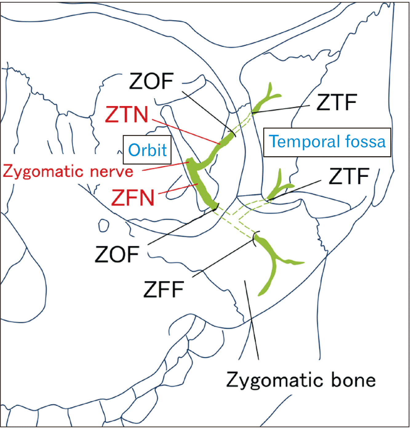
Fig. 2
Micro-CT images of zygomatic foramina. Slices of images obtained at the ZOF (A), ZTF (B), and ZFF levels (C, D), and a 3D image (E) of the zygomatic bone are shown. The ZOF, ZTF, and ZFF are identified on slice images, and the diameter of the ZFF is measured (D). ZOF, zygomaticoorbital foramen; ZTF, zygomaticotemporal foramen; ZFF, zygomaticofacial foramen; CT, computed tomography; 3D, three-dimensional.
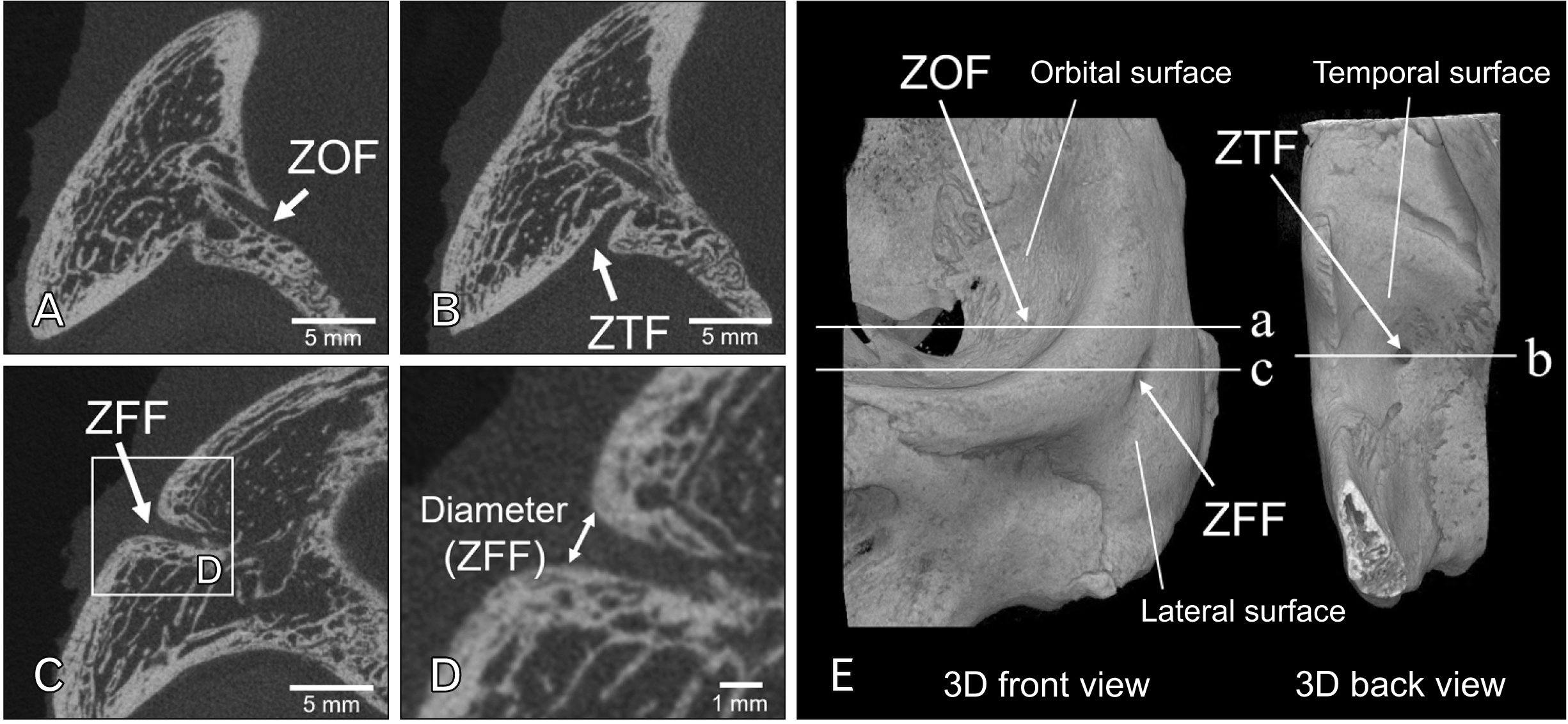
Fig. 3
Assessment of the location of the ZFF. The Ju-Zm line, which connects Ju and Zm, is established as a reference for zygomatic implant placement. Division of the lateral surface into three areas (FPA, ZBA, TPA) allows investigation of the location of the ZFF (A). To determine the anteroposterior location of the ZFF in the ZBA, the zygomatic bone is anteroposteriorly divided into five equal parts (A, AM, M, PM, P) based on the Ju-Zm to facilitate anteroposterior location assessment (B). Determining the location of the ZFF in the ZBA facilitates measuring the distance between the Ju-Zm line and ZFF (C). ZFF, zygomaticofacial foramen; Ju, jugale; Zm, zygomaxillare; FPA, frontal process area; ZBA, zygomatic body area; TPA, temporal process area; A, anterior area; AM, anterior middle area; M, middle area; PM, posterior middle area; P, posterior area.
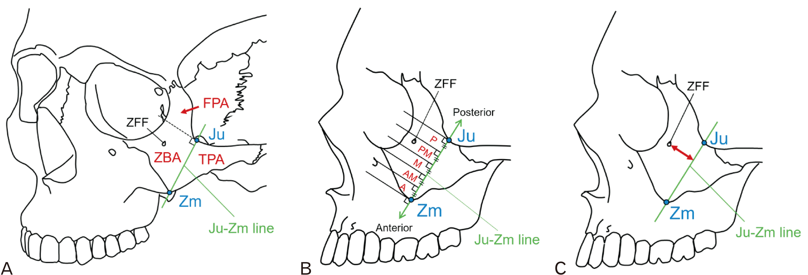
Fig. 4
Assessment of zygomatic canals communicating with ZFFs. A 3D representation of zygomatic canals is shown in (A). The zygomatic canal communicating with the ZFF is the focus of the present study (B). The canal is classified as either type I or II, depending on whether its course communicates with the ZOF. *Type I canals are further classified as type Ia (98 canals) or Ib (60 canals), depending on whether they communicate with the ZTF. The courses of type Ia, Ib, and II canals are shown in (C). ZOF, zygomaticoorbital foramen (green arrowheads); ZTF, zygomaticotemporal foramen (blue arrowheads); ZFF, zygomaticofacial foramen (orange arrowheads); 3D, three-dimensional.
Fig. 5
Diameter of the ZFF. ZFF, zygomaticofacial foramen. There is no significant difference between males and females (P>0.05, t-test).
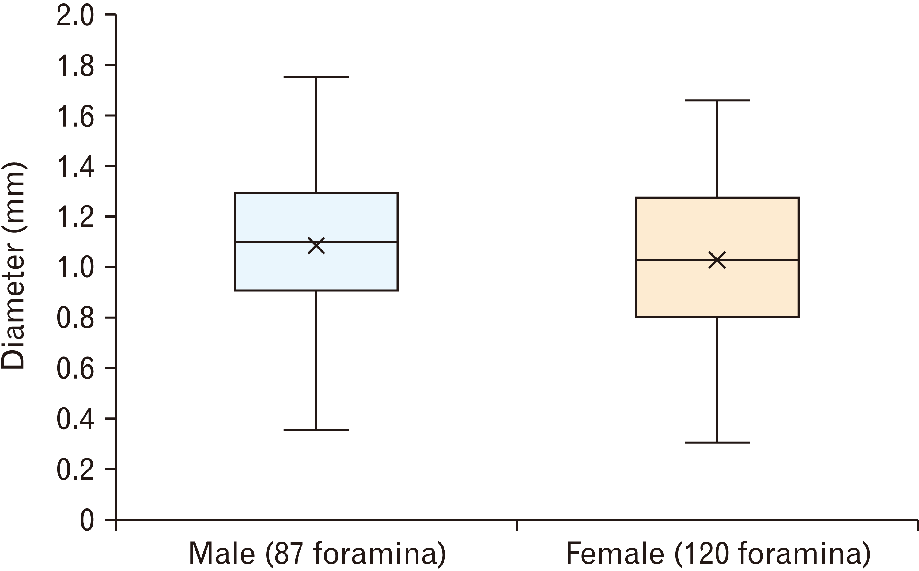
Fig. 6
Location of the ZFF in three zygomatic bone regions: the ZBA, FPA, and TPA (see Fig. 3A). ZFF, zygomaticofacial foramen; ZBA, zygomatic body area; FPA, frontal process area; TPA, temporal process area. No significant difference between males and females is observed (P>0.05, chi-squared test).
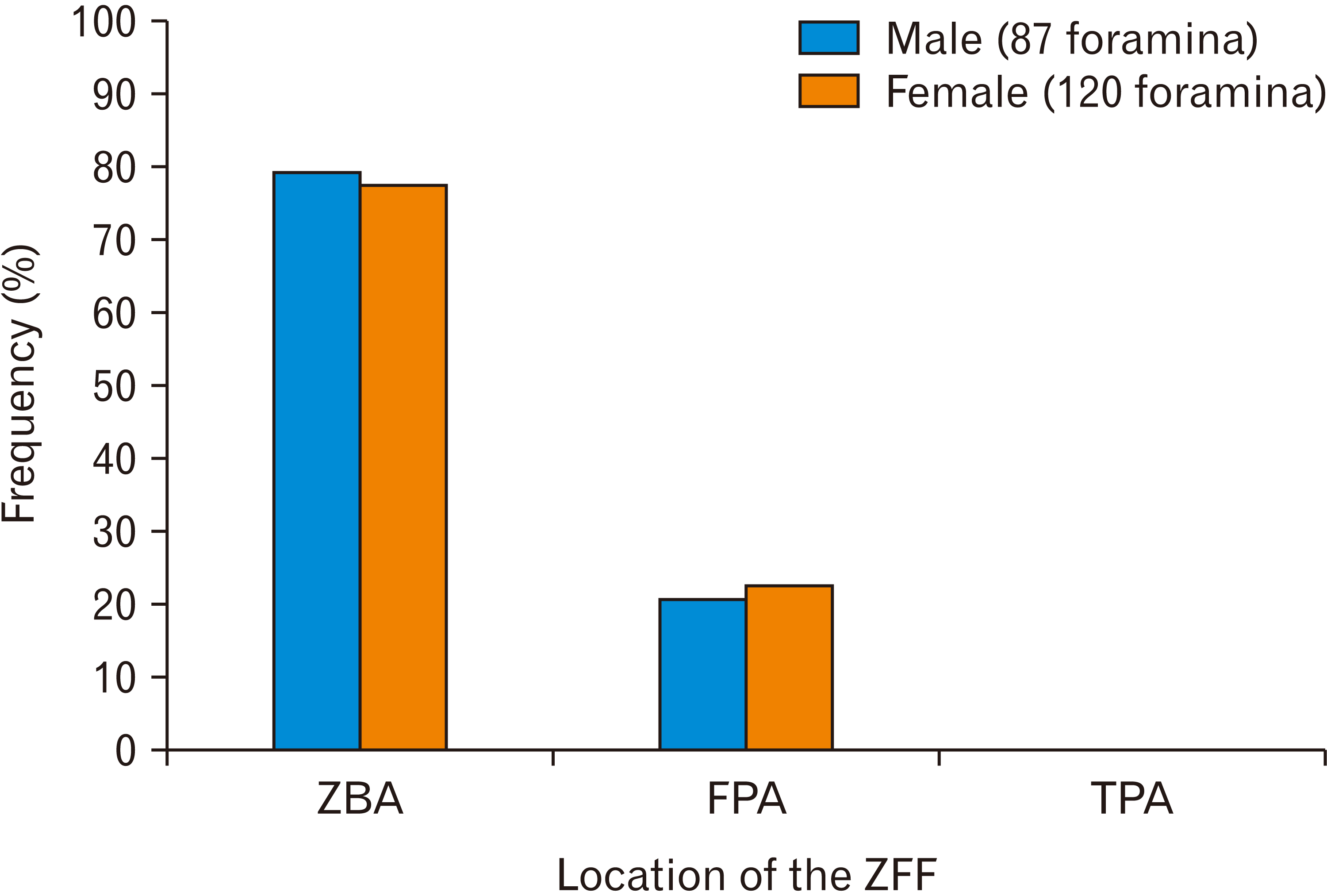
Fig. 7
Anteroposterior location of the ZFF in the ZBA in the following five areas: A, AM, M, PM, and P (see Fig. 3B). ZFF, zygomaticofacial foramen; ZBA, zygomatic body area; A, anterior area; AM, anterior middle area; M, middle area; PM, posterior middle area; P, posterior area. A significant difference between males and females is seen (P<0.05, chi-squared test).
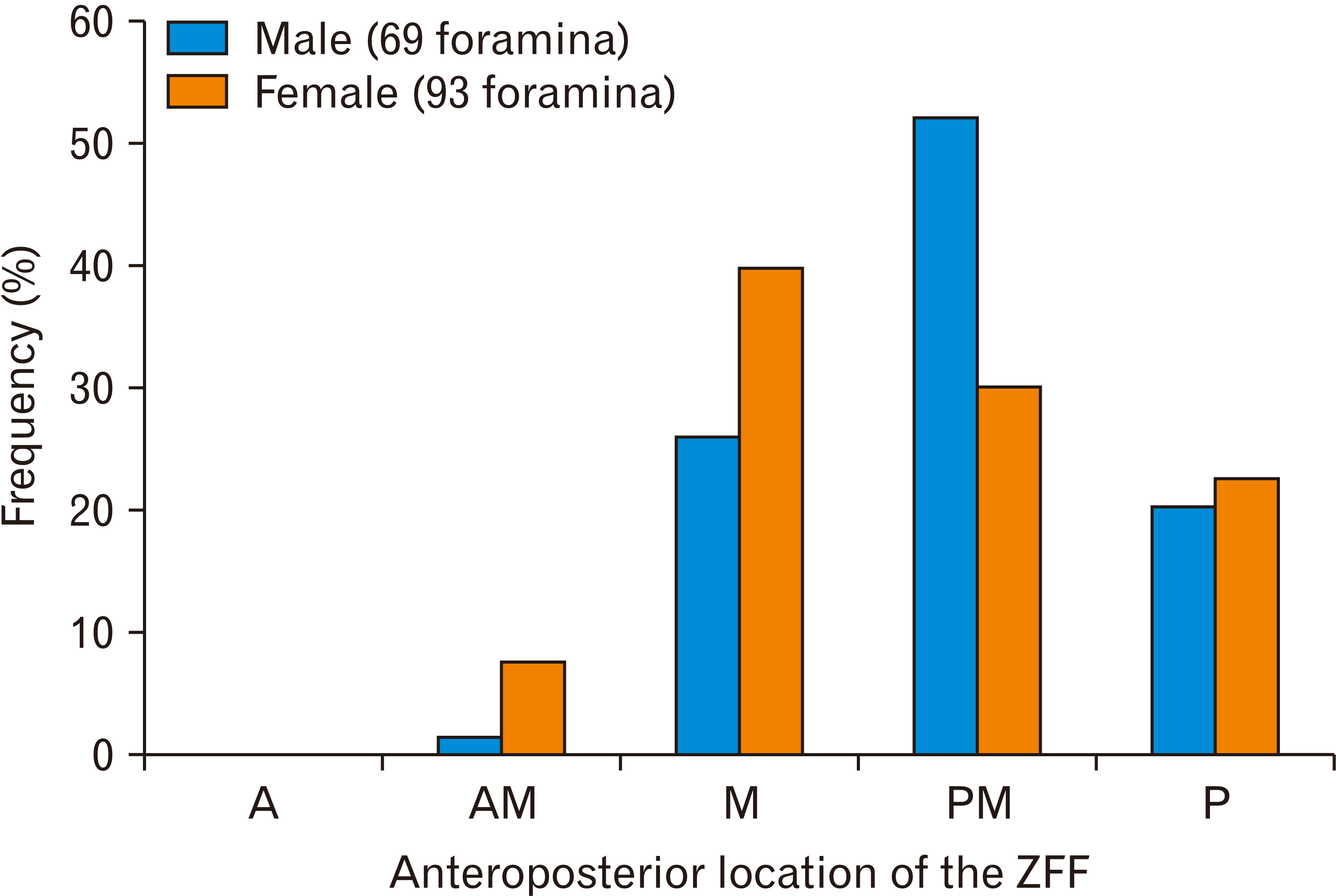
Fig. 8
Distance between the Ju-Zm line and the ZFF in the ZBA (see Fig. 3C). ZFF, zygomaticofacial foramen; Ju, jugale; Zm, zygomaxillare; ZBA, zygomatic body area. *P<0.05 (t-test).
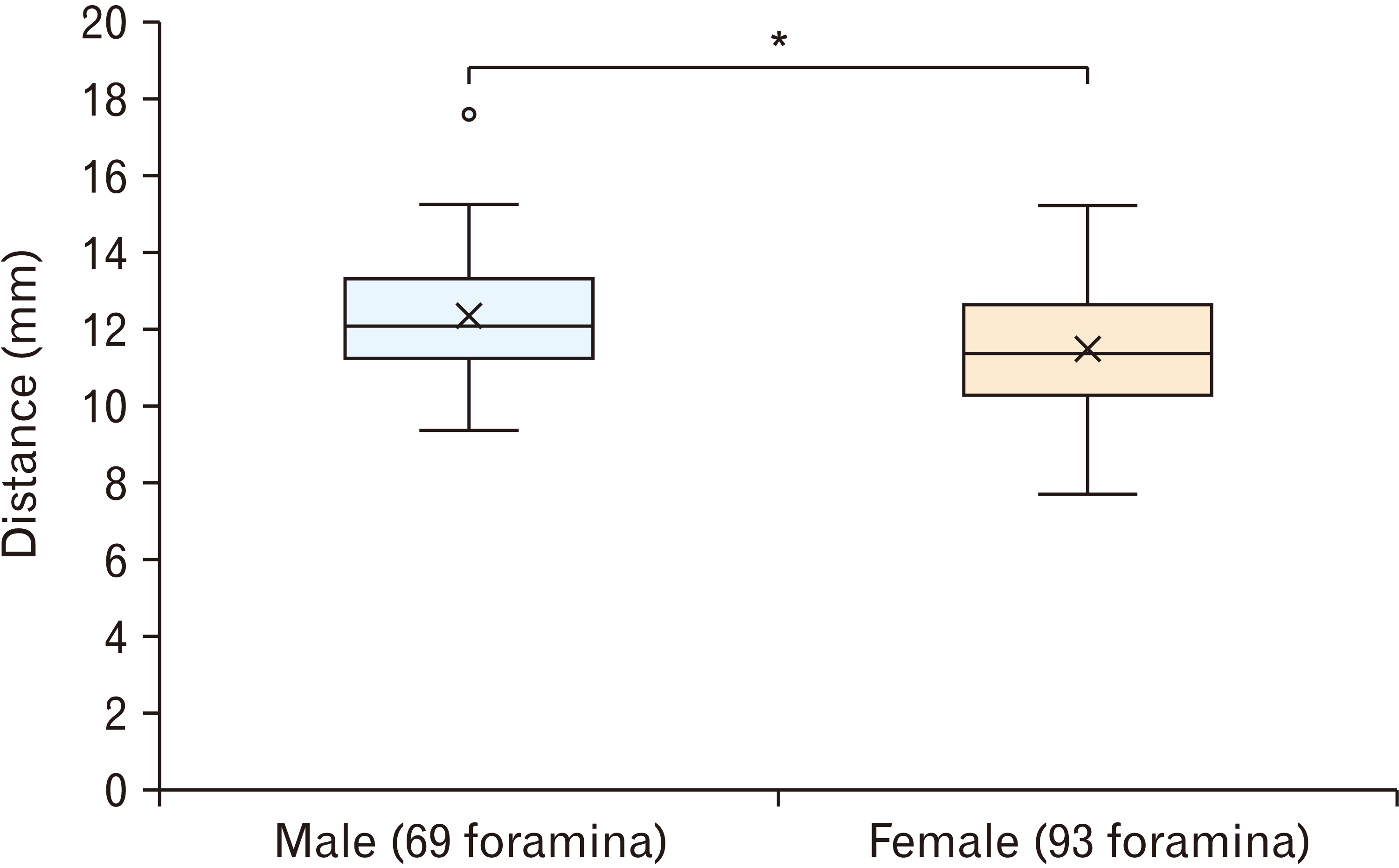
Fig. 9
3D micro-CT images of zygomatic canals communicating with ZFFs in several cases. Zygomaticoorbital foramen (green arrowheads), zygomaticotemporal foramen (blue arrowheads). ZFF, zygomaticofacial foramen (orange arrowheads); 3D, three-dimensional; CT, computed tomography.
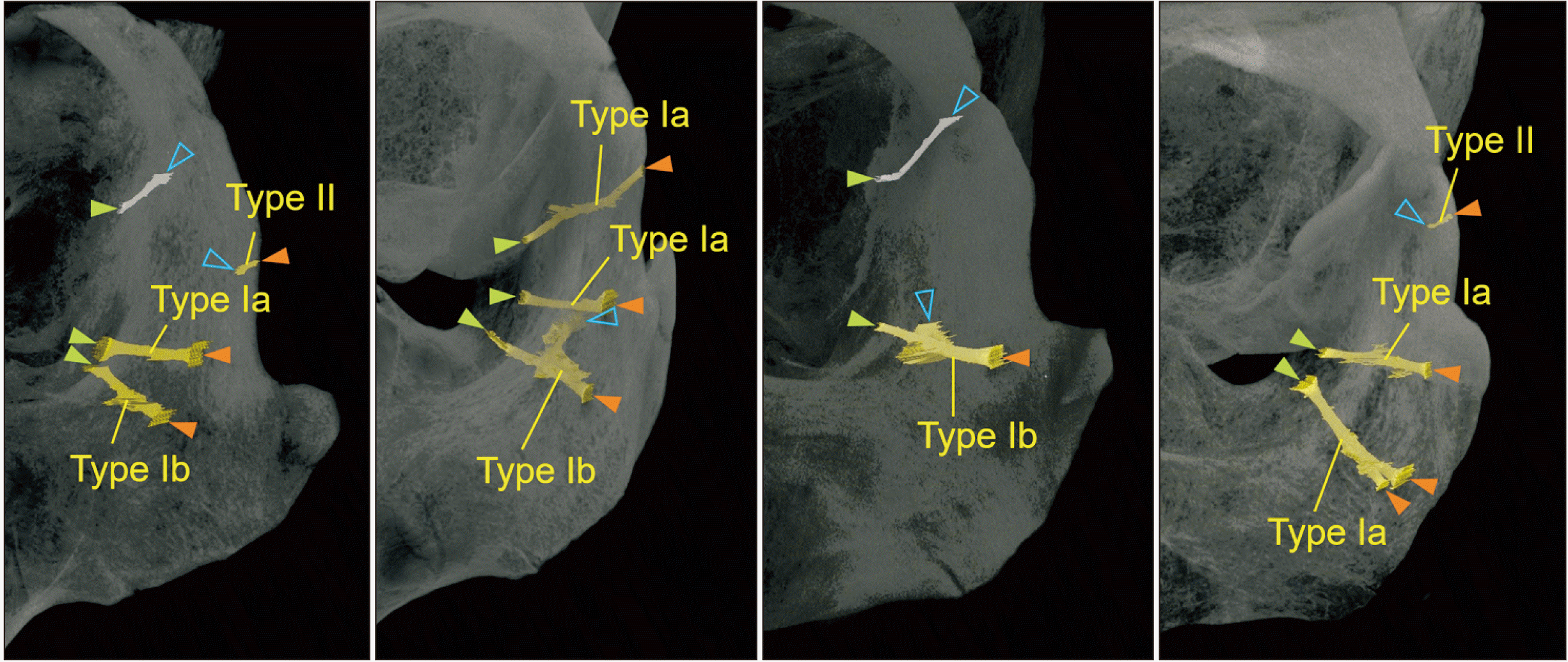
Table 1
Numbers of zygomatic foramina
Values are presented as number (%). Male: 26 cadavers, 52 sides; female: 26 cadavers, 52 sides. ZOF, zygomaticoorbital foramen; ZTF, zygomaticotemporal foramen; ZFF, zygomaticofacial foramen. There were no significant differences between males and females in the ZOF and ZTF (P>0.05, chi-squared test). A significant difference in ZFF between males and females was found (P<0.05, chi-squared test).
Table 2
Numbers of zygomatic canal types and locations of the zygomaticofacial foramen in each type of canal


 PDF
PDF Citation
 Print
Print


 XML Download