Abstract
Undifferentiated carcinoma with osteoclast-like giant cells (UC−OGCs) of the pancreas is a rare neoplasm that accounts for less than 1% of all pancreatic malignancies. The aim of this study was to review the literature regarding UC−OGC, and to highlight its biological behavior, clinicopathologic characteristics, prognosis, and therapeutic options. A systematic review of the literature in PubMed/Medline and Scopus databases was performed (last search October 31st, 2023) for articles concerning pancreatic UC−OGC in the adult population. Fifty-seven studies met the inclusion criteria, involving 69 patients with a male-to-female ratio of 1.1:1 and a mean age of 62.96. Main symptoms included abdominal pain (33.3%), jaundice (14.5%), weight loss (8.7%), while fourteen patients (20.3%) were asymptomatic. Surgical resection was performed in 88.4% of cases. Survival rates at one, three, and five years were 58%, 44.7%, and 37.3% respectively. Sex, age, size (cut-off of 4 cm), location, and adjuvant treatment did not significantly affect patient survival. UC−OGC of the pancreas is a rare subtype of undifferentiated pancreatic carcinoma with a better prognosis than conventional pancreatic ductal adenocarcinoma or undifferentiated carcinoma without giant cells. The establishment of a dedicated patient registry is imperative to further delineate the optimal treatment for this uncommon clinical entity.
Undifferentiated pancreatic carcinoma (UPC) is a rare and aggressive tumor with various histological appearances that include anaplastic, pleomorphic, spindle cell, sarcomatoid carcinoma, and carcinosarcoma. Despite their differences, these entities are collectively named in the current WHO Classification as undifferentiated carcinoma of the pancreas [1]. Undifferentiated carcinoma with osteoclast-like giant cells (UC−OGCs) is a distinctive UPC subtype that is considered as a variant of ductal adenocarcinoma with striking chemotaxis of osteoclastic giant cells [2].
UC−OGC is an extremely rare tumor, accounting for less than 1% of all pancreatic malignancies [3]. Histologically, UC−OGC is composed of both pleomorphic-to-spindle-shaped cells, and the hallmark feature of non-neoplastic OGCs [4]. These OGCs are positive for CD68, vimentin, and leukocyte common antigen, but negative for keratin and p53 antibodies [5]. The origin of OGCs in UC−OGC is not well understood, despite the chemotactic factors produced by cancer cells that have been found responsible so far. UC−OGC could be manifested either alone, or as part of other more common pancreatic tumors, such as pancreatic ductal adenocarcinoma or mucinous cystic neoplasm [6]. It is reportedly found in elderly females, with a mean age of presentation of approximately 63 years, and has similar clinical manifestations to other types of pancreatic tumors, such as upper abdominal pain and weight loss [7].
UC−OGC of the pancreas was initially believed to carry a poorer prognosis than invasive ductal adenocarcinoma of the pancreas [8-10], mainly due to its diagnosis at advanced stages [11], and its tendency for early recurrence, despite complete surgical removal [12,13]. However, when true UC–OGCs are carefully distinguished from other anaplastic carcinomas, it becomes clear that UC−OGCs have a more indolent behavior, especially pure UC−OGCs. Some series reported a significantly better prognosis for UC−OGC compared to conventional ductal adenocarcinoma, with a 5-year survival rate of over 50% [14].
The aim of this systematic literature review was to evaluate the available evidence on the biological behavior and prognosis of UC−OGC, as well as the role of surgical management.
This systematic review was conducted following the Preferred Reporting Items for Systematic Reviews and Meta-Analyses guidelines [15]. Two investigators independently searched PubMed/Medline and Scopus databases reporting on UC−OGC (last search October 31st, 2023), using the following search algorithm: (“undifferentiated” [Title/Abstract] OR “anaplastic” [Title/Abstract]) AND “pancreas” [Title/Abstract] AND (“osteoclast” [Title/Abstract] OR “giant cells” [Title/Abstract]). Any controversy was solved by recourse to a senior investigator.
All articles written in the English language presenting case reports or case series were included in this systematic review, whereas reviews and meta-analyses were excluded. Non-English literature and articles involving children or animals were omitted. Moreover, articles with no clear diagnosis or insufficient data were excluded as irrelevant.
Data extraction of the included studies was performed independently by two of the authors (TSD and PL) using a predefined form. Information regarding sex, age, symptomatology, and medical history was compiled. Additionally, data on tumor location, treatment approach, use of neoadjuvant and adjuvant therapy, and outcome were gathered.
The study has been granted an exemption from requiring ethics approval by the Laikon General Hospital of Athens, Athens, Greece Ethics Committee.
Numerical variables were presented as the mean plus or minus standard deviation (SD), whereas categorical ones as the frequency and valid percentages. Patients included in case series were considered as unique cases, to assess the variables of interest. Several studies lacked data on some variables of interest, and therefore rates were estimated based on the available data. Statistical analysis was carried out using IBM SPSS Statistics for Windows, Version 22.0 (IBM Corp.).
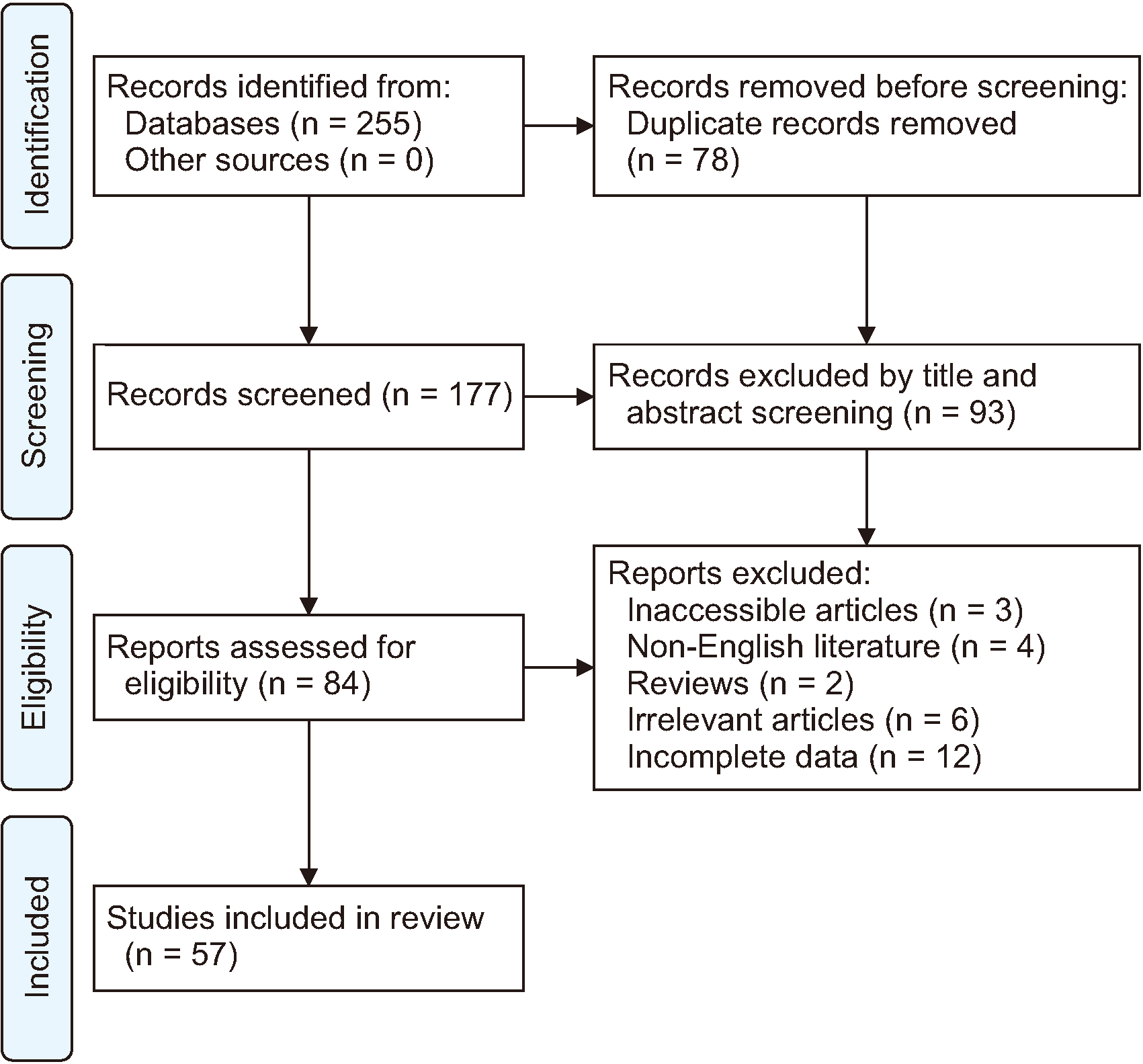
The present systematic review included 57 studies out of 176 unique articles and reports on 69 patients with UC−OGC. Fig. 1 presents the detailed article identification flow. The studies consisted of 54 case reports and 3 case series, including 36 female (52.1%) and 33 male (47.9%) patients, representing a 1.1:1 sex ratio. The mean age of the patients with UC−OGC was 62.96 ± 13.23 years (mean ± SD).
Symptoms of the patients included in this review involved abdominal pain (33.3%), jaundice (14.5%), weight loss (8.7%), and bowel obstruction (4.3%). Fourteen patients (20.3%) were asymptomatic and the lesion was discovered incidentally, while 6 patients (8.7%) presented with an abdominal mass. Other clinical manifestations included complaints of fullness, upper gastrointestinal bleeding, an abnormal lung shadow on chest X-ray, urinary tract infection, and functional bowel disorders. Table 1 shows a detailed clinical presentation of the patients.
The majority of the neoplasms were located in the head (36.2%), followed by the tail (27.5%) and the body (15.9%) of the pancreas. The mean size of the lesion was 74.5 ± 61.1 mm (mean ± SD), with a tumor size range, 3−280 mm. Table 2 shows the size in relation to tumor location. At the time of diagnosis, 8 patients (11.6%) presented with locally advanced disease due to the invasion of adjacent organs [16-21], vessels [22], or lymph nodes [23]. At the same time, 5 patients (7.2%) exhibited metastatic disease [24-28], primarily involving the liver [26-28], and the lungs [25].
Surgical resection of the pancreatic neoplasm was the therapeutic treatment in the majority of the patients (61 patients, 88.4%). Bypass surgery was offered to 2 patients, due to extended disease and poor performance status of the patients [23,24], while palliative stent placement was the therapeutic approach in 2 additional patients presenting with concurrent liver metastases [26,27]. Best supportive care was provided to 4 patients, based on their poor performance status [29], disease burden [28], and patient preference [24].
Concerning surgical operations, distal pancreatectomy and splenectomy was the most common procedure, accounting for 31 cases (50.8%). Among these, resection of adjacent organs was performed in 11 cases, with the stomach and the transverse colon being the most common resected organs. Whipple’s procedure followed as the second most common procedure, carried out in 25 cases (40.9%). Spleen-preserving distal pancreatectomy was performed in 4 cases (6.5%), while one patient (1.6%) underwent total pancreatectomy and splenectomy. The mean length of stay for patients was 14.15 days ranging 4 to 49 days. Two postoperative complications classified as Grade 3 or higher according to the Clavien–Dindo classification were reported. The first was a case of small bowel obstruction leading to sepsis, necessitating an exploratory laparotomy, after which a satisfactory recovery of the patient was reported. The second complication was a progressively enlarging pancreatic pseudocyst, successfully addressed with a second laparotomy for pseudocyst–jejunostomy, performed three months following the initial surgical intervention. Table 3 shows the detailed surgical treatment of the patients.
Pathologic staging reports were available for 42 patients, classified according to the AJCC Cancer Staging Manual (8th edition) [30]. Stage distribution among patients with UC−OGC was as follows: 2 patients (4.8%) were at stage IA (T1, N0, M0), 7 patients (16.7%) at stage IB (T2, N0, M0), 12 patients (28.6%) at stage IIA (T3, N0, M0), 2 patients (4.8%) at stage IIB (varying T1−T3, N1, M0), 14 patients (33.3%) at stage III (varying T1−T4, N2 or T4, Any N, M0), and 5 (11.9%) patients at stage IV (Any T, Any N, M1).
Following surgical resection, adjuvant chemotherapy was provided to 19 patients. The majority of patients receiving adjuvant therapy were treated with gemcitabine, or with leucovorin–fluorouracil–irinotecan–oxaliplatin (FOLFIRINOX) regimen.
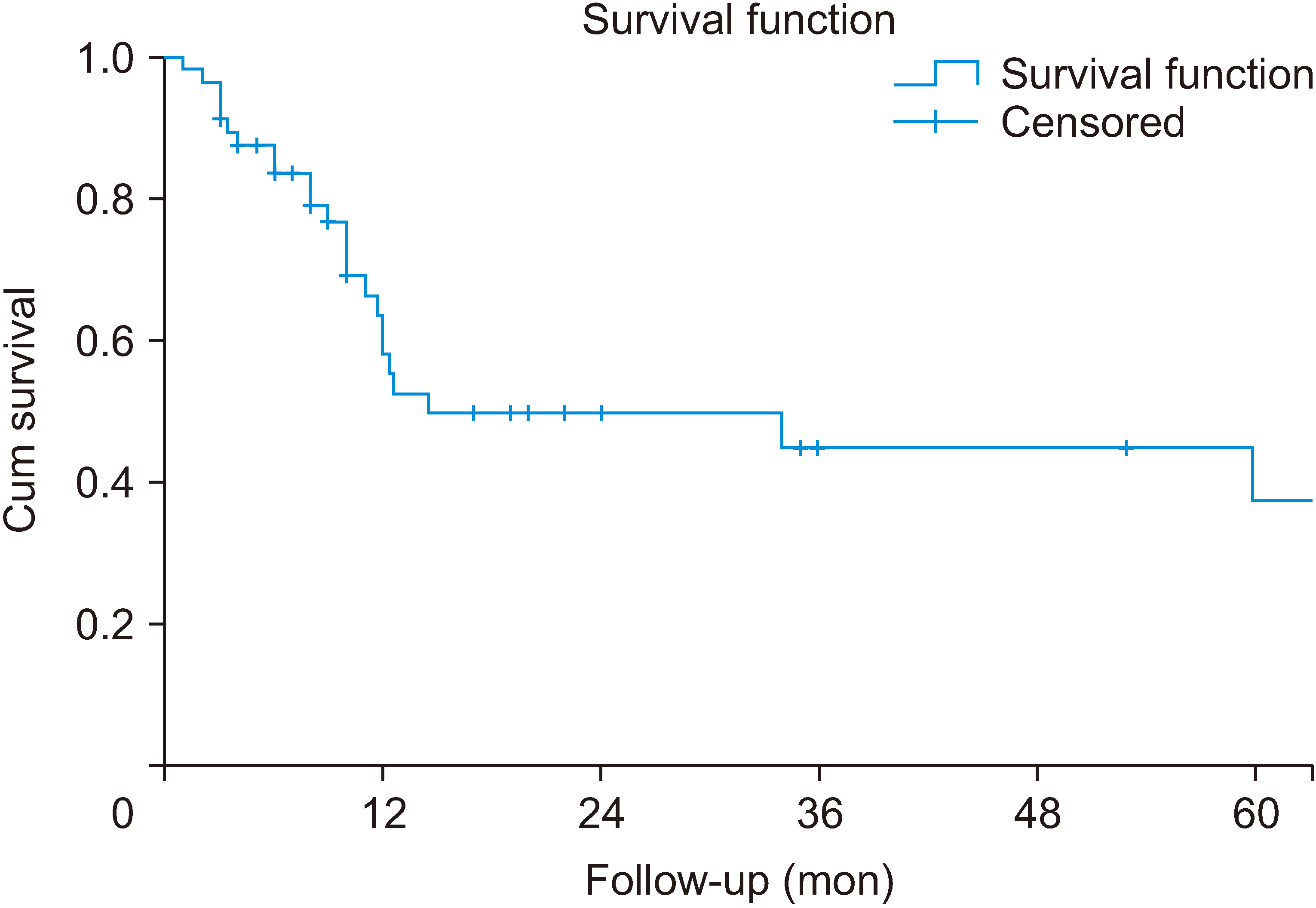
Patients’ follow-up was reported in 57 cases, and the follow-up period ranged 1 month to 14 years, with a median of 20.6 months. The longest reported survival was 14 years, while 24 deaths were reported during the follow-up period. The mean survival of the entire group of patients was 14.5 months. One-year, three-year, and five-year survival rates were 58% (95% confidence interval [CI], 42.8%−73.2%), 44.7% (95% CI, 27.7%−61.7%), and 37.3% (95% CI, 17.7%−56.9%), respectively. Fig. 2 shows the cumulative survival.
Unifactorial Cox’s proportional hazards model analysis showed that sex, age, size (setting a cut-off of 4 cm), location, and adjuvant treatment did not significantly affect patient survival (Table 4).
UPC is a rare type of pancreatic adenocarcinoma that usually exhibits perineural invasion, as well as infiltration of local lymph nodes and blood vessels [31]. UC−OGC is a very rare subtype of UPC, accounting for less than 1% of all pancreatic malignancies [3], and was first described by Maier and Sommers in 1987 [32]. It is characterized by the presence of undifferentiated carcinoma cells and multinucleated OGCs, resembling giant cell tumor of the bone [33]. Such cells have been reported in tumors in a variety of organs, such as the kidney, breast, heart, parotid, and skin [34].
The possible pathogenetic mechanisms of UC−OGC involve a complex interplay of genetic alterations and cellular changes. Whole exome sequencing has revealed that UC−OGC shares a similar genetic background to pancreatic ductal adenocarcinoma, including mutations in key genes, such as KRAS, CDKN2A, TP53, and SMAD4 [35]. Additional mutations in SERPINA3 [35] and GLI3 [35,36] have been detected, suggesting their role as oncogenes and drivers of UC−OGC, respectively. Furthermore, mutations in TTN, MAGEB4, and MEGF8 have also been described; thus, their functional importance remains unclear [35]. The pleiomorphic phenotype of UC−OGC cells may result from epithelial-to-mesenchymal transition (EMT), characterized by loss of E–cadherin [37], deregulation of β–catenin [38], and strong expression of vimentin and ZEB1 [39]. However, EMT activation appears to be more frequent in undifferentiated carcinoma than in UC−OGC, and its presence and significance may be affected by neoadjuvant chemotherapy [31]. Lastly, pleiomorphic tumor cells produce granulocyte colony-stimulating factor, which recruits non-neoplastic OGCs [40], playing a key role in the unique features of UC−OGC.
The diagnosis of UC−OGC is made through a combination of clinical, imaging, and histopathological evaluation. Depending on the location of the tumor, patients may present with abdominal pain, jaundice, weight loss, or bowel obstruction. The findings of the present study suggest that approximately one in six patients with UC−OGC remains asymptomatic until incidentally diagnosed.
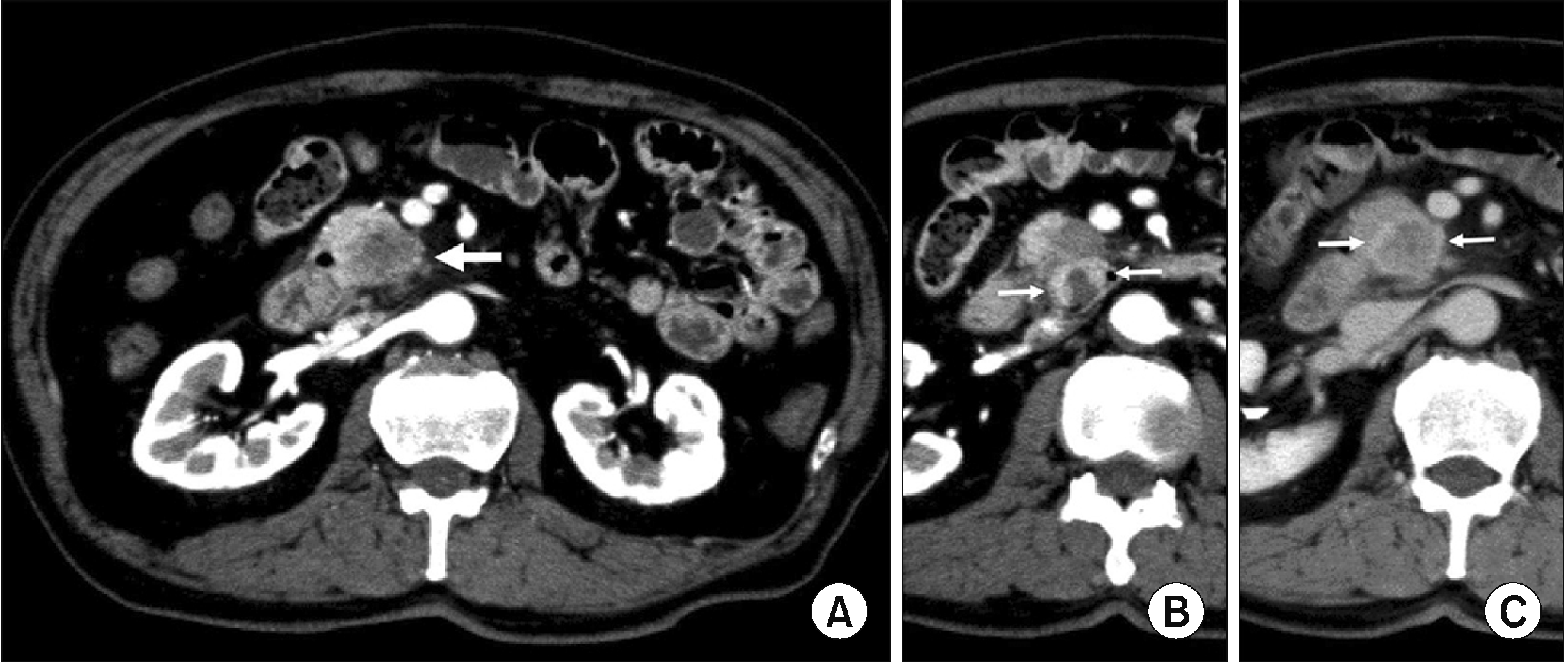
The imaging characteristics of UC–OGC are not well established, due to the rarity of the disease. It typically appears as a well-defined lesion with or without a cystic component, often including areas of hemorrhage and/or necrosis [41] and causing bile duct or pancreatic duct dilatation [42,43]. On contrast-enhanced computed tomography (CT), the tumor appears as a heterogeneous well-defined hypervascular mass, possibly related to its rapid growth of UC−OGC, or the associated inflammatory reaction. In contrast, the typical ductal pancreatic adenocarcinoma appears hypovascular on contrast CT scans. On magnetic resonance imaging, UC−OGC typically displays low-to-dark signal intensity on T1- and T2-weighted images. This characteristic is attributed to multifocal hemorrhage within the lesion, or the formation of bony and cartilaginous tissue by recruited osteoclasts [44]. Differentiating it from other cystic diseases of the pancreas can be challenging, due to the wide variety of features observed in UC−OGC [41]. Endoscopic ultrasound with fine needle aspiration or biopsy can provide cytological or histological material for the definitive diagnosis of UC−OGC. However, giant cells may also be present in cases of pancreatitis, thus representing a challenge for differential diagnosis [45]. Fig. 3, 4 present typical radiological images of UC−OGC.
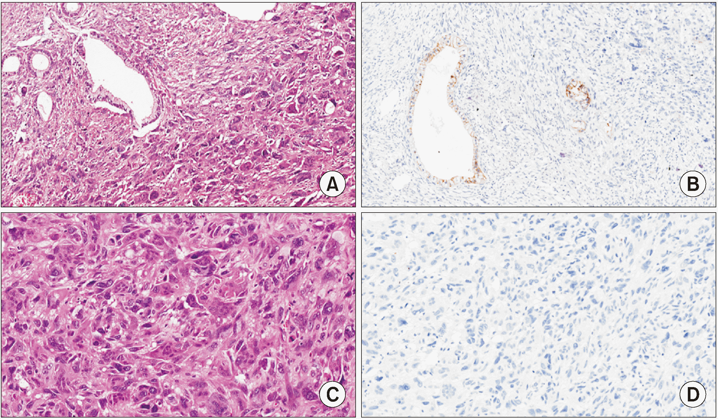
As previously mentioned, the histopathological appearance of UC−OGC is characterized by the coexistence of three distinct cell populations: non-neoplastic osteoclast-like multinucleated giant cells, mononuclear histiocytes, and neoplastic mononuclear cells [1]. The OGCs are often found contiguous to hemorrhage or necrosis, and may also be accompanied by osteochondroid differentiation, osteoid development, and bone formation [29,46,47]. Along with mononuclear histiocytes, giant cells are positive for histiocytic markers and negative for keratin staining, which differentiates them from the bizarre-appearing giant neoplastic cells seen in anaplastic undifferentiated carcinomas [1,48]. The neoplastic cells in UC−OGC range in morphology from spindle to epithelioid, and can be very large and pleomorphic [1]. Most of them have a high Ki–67 proliferation index and express vimentin, while they can show keratin and p53 positivity [1,9]. Additionally, in UC−OGC, the cancer cells typically lack cohesive organization, and instead present as individual cells, or in loose aggregates. These cells are frequently observed within the cytoplasm of OGCs. This differs markedly from the glandular, duct-like structures commonly seen in conventional ductal adenocarcinomas [1,49]. In general, UC−OGC can be pure, or may arise in association with conventional ductal adenocarcinoma or its precursors, such as intraductal papillary mucinous neoplasm and mucinous cystic neoplasm of the pancreas [12,14,50]. Fig. 5 presents representative histological images of UC−OGC.
Treatment approach for UC−OGC mainly involves en bloc surgical excision of the tumor, in accordance with the principles of pancreatic adenocarcinoma treatment. The role of neo-adjuvant chemotherapy is not yet determined, considering that out of the 67 patients, the use of neoadjuvant chemotherapy was reported in only three cases (4.4%). As far as adjuvant therapy is concerned, treatment regiments similar to those applied for pancreatic adenocarcinoma are offered, using FOLFIRINOX schemes in fit patients, and gemcitabine-based regiments in frailer patients. In contrast to the limited use of neoadjuvant chemotherapy, adjuvant chemotherapy was offered to 39% of the reported patients. In addition to the chemotherapy, Obayashi et al. [25] reported a case of UC−OGC with distant metastasis, where the use of anti-PD−1 antibody therapy was effective against a lung metastasis, reflecting the potential benefit of immunotherapy in these tumors.
Surgical resection is considered to carry the best chance for cure, but the extent of resection required is not well established, due to the rarity of the disease, and the limited data available [4]. In general, the location of the primary lesion dictates the type and extent of surgical resection. For tumors located in the head of the pancreas, traditional or pylorus-preserving pancreatoduodenectomy is performed, whereas distal pancreatectomy is conducted for carcinomas of the pancreatic body or tail. In some cases, UC−OGC can invade adjacent organs, such as the stomach, jejunum, colon, left kidney, and diaphragm, and composite resection may be necessary [51].
UC−OGC is a specific type of pancreatic cancer that presents with unique clinicopathological characteristics and varying prognosis. Recent research has shown that patients with UC−OGC tend to have a better prognosis, compared to those with conventional pancreatic ductal adenocarcinoma, or those with UPC with pleomorphic/sarcomatoid giant cell tumors [52]. The improved survival of patients with pure UC−OGC may suggest that its unique morphology is a result of the immune response to the classical pancreatic ductal adenocarcinoma, potentially leading to the elimination of the pancreatic ductal adenocarcinoma component via a strong immune response [35].
While generally displaying a slower metastatic progression and a reduced rate of lymph node metastasis, compared to carcinoma without giant cells [14], cases of UC−OGC exhibiting aggressive behavior are not uncommon, reported mean survival varies among studies, with documented durations of 13 months [53], 19.6 months [41], and 20.4 months [51]. Several demographic and clinical factors have been associated with poorer prognosis, including the presence of a concurrent ductal adenocarcinoma component, older age, male sex, expression of PD−L1, and positive lymph node metastasis [24,27,28,53,54]. Conversely, indications of a more favorable prognosis have been associated with the identification of pure UC−OGC histology and surgical intervention for tumor removal [2,53]. In the present study, it was demonstrated that the median survival rate of patients with UC−OGC was 14.5 months, with one-, three-, and five-year survival rates of 58%, 44.7%, and 37.3%, respectively. The hazards model analysis of the present work revealed that sex, age, tumor size (using a cutoff of 4 cm), location, and adjuvant treatment did not significantly affect patient survival.
To the best of the authors’ knowledge, this is the first systematic review of the literature concerning the epidemiology, clinical manifestation, treatment approach, and prognosis of patients with pancreatic UC−OGC. However, the present study presents some limitations. First, the data presented in this study were obtained from case reports and case series, which implies that the accuracy and completeness of the data depend on the authors of those reports. Additionally, the data came from various surgical centers with different diagnostic, treatment, and follow-up algorithms, leading to heterogeneity and potential biases in the present analysis. Furthermore, this study may have been affected by methodological limitations, such as the small sample size, and the unavailability of full data. Another possible limitation of the present work could be the infrequent reporting of neoadjuvant and adjuvant therapies, and the use of a variety of therapeutic regimens, which may have impacted the prognosis of the cases under analysis. Lastly, there is a potential for publication bias, where cases with poorer outcomes may have been underreported, leading to a potential limitation in the generalizability of our findings.
Given the rare nature and distinctive features of UC−OGC, it is prudent to establish a dedicated patient registry to comprehensively document cases, thereby facilitating a collaborative and detailed analysis. Furthermore, research into the molecular foundations of these tumors is crucial to elucidate the exact mechanisms underlying their development. Importantly, investigations into UC−OGC promise significant advancements in understanding and treating this specific cancer type, while also potentially providing vital insights into pancreatic adenocarcinoma. It is hypothesized that the enhanced survival rates observed in UC−OGC cases may be indicative of an immune response that is capable of effectively targeting cells associated with classical pancreatic ductal adenocarcinoma, potentially orchestrating their elimination through a potent immune response [35].
From a surgical perspective, the notably better prognosis associated with UC−OGC, compared to conventional pancreatic ductal adenocarcinoma, strongly advocates pursuing surgical resection whenever feasible. Simultaneously, it is imperative for the oncology community to pioneer innovative treatment strategies, aiming to identify the most efficacious therapies for these tumors. A flexible approach, possibly modifying the treatment plans based on the presence or absence of a ductal component, might pave the way for more personalized therapies, potentially enhancing patient outcomes and extending survival rates.
UC−OGC of the pancreas is a rare subtype of UPC characterized by the presence of non-neoplastic OGCs. Depending on the location of the tumor, patients can present with abdominal pain, jaundice, weight loss, or remain asymptomatic until the diagnosis is made incidentally. Surgical resection of the malignancy along with involved adjacent organs constitutes the mainstay of treatment. UC−OGC appears to convey a better prognosis than conventional pancreatic ductal adenocarcinoma or Undifferentiated Carcinoma without giant cells. Further research on pathogenesis, as well as the establishment of a patient registry, are required to investigate potential prognostic factors for this tumor type; the exploration of possible therapeutic approaches that may improve patient outcomes is also needed. The identification and understanding of the unique characteristics of UC−OGC are crucial for appropriate clinical management, and for the development of potential targeted therapies.
References
1. Nagtegaal ID, Odze RD, Klimstra D, Paradis V, Rugge M, Schirmacher P, et al. 2020; The 2019 WHO classification of tumours of the digestive system. Histopathology. 76:1821188. DOI: 10.1111/his.13975. PMID: 31433515. PMCID: PMC7003895.
2. Demetter P, Maréchal R, Puleo F, Delhaye M, Debroux S, Charara F, et al. 2021; Undifferentiated pancreatic carcinoma with osteoclast-like giant cells: what do we know so far? Front Oncol. 11:630086. DOI: 10.3389/fonc.2021.630086. PMID: 33747949. PMCID: PMC7973287.
3. Maksymov V, Khalifa MA, Bussey A, Carter B, Hogan M. 2011; Undifferentiated (anaplastic) carcinoma of the pancreas with osteoclast-like giant cells showing various degree of pancreas duct involvement. A case report and literature review. JOP. 12:170–176.
4. Gao HQ, Yang YM, Zhuang Y, Liu P. 2015; Locally advanced undifferentiated carcinoma with osteoclast-like giant cells of the pancreas. World J Gastroenterol. 21:694–698. DOI: 10.3748/wjg.v21.i2.694. PMID: 25593500. PMCID: PMC4292306.
5. Loya AC, Ratnakar KS, Shastry RA. 2004; Combined osteoclastic giant cell and pleomorphic giant cell tumor of the pancreas: a rarity. An immunohistochemical analysis and review of the literature. JOP. 5:220–224.
6. Ashfaq A, Thalambedu N, Atiq MU. 2022; A rare case of pancreatic cancer: undifferentiated carcinoma of the pancreas with osteoclast-like giant cells. Cureus. 14:e25118. DOI: 10.7759/cureus.25118.
7. Guo YL, Ruan LT, Wang QP, Lian J. 2018; Undifferentiated carcinoma with osteoclast-like giant cells of pancreas: a case report with review of the computed tomography findings. Medicine (Baltimore). 97:e13516. DOI: 10.1097/MD.0000000000013516. PMID: 30508980. PMCID: PMC6283196.
8. Zou XP, Yu ZL, Li ZS, Zhou GZ. 2004; Clinicopathological features of giant cell carcinoma of the pancreas. Hepatobiliary Pancreat Dis Int. 3:300–302.
9. Lukás Z, Dvorák K, Kroupová I, Valásková I, Habanec B. 2006; Immunohistochemical and genetic analysis of osteoclastic giant cell tumor of the pancreas. Pancreas. 32:325–329. DOI: 10.1097/01.mpa.0000202951.10612.fa. PMID: 16628090.
10. Paal E, Thompson LD, Frommelt RA, Przygodzki RM, Heffess CS. 2001; A clinicopathologic and immunohistochemical study of 35 anaplastic carcinomas of the pancreas with a review of the literature. Ann Diagn Pathol. 5:129–140. DOI: 10.1053/adpa.2001.25404. PMID: 11436166.
11. Bergmann F, Esposito I, Michalski CW, Herpel E, Friess H, Schirmacher P. 2007; Early undifferentiated pancreatic carcinoma with osteoclastlike giant cells: direct evidence for ductal evolution. Am J Surg Pathol. 31:1919–1925. DOI: 10.1097/PAS.0b013e318067bca8. PMID: 18043049.
12. Wada T, Itano O, Oshima G, Chiba N, Ishikawa H, Koyama Y, et al. 2011; A male case of an undifferentiated carcinoma with osteoclast-like giant cells originating in an indeterminate mucin-producing cystic neoplasm of the pancreas. A case report and review of the literature. World J Surg Oncol. 9:100. DOI: 10.1186/1477-7819-9-100. PMID: 21902830. PMCID: PMC3186749.
13. Hirano H, Morita K, Tachibana S, Okimura A, Fujisawa T, Ouchi S, et al. 2008; Undifferentiated carcinoma with osteoclast-like giant cells arising in a mucinous cystic neoplasm of the pancreas. Pathol Int. 58:383–389. DOI: 10.1111/j.1440-1827.2008.02240.x. PMID: 18477218.
14. Muraki T, Reid MD, Basturk O, Jang KT, Bedolla G, Bagci P, et al. 2016; Undifferentiated carcinoma with osteoclastic giant cells of the pancreas: clinicopathologic analysis of 38 cases highlights a more protracted clinical course than currently appreciated. Am J Surg Pathol. 40:1203–1216. DOI: 10.1097/PAS.0000000000000689. PMID: 27508975. PMCID: PMC4987218.
15. Page MJ, McKenzie JE, Bossuyt PM, Boutron I, Hoffmann TC, Mulrow CD, et al. 2021; The PRISMA 2020 statement: an updated guideline for reporting systematic reviews. BMJ. 372:n71. DOI: 10.1136/bmj.n71. PMID: 33782057. PMCID: PMC8005924.
16. Yazawa T, Watanabe A, Araki K, Segawa A, Hirai K, Kubo N, et al. 2017; Complete resection of a huge pancreatic undifferentiated carcinoma with osteoclast-like giant cells. Int Cancer Conf J. 6:193–196. DOI: 10.1007/s13691-017-0305-y. PMID: 31149501. PMCID: PMC6498254.
17. Olayinka O, Kaur G, Gupta G. 2021; Undifferentiated pancreatic carcinoma with osteoclast-like giant cells and associated ductal adenocarcinoma with focal signet-ring features. Cureus. 13:e14988. DOI: 10.7759/cureus.14988.
18. Hur YH, Kim HH, Seoung JS, Seo KW, Kim JW, Jeong YY, et al. 2011; Undifferentiated carcinoma of the pancreas with osteoclast-like giant cells. J Korean Surg Soc. 81:146–150. DOI: 10.4174/jkss.2011.81.2.146. PMID: 22066115. PMCID: PMC3204565.
19. Vnuk K, Pavić I, Brletić D, Zovak M, Krušlin B. 2022; Undifferentiated carcinoma of the pancreas with osteoclast-like giant cells: report of two cases. Lib Oncol. 50:39–43. DOI: 10.20471/LO.2022.50.01.07.
20. Matsubayashi H, Kaneko J, Sato J, Satoh T, Ishiwatari H, Sugiura T, et al. 2019; Osteoclast-like giant cell-type pancreatic anaplastic carcinoma presenting with a duodenal polypoid lesion. Intern Med. 58:3545–3550. DOI: 10.2169/internalmedicine.3271-19. PMID: 31462592. PMCID: PMC6949449.
21. Osaka H, Yashiro M, Nishino H, Nakata B, Ohira M, Hirakawa K. 2004; A case of osteoclast-type giant cell tumor of the pancreas with high-frequency microsatellite instability. Pancreas. 29:239–241. DOI: 10.1097/00006676-200410000-00010. PMID: 15367891.
22. Igarashi Y, Gocho T, Taniai T, Uwagawa T, Hamura R, Shirai Y, et al. 2022; Conversion surgery for undifferentiated carcinoma with osteoclast-like giant cells of the pancreas: a case report. Surg Case Rep. 8:42. DOI: 10.1186/s40792-022-01385-x. PMID: 35286506. PMCID: PMC8921425.
23. Kharkhach A, Bouhout T, Serji B, El Harroudi T. 2021; Undifferentiated pancreatic carcinoma with osteoclast-like giant cells: a review and case report analysis. J Gastrointest Cancer. 52:1106–1113. DOI: 10.1007/s12029-021-00583-4. PMID: 33447945.
24. Lahiff C, Swan N, Conlon K, Malone D, Maguire D, Hoti E, et al. 2016; Osteoclastic-type giant cell tumours of the pancreas: a homogenous series of rare tumours diagnosed by endoscopic ultrasound. Dig Surg. 33:401–405. DOI: 10.1159/000445303. PMID: 27160213.
25. Obayashi M, Shibasaki Y, Koakutsu T, Hayashi Y, Shoji T, Hirayama K, et al. 2020; Pancreatic undifferentiated carcinoma with osteoclast-like giant cells curatively resected after pembrolizumab therapy for lung metastases: a case report. BMC Gastroenterol. 20:220. DOI: 10.1186/s12876-020-01362-4. PMID: 32652936. PMCID: PMC7353752.
26. Speisky D, Villarroel M, Vigovich F, Iotti A, García TA, Quero LB, et al. 2020; Undifferentiated carcinoma with osteoclast-like giant cells of the pancreas diagnosed by endoscopic ultrasound guided biopsy. Ecancermedicalscience. 14:1072. DOI: 10.3332/ecancer.2020.1072. PMID: 32863866. PMCID: PMC7434513.
27. Swaid MB, Vitale E, Alatassi N, Siddiqui H, Yazdani H. 2022; Metastatic undifferentiated osteoclast-like giant cell pancreatic carcinoma. Cureus. 14:e27586. DOI: 10.7759/cureus.27586.
28. Pop RM, Diaconu CI, Rimbaş M, Mateescu RB, Rouhani F, Popp C, et al. 2023; EUS-guided fine needle biopsy is able to provide diagnosis in rare osteoclast-like giant cells undifferentiated carcinoma of the pancreas: report of two cases. Rom J Intern Med. 61:116–124. DOI: 10.2478/rjim-2023-0008. PMID: 36884386.
29. Molberg KH, Heffess C, Delgado R, Albores-Saavedra J. 1998; Undifferentiated carcinoma with osteoclast-like giant cells of the pancreas and periampullary region. Cancer. 82:1279–1287. DOI: 10.1002/(SICI)1097-0142(19980401)82:7<1279::AID-CNCR10>3.0.CO;2-3.
30. Amin MB, Greene FL, Edge SB, Compton CC, Gershenwald JE, Brookland RK, et al. 2017; The Eighth Edition AJCC Cancer Staging Manual: continuing to build a bridge from a population-based to a more 'personalized' approach to cancer staging. CA Cancer J Clin. 67:93–99. DOI: 10.3322/caac.21388. PMID: 28094848.
31. Mattiolo P, Fiadone G, Paolino G, Chatterjee D, Bernasconi R, Piccoli P, et al. 2021; Epithelial-mesenchymal transition in undifferentiated carcinoma of the pancreas with and without osteoclast-like giant cells. Virchows Arch. 478:319–326. DOI: 10.1007/s00428-020-02889-3. PMID: 32661742. PMCID: PMC7969490.
32. Maier HC, Sommers SC. 1987; Recurrent and metastatic pulmonary fibrous histiocytoma/plasma cell granuloma in a child. Cancer. 60:1073–1076. DOI: 10.1002/1097-0142(19870901)60:5<1073::AID-CNCR2820600524>3.0.CO;2-O. PMID: 3038296.
33. Jo S. 2014; Huge undifferentiated carcinoma of the pancreas with osteoclast-like giant cells. World J Gastroenterol. 20:2725–2730. DOI: 10.3748/wjg.v20.i10.2725. PMID: 24627610. PMCID: PMC3949283.
34. Sakhi R, Hamza A, Khurram MS, Ibrar W, Mazzara P. 2017; Undifferentiated carcinoma of the pancreas with osteoclast-like giant cells reported in an asymptomatic patient: a rare case and literature review. Autops Case Rep. 7:51–57. DOI: 10.4322/acr.2017.042. PMID: 29259932. PMCID: PMC5724056.
35. Luchini C, Pea A, Lionheart G, Mafficini A, Nottegar A, Veronese N, et al. 2017; Pancreatic undifferentiated carcinoma with osteoclast-like giant cells is genetically similar to, but clinically distinct from, conventional ductal adenocarcinoma. J Pathol. 243:148–154. DOI: 10.1002/path.4941. PMID: 28722124. PMCID: PMC6664430.
36. Matissek SJ, Elsawa SF. 2020; GLI3: a mediator of genetic diseases, development and cancer. Cell Commun Signal. 18:54. DOI: 10.1186/s12964-020-00540-x. PMID: 32245491. PMCID: PMC7119169.
37. Yonemasu H, Takashima M, Nishiyama KI, Ueki T, Yao T, Tanaka M, et al. 2001; Phenotypical characteristics of undifferentiated carcinoma of the pancreas: a comparison with pancreatic ductal adenocarcinoma and relevance of E-cadherin, alpha catenin and beta catenin expression. Oncol Rep. 8:745–752. DOI: 10.3892/or.8.4.745. PMID: 11410776.
38. Sano M, Homma T, Hayashi E, Noda H, Amano Y, Tsujimura R, et al. 2014; Clinicopathological characteristics of anaplastic carcinoma of the pancreas with rhabdoid features. Virchows Arch. 465:531–538. DOI: 10.1007/s00428-014-1631-5. PMID: 25031015.
39. Naito Y, Kinoshita H, Okabe Y, Arikawa S, Higaki K, Morimitsu Y, et al. 2009; Pathomorphologic study of undifferentiated carcinoma in seven cases: relationship between tumor and pancreatic duct epithelium. J Hepatobiliary Pancreat Surg. 16:478–484. DOI: 10.1007/s00534-009-0078-6. PMID: 19367361.
40. Kubota N, Naito Y, Kawahara A, Taira T, Yamaguchi T, Yoshida T, et al. 2017; Granulocyte colony-stimulating factor-producing pancreatic anaplastic carcinoma in ascitic fluid at initial diagnosis: a case report. Diagn Cytopathol. 45:463–467. DOI: 10.1002/dc.23682. PMID: 28185423.
41. Togawa Y, Tonouchi A, Chiku T, Sano W, Doki T, Yano K, et al. 2010; A case report of undifferentiated carcinoma with osteoclast-like giant cells of the pancreas and literature review. Clin J Gastroenterol. 3:195–203. DOI: 10.1007/s12328-010-0160-2. PMID: 26190247.
42. Fukukura Y, Kumagae Y, Hirahara M, Hakamada H, Nagano H, Nakajo M, et al. 2019; CT and MRI features of undifferentiated carcinomas with osteoclast-like giant cells of the pancreas: a case series. Abdom Radiol (NY). 44:1246–1255. DOI: 10.1007/s00261-019-01958-9. PMID: 30815714.
43. Shindoh N, Ozaki Y, Kyogoku S, Nakanishi A, Sumi Y, Katayama H. 1998; Osteoclast-type giant cell tumor of the pancreas: helical CT scans. AJR Am J Roentgenol. 170:653–654. DOI: 10.2214/ajr.170.3.9490947. PMID: 9490947.
44. Yang KY, Choi JI, Choi MH, Park MY, Rha SE, Byun JY, et al. 2015; Magnetic resonance imaging findings of undifferentiated carcinoma with osteoclast-like giant cells of pancreas. Clinical Imaging. 40:148–151. DOI: 10.1016/j.clinimag.2015.09.013. PMID: 26520702.
45. Brosens LA, Leguit RJ, Vleggaar FP, Veldhuis WB, van Leeuwen MS, Offerhaus GJA. 2015; EUS-guided FNA cytology diagnosis of paraduodenal pancreatitis (groove pancreatitis) with numerous giant cells: conservative management allowed by cytological and radiological correlation. Cytopathology. 26:122125. DOI: 10.1111/cyt.12140. PMID: 24650015.
46. Luchini C. 2021; Undifferentiated carcinoma of the pancreas with osteoclast-like giant cells: a challenging cancer with new horizons? Virchows Archiv. 478:595–596. DOI: 10.1007/s00428-021-03021-9. PMID: 33447898.
47. Manduch M, Dexter DF, Jalink DW, Vanner SJ, Hurlbut DJ. 2009; Undifferentiated pancreatic carcinoma with osteoclast-like giant cells: report of a case with osteochondroid differentiation. Pathol Res Pract. 205:353–359. DOI: 10.1016/j.prp.2008.11.006. PMID: 19147301.
48. Oymaci E, Yakan S, Yildirim M, Argon A, Namdaroglu O. 2017; Anaplastic carcinoma of the pancreas: a rare clinical entity. Cureus. 9:e1782. DOI: 10.7759/cureus.1782. PMID: 29279808. PMCID: PMC5736168.
49. Ren B, Liu X, Suriawinata AA. 2019; Pancreatic ductal adenocarcinoma and its precursor lesions: histopathology, cytopathology, and molecular pathology. Am J Pathol. 189:9–21. DOI: 10.1016/j.ajpath.2018.10.004. PMID: 30558727.
50. Jang KT, Park SM, Basturk O, Bagci P, Bandyopadhyay S, Stelow EB, et al. 2015; Clinicopathologic characteristics of 29 invasive carcinomas arising in 178 pancreatic mucinous cystic neoplasms with ovarian-type stroma: implications for management and prognosis. Am J Surg Pathol. 39:179–187. DOI: 10.1097/PAS.0000000000000357. PMID: 25517958. PMCID: PMC4460193.
51. Shiozawa M, Imada T, Ishiwa N, Rino Y, Hasuo K, Takanashi Y, et al. 2002; Osteoclast-like giant cell tumor of the pancreas. Int J Clin Oncol. 7:376–380. DOI: 10.1007/s101470200059. PMID: 12494256.
52. Strobel O, Hartwig W, Bergmann F, Hinz U, Hackert T, Grenacher L, et al. 2011; Anaplastic pancreatic cancer: presentation, surgical management, and outcome. Surgery. 149:200–208. DOI: 10.1016/j.surg.2010.04.026. PMID: 20542529.
53. Xu M, Chen W, Wang D, Nie M. 2020; Clinical characteristics and prognosis of osteoclast-like giant cell tumors of the pancreas compared with pancreatic adenocarcinomas: a population-based study. Med Sci Monit. 26:e922585. DOI: 10.12659/MSM.922585.
54. Hrudka J, Lawrie K, Waldauf P, Ciprová V, Moravcová J, Matěj R. 2020; Negative prognostic impact of PD-L1 expression in tumor cells of undifferentiated (anaplastic) carcinoma with osteoclast-like giant cells of the pancreas: study of 13 cases comparing ductal pancreatic carcinoma and review of the literature. Virchows Arch. 477:687–696. DOI: 10.1007/s00428-020-02830-8. PMID: 32424767.