Abstract
Purpose
This study aimed to assess the marginal and internal fit of 3-unit monolithic zirconia fixed partial dentures (FPDs) fabricated via computer-aided design and computer-aided manufacturing (CAD/CAM) from solid working casts and removable die system.
Materials and Methods
The tooth preparation protocol for a zirconia crown was executed on the mandibular right first premolar and mandibular right first molar, with the creation of a reference cast featuring an absent mandibular right second premolar. The reference cast was duplicated using polyvinyl siloxane impression, from which 20 working casts were fabricated following typical dental laboratory procedures. For comparative analysis, 10 FPDs were produced from a removable die system (RD group) and the remaining 10 FPDs from the solid working casts (S group). The casts were digitized using a dental desktop scanner to establish virtual casts and design the FPDs using CAD. The definitive 3-unit monolithic zirconia FPDs were fabricated via a CAM milling process. The seated FPDs on the reference cast underwent digital evaluation for marginal and internal fit. The Mann-Whitney U test was applied for statistical comparison between the two groups (α = 0.05).
Results
The RD group showed significantly higher discrepancies in fit for both premolars and molars compared to the S group (P < 0.05), particularly in terms of marginal and occlusal gaps. Color mapping also highlighted more significant deviations in the RD group, especially in the marginal and occlusal regions.
초록
목적
본 연구의 목적은 가철성 다이 시스템으로 제작된 작업 모형과 솔리드 작업 모형을 이용해 치과용 캐드캠 시스템(CAD/CAM)으로 제작된 지르코니아 3본 고정성 치과 보철물의 변연 및 내부 적합성을 평가하고자 하였다.
연구 재료 및 방법
하악 우측 제1소구치와 하악 우측 제1대구치에 지르코니아 크라운을 위한 치아 삭제 프로토콜을 수행하고, 하악 우측 제2소구치가 없는 레퍼런스 모델을 만들었다. 레퍼런스 모델은 폴리비닐 실록산 인상체를 사용하여 복제되었고, 일반적인 치과 기공 절차에 따라 20개의 작업 모형이 제작되었다. 비교 분석을 위해, 10개의 지르코니아 3본 고정성 치과 보철물은 가철성 다이 시스템에서, 나머지 10개는 솔리드 작업 모형에서 제작되었다. 모든 작업 모형은 치과용 데스크탑 스캐너를 사용하여 디지털화되었고, 캐드 소프트웨어에서 보철물을 설계하였다. 최종 3본 고정성 치과 보철물은 밀링 과정을 통해 제작되었다. 변연 및 내부 적합도 평가는 레퍼런스 모델에 제작된 보철물을 위치시키고, 디지털 평가 방법으로 적합도가 측정되었다. 두 그룹 간의 통계 비교를 위해 Mann-Whitney U 검정이 적용되었다(α = 0.05).
Traditional dental CAD/CAM protocols require the inclusion of physical working cast fabrication.1-5 Numerous intraoral constraints, limiting the efficacy of intraoral scanners, rely on impression taking and working cast fabrication. Intraoral scanners have demonstrated inaccuracies in complete arch scans for fixed partial dentures (FPDs) and are often inaccurate in confined interdental spaces.6,7 External factors such as humidity, temperature, and ambient lighting also influence scanning accuracy.8-10 Despite the potential shift toward a fully digital workflow concomitant with advancements in intraoral scanning technology, conventional methodologies involving working cast fabrication remain integral in dental practice.11,12
Historically, the development of working casts in conventional workflows has evolved through various methodologies.13,14 Early research predominantly focused on selecting materials for working cast fabrication, developing techniques for creating removable dies, and balancing considerations of practicality, cost-effectiveness, and accuracy of FPDs.15,16 At present, dental stone materials are employed to fabricate working casts with removable dies for the fabrication of FPDs.17 However, previous studies have indicated that the use of removable dies could lead to inaccuracies in FPD outcomes.18-20 As a result, a specialized technique exists for fabricating solid working casts without removable dies while dealing with a supragingival finish line condition, thereby enhancing accuracy in dental prosthetics.21 Nonetheless, studies evaluating the effect of these working cast fabrication methodologies on the accuracy of FPDs in the context of dental CAD/CAM processes are limited.
In the dental CAD/CAM workflow, scan data acquisition typically involves either intraoral scanning or scanning of working casts. The limitations inherent in many intraoral scans often necessitate the use of working casts for scanning. Previous studies have indicated that the use of working casts with removable dies might lead to variations in die positioning depending on the fabrication methods employed.18-20 However, in the CAD/CAM process, removable dies are digitally scanned as virtual entities, and these scanned dies must be precisely aligned within the entire virtual model.15 A previous study reported errors associated with this alignment process.15 Removable dies in working casts are produced predominantly for the ease of fabricating FPDs, particularly when the finish line is situated subgingivally, posing challenges in managing the crown’s marginal area during scanning and other fabrication stages.18 Consequently, pins are positioned below the die in the working cast for separation, leading to the production of detachable individual dies. The gingival and lower areas of the die’s finish line are meticulously trimmed, preserving only the finish line, completing the creation of a removable die.18,19 These processes simplify handling throughout the stages of manufacturing, including scanning.15 In dental laboratory practice, the development of removable dies has thus been integrated as a routine part of working cast production. However, as the field has transitioned into dental CAD/CAM production, the precision of fabrications has improved. The need for removable dies has reduced, except in situations where the finish line has a subgingival location, owing to the feasibility of scanning and designing the die’s adjacent regions.21 Previous studies have highlighted that errors associated with removable die production are inevitable.18-20 Traditional methods involving wax pattern creation and casting have various inherent errors in each step, necessitating adjustments during the dental laboratory phase when using removable dies.
Therefore, this study aimed to assess the marginal and internal fit of 3-unit monolithic zirconia FPDs produced from solid working casts and working casts with removable die. The null hypothesis was that the two working cast fabrication techniques do not considerably influence the marginal and internal fit of 3-unit FPDs.
This study was performed as outlined in the flow diagram depicted in Fig. 1. In this study, a mandibular typodont (ANA-4 Z; Frasaco GmbH, Tettnang, Germany) was used for the fabrication of a reference cast. The tooth preparations for the mandibular right first premolar and first molar involved axial reduction by 1.5 mm and the formation of a supragingival chamfer finish line, which was tailored for the production of zirconia FPDs. The mandibular right second premolar was excluded to enable restoration with a 3-unit zirconia FPD, encompassing the first premolar and first molar. A reference stone cast was fabricated via the replication of a mandibular study model.
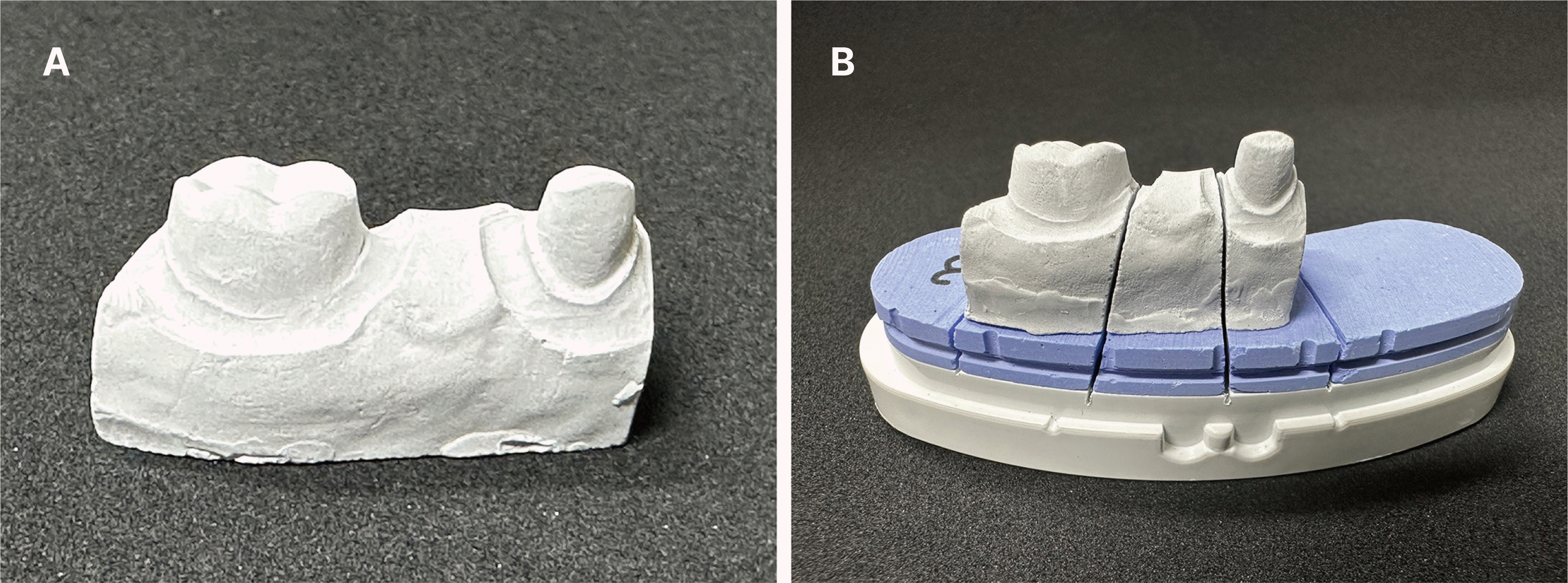
The reference stone casts were fabricated using polyvinyl siloxane impression material (Medium body; 3M, St. Paul, USA) and an impression tray (Impression tray; 3M), resulting in 20 impressions. Subsequently, these impressions were cast into 20 stone working casts employing type IV dental stone (FUGIROCK; GC, Leuven, Belgium). The working casts were allocated into two distinct groups: the solid working cast (S) group and the removable die (RD) group (Fig. 2). In the S group, 10 stone working casts had no further modifications (Fig. 2A). Conversely, in the RD group, 10 stone working casts were converted into working casts with removable dies, adhering to typical dental laboratory protocols (Fig. 2B). The working casts with removable dies were fabricated through a removable die system (Pindex system; Coltene/Whaldent AG, Altstätten, Switzerland). All working casts in both groups were fabricated by a clinical expert (W.-S.L.) who had over three decades of experience in dental prosthetic fabrication. The working casts for the RD group were prepared using a conventional method with die stone (FUGIROCK; GC). After setting, the casts were trimmed and drilled to insert standardized pins and metal sleeves, secured with adhesive. A layer of petrolatum was applied for easy separation, and type III dental stone (FUGIROCK; GC) was used to complete the base.
To create virtual models, the working casts were digitized using a dental desktop scanner (E3; 3Shape A/S, Copenhagen, Denmark). The scanner was calibrated according to the manufacturer’s recommendations. For the S group, complete scanning was performed to include the dies of the mandibular right first premolar and first molar, ensuring the inclusion of adjacent surfaces and finish lines, along with the adjacent teeth. In the RD group, a working cast was initially scanned in its entirety, including the removable die and adjacent teeth, without detaching the dies. Subsequently, the dies for the mandibular right first premolar and first molar were individually removed and scanned with precision. Then, these individually scanned dies were automatically aligned within the comprehensive virtual cast. All scanning procedures were meticulously reviewed by a clinical expert (W.-S.L.).
The CAD protocol for the 3-unit zirconia FPDs was uniformly applied across the two groups using dental CAD software (3Shape Dental System; 3Shape A/S). The dimensions of the 3-unit FPDs were established, referencing the size of the adjacent teeth and their contact points. Regarding design parameters, no cement space was allocated for the first 1 mm above the finish line in the axial direction, whereas a 60-µm cement space was designated beyond this 1-mm threshold. In addition, the FPDs were designed with a minimum thickness of 1 mm.
In this study, the evaluation of marginal and internal fit was conducted using the same methodology as validated in previous studies.22-25 The stereolithography (STL) files of the designed FPDs were uploaded into the CAM software, where optimized toolpaths were implemented, followed by milling the definitive 3-unit monolithic zirconia FPDs using unused milling burs and presintered monolithic zirconia blocks (Zirconia block; Luxen, Dentalmax, Seoul, Republic of Korea). A milling equipment was used. After milling, the zirconia FPDs underwent a postsintering process in compliance with the manufacturer’s guidelines to finalize the production of the zirconia restorations.
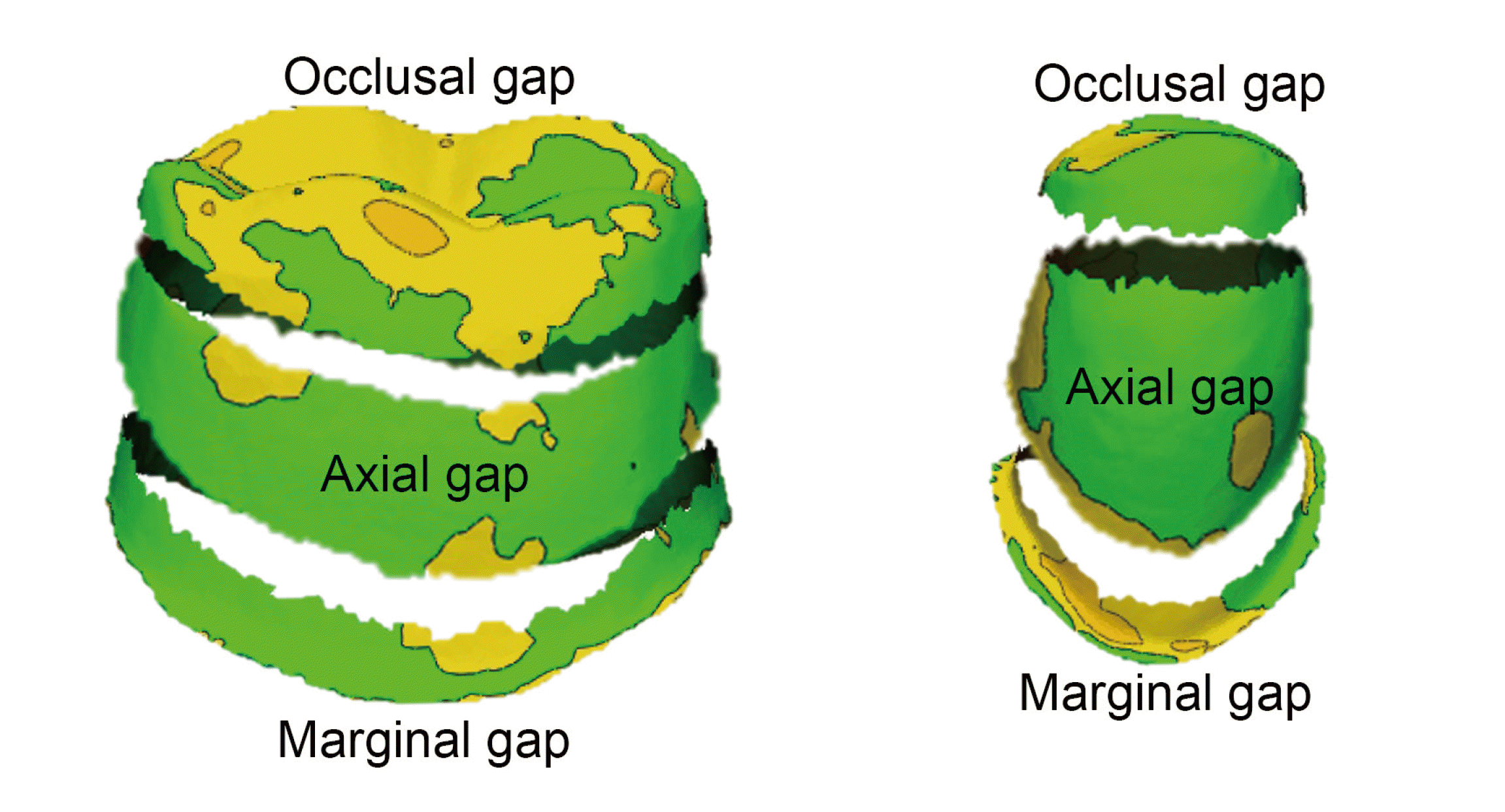
The definitive 3-unit monolithic zirconia FPDs were evaluated for marginal and internal fit without any internal or external modifications. Drawing on established literature,22-24 a digital assessment strategy was adopted. Initially, the FPDs were adapted onto a reference cast, and the assembly was scanned with a desktop scanner to generate STL files (adaptation STL file). The intaglio and external surfaces of the definitive FPDs were scanned to acquire comprehensive STL files, whereas the reference cast, inclusive of the die and adjacent teeth, was also fully scanned. The STL files of the FPD and reference cast were then aligned based on an adaptation STL file using 3D inspection software (Geomagic control X; 3D Systems, Rock Hill, USA). The gap between the aligned definitive FPD and the reference cast was quantified as the measure for marginal and internal fit. The marginal gap was assessed within 1 mm above the finish line of the die (Fig. 3). The occlusal gap for the internal fit was evaluated within 1 mm below the line angle of the occlusal surface (Fig. 3). Furthermore, the axial gap for the internal fit was defined as the area beyond the regions of the marginal and occlusal gaps (Fig. 3). The formula used to calculate the gap between the definitive FPD and the reference cast is as follows:
In this formula, X1,i denotes the i-th measurement point on the die surface of the reference cast, whereas X2,i corresponds to the i-th measurement point on the intaglio surface of the definitive FPD. The variable n represents the total count of points measured in each analysis. The marginal, axial, and occlusal gaps were consistently assessed at identical measurement points within the specified regions across all samples. For visual verification, a color map was created to highlight any deviations exceeding 50 µm with a yellow coloration (Fig. 3).
A suitable sample size, adequate to reveal statistical significance, was established for this study. Pilot experiments were performed three times, which employed methodology identical to the present study, and a power analysis software program (G*Power v3.1.9.2; Heinrich-Heine-Universität Düsseldorf, Germany) was utilized to ascertain a sample size of 10 for each group (actual power = 98.1%; power = 96.9%; α = 0.05). All data were analyzed using IBM SPSS version 27.0 (IBM Corp., Armonk, USA) (α = 0.05). Data normality was assessed using the Shapiro-Wilk test. Upon determining that the data did not follow a normal distribution, nonparametric statistical methods were employed. This study involved a comparative analysis of the two groups, and the Mann-Whitney U test was utilized to determine statistical significance.
The RD group exhibited significantly higher overall fit values than the S group for premolars and molars (P = 0.005), premolars (P = 0.01), and molars (P = 0.003) (Table 1, Fig. 4A). A significant difference in the marginal gap was observed for premolars and molars in the RD group (P = 0.038) (Table 1, Fig. 4B). However, in a separate analysis, no significant differences in the marginal gap were detected between the two groups for the premolars (P = 0.161) or molars (P = 0.195) (Table 1, Fig. 4B). Similarly, no significant differences were noted for the axial gaps in any of the comparisons (P = 0.195, Table 1, Fig. 4C). The RD group also demonstrated a significantly higher occlusal gap for premolars and molars (P = 0.002), premolars (P = 0.021), and molars (P = 0.002) (Table 1, Fig. 4D).
Consistent with the majority of marginal and internal fit outcomes in the RD group, the distribution of colors indicating higher deviations in the color map analysis was markedly evident in the RD group (Fig. 5). Notably, the presence of yellow deviations, indicating discrepancies > 50 µm, particularly in the marginal and occlusal regions within the RD group, was prominent (Fig. 5B).
This study evaluated the fit of 3-unit zirconia FPDs fabricated through the CAD/CAM process and established that FPDs fabricated from working casts without removable dies exhibited superior fit. Contrary to this hypothesis, significant discrepancies in fit were observed between the two methods, with FPDs fabricated from working casts with removable dies demonstrating reduced accuracy in fit, leading to the rejection of the null hypothesis (P < 0.05). Thus, dental technicians should judiciously reassess the routine fabrication of removable dies during the CAD/CAM production of FPDs, particularly in the absence of specific grounds for their necessity.
Removable dies are created through methodologies that affect the precision of die placement. Recent trends highlight the use of 3D printing in working cast fabrication, potentially introducing errors between 30 and 60 µm because of factors such as layer thickness and die design.13,14 Previous studies have clarified that the number of dies in a working cast, when scanned using a dental scanner, does not significantly affect the results.15 Nevertheless, numerous studies have historically reported die position errors of > 30 µm.19,20 In addition, the accuracy of die placement varies depending on the fabrication system used, with the Pindex system exhibiting the highest accuracy in working models.18-20 Accordingly, the Pindex system was used in this study for removable die production. Another study pointed out potential inaccuracies stemming from stone expansion at the stone-resin interface in the Pindex system, leading to postcutting inaccuracies.21 Hoffman et al. assessed the accuracy of interproximal distances in working casts fabricated using two methods, with and without removable dies.21 Their findings indicated that single-die working casts showed no significant difference in accuracy regardless of the presence of removable dies. However, two-die working casts demonstrated significantly decreased accuracy in the removable die group.21 This decrease in accuracy was pronounced when using 3D printers for fabricating working casts with removable dies.21 Based on these previous studies, the present study comparing the marginal and internal fit of 3-unit zirconia FPDs corroborates the observation that removable dies affect outcomes similarly.
In the present study, significant differences were observed in the marginal and occlusal gaps between the two groups, whereas no significant difference was found in the axial gap. Previous research has indicated that working casts fabricated using a removable die system may exhibit greater vertical errors than horizontal errors.19,20 The primary reason for these increased vertical errors is the process involved in creating holes on the base of the working cast and inserting pins to construct a removable die.19,20 Furthermore, repeated attachment and detachment of the die can amplify these vertical errors. In the present study, it is hypothesized that the larger deviation in the occlusal gap within the RD group can be attributed to vertical errors, which also likely influenced the marginal gap variation. Conversely, due to the lesser horizontal errors, it is presumed that they had a minimal impact on the axial gap errors.
This study had minor limitations, particularly its exclusive focus on tooth preparations featuring supragingival finish lines. The study aimed to evaluate prostheses fabricated from working casts without removable dies in manageable conditions similar to those with supragingival finish lines, regardless of the position of the actual finish line. However, scenarios requiring the fabrication of removable dies even under supragingival finish line conditions may arise. Previous studies have indicated that the scanning accuracy degrades when the distance between interproximal surfaces is < 1 mm. Moreover, the accuracy of the final prosthesis can vary based on the diverse designs of the tooth preparation finish lines. Consequently, further research is essential to evaluate prostheses fabricated from working casts in diverse conditions. Therefore, to approach a clinically more significant topic, additional experiments with varied methodologies and group settings are necessary.
The result of the present study revealed that definitive 3-unit monolithic zirconia FPDs fabricated from working casts with removable dies had less accurate marginal and internal fit than those fabricated from solid working casts without removable dies. These results indicate that errors inherent in the production of removable dies may compromise the marginal and internal fit of 3-unit monolithic zirconia FPDs.
References
1. Pereira ALC, de Medeiros AKB, de Sousa Santos K, de Almeida ÉO, Barbosa GAS, da Fonte Porto Carreiro A. 2021; Accuracy of CAD-CAM systems for removable partial denture framework fabrication: A systematic review. J Prosthet Dent. 125:241–8. DOI: 10.1016/j.prosdent.2020.01.003. PMID: 32147252.

2. Lee YS, Kim SY, Oh KC, Moon HS. 2022; A comparative study of the accuracy of dental CAD programs in designing a fixed partial denture. J Prosthodont. 31:215–20. DOI: 10.1111/jopr.13406. PMID: 34310790.

3. Kim SH, Choi YS, Kang KH, Att W. 2022; Effects of thermal and mechanical cycling on the mechanical strength and surface properties of dental CAD-CAM restorative materials. J Prosthet Dent. 128:79–88. DOI: 10.1016/j.prosdent.2020.11.014. PMID: 33546857.

4. Parize H, Tardelli JDC, Bohner L, Sesma N, Muglia VA, Dos Reis AC. 2022; Digital versus conventional workflow for the fabrication of physical casts for fixed prosthodontics: A systematic review of accuracy. J Prosthet Dent. 128:25–32. DOI: 10.1016/j.prosdent.2020.12.008. PMID: 33551140.

5. Takaichi A, Fueki K, Murakami N, Ueno T, Inamochi Y, Wada J, Wakabayashi N. 2022; A systematic review of digital removable partial dentures. Part II: CAD/CAM framework, artificial teeth, and denture base. J Prosthodont Res. 66:53–67. DOI: 10.2186/jpr.JPR_D_20_00117. PMID: 33504722.

6. Revilla-León M, Kois DE, Kois JC. 2023; A guide for maximizing the accuracy of intraoral digital scans: Part 2 - Patient factors. J Esthet Restor Dent. 35:241–9. DOI: 10.1111/jerd.12993. PMID: 36639916.
7. Baghani MT, Shayegh SS, Johnston WM, Shidfar S, Hakimaneh SMR. 2021; In vitro evaluation of the accuracy and precision of intraoral and extraoral complete-arch scans. J Prosthet Dent. 126:665–70. DOI: 10.1016/j.prosdent.2020.08.017. PMID: 33070974.
8. Huang MY, Son K, Lee KB. 2021; Effect of distance between the abutment and the adjacent teeth on intraoral scanning: An in vitro study. J Prosthet Dent. 125:911–7. DOI: 10.1016/j.prosdent.2020.02.034. PMID: 32473732.
9. Son K, Lee KB. 2021; Effect of finish line locations of tooth preparation on the accuracy of intraoral scanners. Int J Comput Dent. 24:29–40.
10. Chen Y, Zhai Z, Li H, Yamada S, Matsuoka T, Ono S, Nakano T. 2022; Influence of liquid on the tooth surface on the accuracy of intraoral scanners: an in vitro study. J Prosthodont. 31:59–64. DOI: 10.1111/jopr.13358. PMID: 33829613.

11. Rekow ED. 2020; Digital dentistry: The new state of the art - Is it disruptive or destructive? Dent Mater. 36:9–24. DOI: 10.1016/j.dental.2019.08.103. PMID: 31526522.

12. AlRumaih HS. 2021; Clinical applications of intraoral scanning in removable prosthodontics: a literature review. J Prosthodont. 30:747–62. DOI: 10.1111/jopr.13395. PMID: 34043266.

13. Yilmaz B, Donmez MB, Kahveci Ç, Cuellar AR, de Paula MS, Schimmel M, Abou-Ayash S, Çakmak G. 2022; Effect of printing layer thickness on the trueness and fit of additively manufactured removable dies. J Prosthet Dent. 128:1318.e1–9. DOI: 10.1016/j.prosdent.2022.10.011. PMID: 36435670.

14. Azpiazu-Flores FX, Johnston WM, Mata-Mata SJ, Yilmaz B. 2023; Positional trueness of three removable die designs with different root geometries manufactured using stereolithographic 3D printing. J Prosthet Dent. S0022-3913(23)00606-6. DOI: 10.1016/j.prosdent.2023.08.036.

15. Yu BY, Son K, Lee KB. 2019; Effect of abutment superimposition process of dental model scanner on final virtual model. J Korean Acad Prosthodont. 57:203–10. DOI: 10.4047/jkap.2019.57.3.203.

16. Conrad HJ, Seong WJ, Pesun IJ. 2007; Current ceramic materials and systems with clinical recommendations: a systematic review. J Prosthet Dent. 98:389–404. DOI: 10.1016/S0022-3913(07)60124-3. PMID: 18021828.

17. Ceylan G, Emir F. 2023; Evaluating the accuracy of CAD/CAM optimized stones compared to conventional type IV stones. PLoS One. 18:e0282509. DOI: 10.1371/journal.pone.0282509. PMID: 36877717. PMCID: PMC9987827.

18. Sivakumar I, Mohan J, Arunachalam KS, Zankari V. 2013; A comparison of the accuracy of three removable die systems and two die materials. Eur J Prosthodont Restor Dent. 21:115–9.
19. Serrano JG, Lepe X, Townsend JD, Johnson GH, Thielke S. 1998; An accuracy evaluation of four removable die systems. J Prosthet Dent. 80:575–86. DOI: 10.1016/S0022-3913(98)70035-6. PMID: 9813809.

20. Covo LM, Ziebert GJ, Balthazar Y, Christensen LV. 1988; Accuracy and comparative stability of three removable die systems. J Prosthet Dent. 59:314–8. DOI: 10.1016/0022-3913(88)90180-1. PMID: 3279188.

21. Hoffman M, Cho SH, Bansal NK. 2017; Interproximal distance analysis of stereolithographic casts made by CAD-CAM technology: An in vitro study. J Prosthet Dent. 118:624–30. DOI: 10.1016/j.prosdent.2017.01.027. PMID: 28477918.
22. Loetzerich JM, Raith S, Reich S. 2023; Verification of a digital approach for three-dimensional evaluation of marginal and internal fit. J Prosthet Dent. S0022-3913(23)00618-2. DOI: 10.1016/j.prosdent.2023.09.004. PMID: 37852857.

23. Holst S, Karl M, Wichmann M, Matta RE. 2011; A new triple-scan protocol for 3D fit assessment of dental restorations. Quintessence Int. 42:651–7. DOI: 10.1177/1758736012452181. PMID: 22924063. PMCID: PMC3425397.
24. Dahl BE, Rønold HJ, Dahl JE. 2017; Internal fit of single crowns produced by CAD-CAM and lost-wax metal casting technique assessed by the triple-scan protocol. J Prosthet Dent. 117:400–4. DOI: 10.1016/j.prosdent.2016.06.017. PMID: 27692584.

25. Park JM, Hämmerle CHF, Benic GI. 2017; Digital technique for in vivo assessment of internal and marginal fit of fixed dental prostheses. J Prosthet Dent. 118:452–4. DOI: 10.1016/j.prosdent.2016.12.016. PMID: 28343673.
Fig. 1
Study diagram.
FPD: fixed partial denture; CAD/CAM: computer-aided design and computer-aided manufacturing.
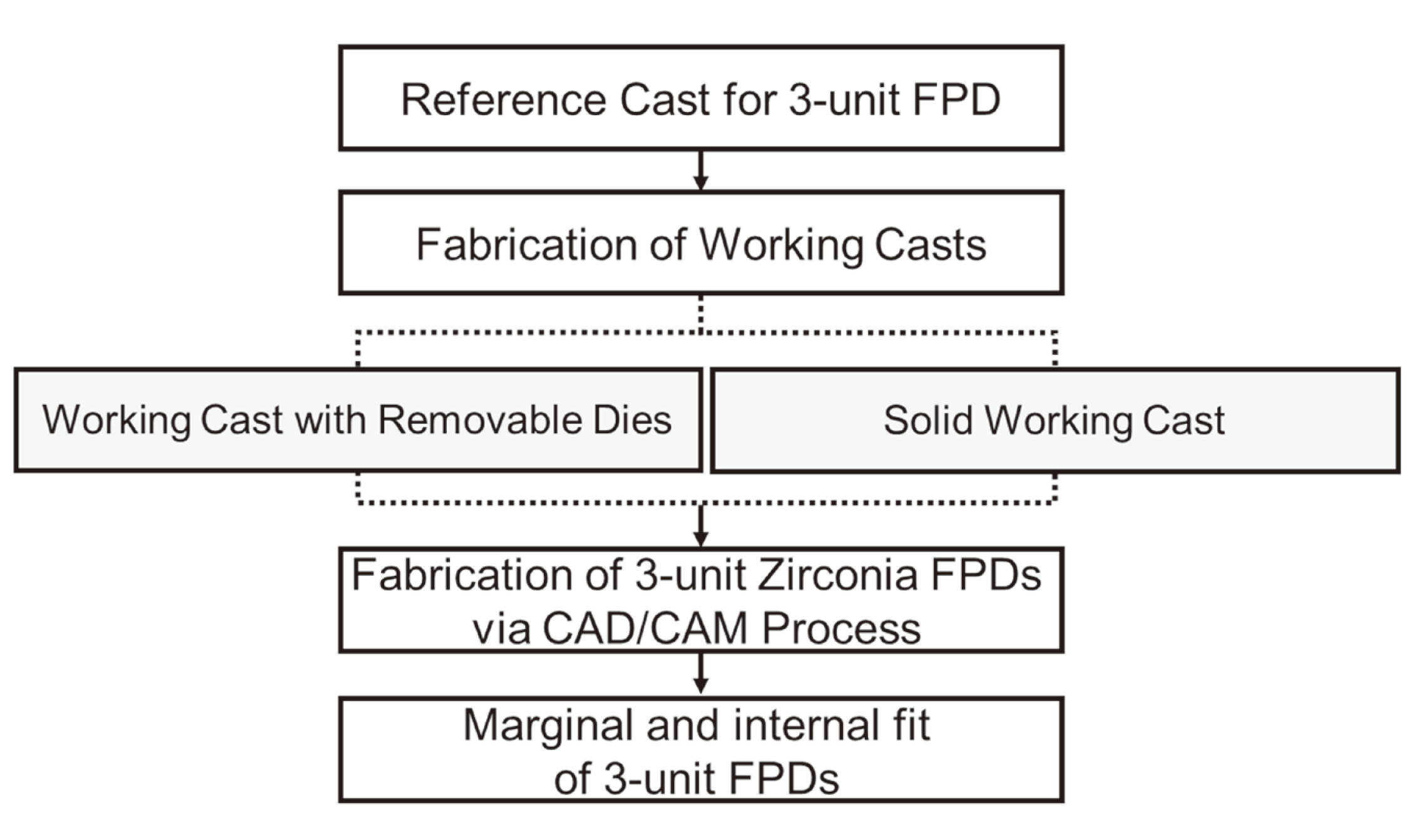
Fig. 4
Comparison of the marginal and internal fit of the three-unit monolithic zirconia FPDs fabricated via solid working casts and removable die system. (A) Overall region, (B) Marginal gap, (C) Axial gap, (D) Occlusal gap.
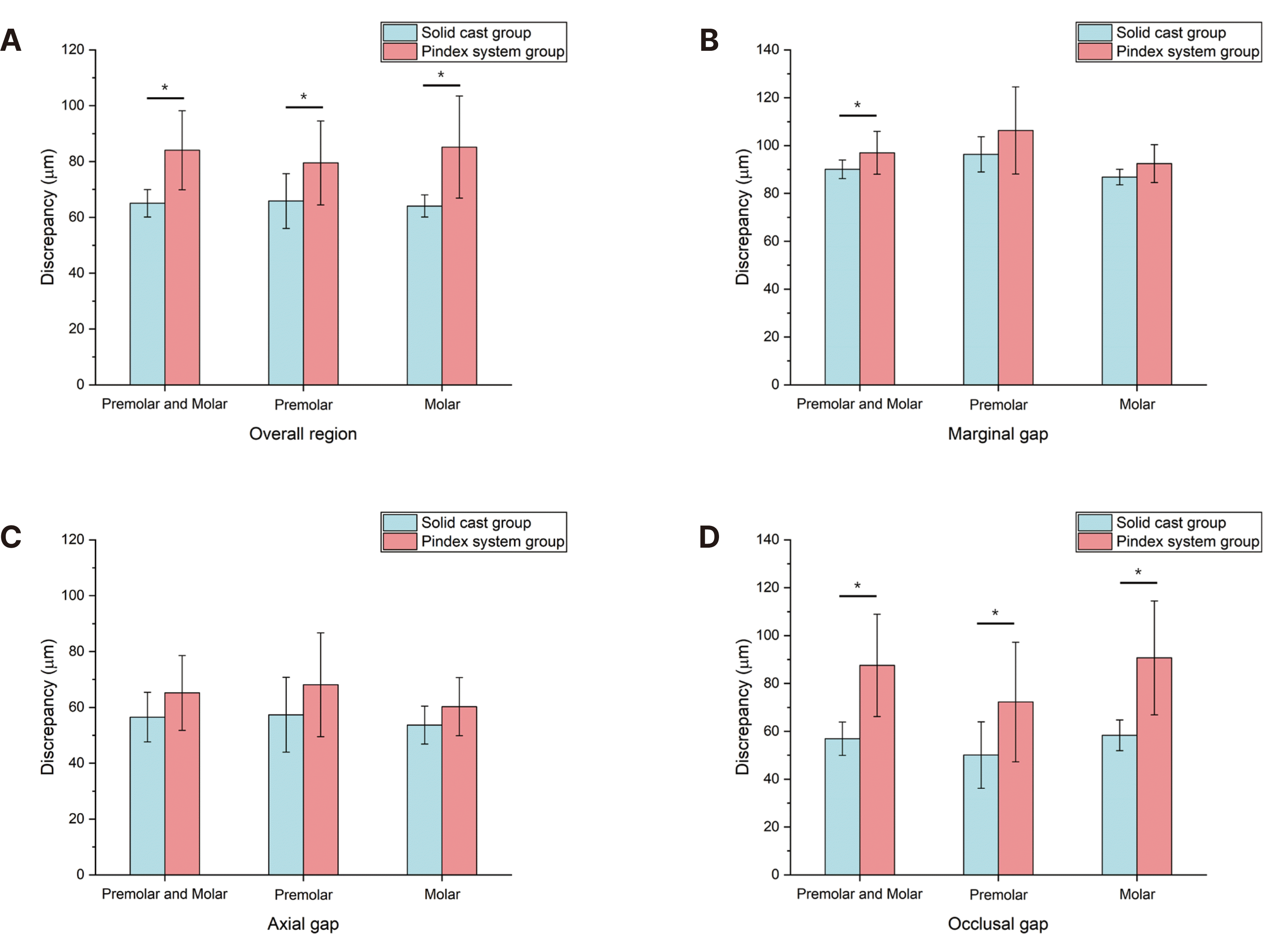
Fig. 5
Comparison of deviations in the color difference map of the 3-unit monolithic zirconia FPDs fabricated via solid working casts (A) and removable die system (B).
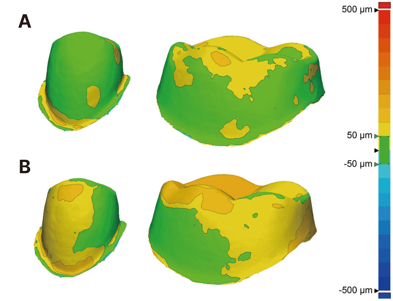
Table 1
Comparison of the marginal and internal fit of 3-unit monolithic zirconia FPDs fabricated via solid working casts and working casts with removable dies
| Measurement region | Tooth type | Group | Mean | SD | P |
|---|---|---|---|---|---|
| Overall | Premolar and molar | S | 65.1 | 4.9 | 0.005* |
| RD | 84.1 | 14.2 | |||
| Premolar | S | 65.9 | 9.8 | 0.01* | |
| RD | 79.5 | 15.0 | |||
| Molar | S | 64.1 | 4.0 | 0.003* | |
| RD | 85.2 | 18.3 | |||
| Marginal gap | Premolar and molar | S | 90.1 | 3.9 | 0.038* |
| RD | 97.0 | 9.0 | |||
| Premolar | S | 96.4 | 7.4 | 0.161 | |
| RD | 106.3 | 18.2 | |||
| Molar | S | 86.8 | 3.2 | 0.195 | |
| RD | 92.5 | 7.9 | |||
| Axial gap | Premolar and molar | S | 56.5 | 8.9 | 0.195 |
| RD | 65.2 | 13.4 | |||
| Premolar | S | 57.4 | 13.4 | 0.195 | |
| RD | 68.1 | 18.6 | |||
| Molar | S | 53.7 | 6.8 | 0.195 | |
| RD | 60.3 | 10.4 | |||
| Occlusal gap | Premolar and molar | S | 56.9 | 6.9 | 0.002* |
| RD | 87.6 | 21.4 | |||
| Premolar | S | 50.1 | 13.9 | 0.021* | |
| RD | 72.2 | 25.0 | |||
| Molar | S | 58.3 | 6.4 | 0.002* | |
| RD | 90.7 | 23.8 |


 PDF
PDF Citation
 Print
Print


 XML Download