Journal List > Diabetes Metab J > v.48(3) > 1516087294

Bu, Sun, Pan, Deng, and Yuan: Glucagon-Like Peptide-1: New Regulator in Lipid Metabolism

Abstract

Glucagon-like peptide-1 (GLP-1) is a 30-amino acid peptide hormone that is mainly expressed in the intestine and hypothalamus. In recent years, basic and clinical studies have shown that GLP-1 is closely related to lipid metabolism, and it can participate in lipid metabolism by inhibiting fat synthesis, promoting fat differentiation, enhancing cholesterol metabolism, and promoting adipose browning. GLP-1 plays a key role in the occurrence and development of metabolic diseases such as obesity, nonalcoholic fatty liver disease, and atherosclerosis by regulating lipid metabolism. It is expected to become a new target for the treatment of metabolic disorders. The effects of GLP-1 and dual agonists on lipid metabolism also provide a more complete treatment plan for metabolic diseases. This article reviews the recent research progress of GLP-1 in lipid metabolism.

KEY FIGURE

Highlights

• GLP-1 increases ABCA1 mRNA and apo AI, mediating intracellular cholesterol efflux.
• GLP-1 induces FNDC5 expression in β-cells via CREB, promoting WAT to BAT conversion.
• GLP-1 oxidates fatty acids and reduces its influx into hepatocytes, inhibiting NAFLD.
• GLP-1R/GIPR dual agonists improve lipid metabolism, with greater potential than GLP-1RAs.

The DEVELOPMENT AND USE OF GLUCAGON-LIKE PEPTIDE-1

Glucagon-like peptide-1 (GLP-1) is a 30-amino acid peptide hormone produced by intestinal L cells through differential processing of the proglucagon (PG) gene [1]. Kimball and Murlin [2] discovered PG in 1923. GLP-1 was discovered in 1983 when Bell et al. [3] analyzed the genetic sequence of glucagon. PG gene sequence consists of six exons and five introns, and GLP-1 (72–108) is one of the main domains. PG gene is expressed in pancreatic α-cells, intestinal L cells, and some nerve cells, and through tissue-specific translation modification, different end products are finally formed. While GLP-1 is mainly expressed and translated in intestinal L cells. In 1987, GLP-1 (7–36) was recognized as a physiological incretin in the studies of Kreymann et al. [4]. GLP-1 is derived from the PG precursor, which is a class of highly efficient incretins released by the body in response to nutrient intake. Such as the release of GLP-1 can be directly stimulated by nutrients such as glucose and fat in the intestinal lumen [5]. In 2005, exenatide was approved as a kind of GLP-1 receptor agonists (GLP-1RAs) for the clinical treatment of type 2 diabetes mellitus (T2DM) as the first international incretin drug [6]. Following the application of exenatide, other GLP-1 receptor (GLP-1R) activators were developed such as liraglutide (Lira). Meanwhile, the application of GLP-1RAs in non-diabetic diseases has also been launched.

STRUCTURE, DISTRIBUTION, AND PHYSIOLOGICAL FUNCTION OF GLP-1

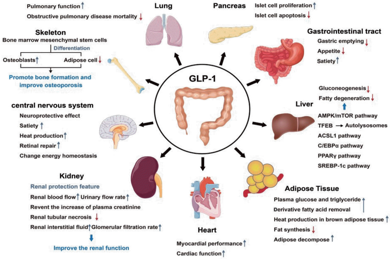
GLP-1 is composed of 30 amino acid residues, which primary structure begins with histidine and ends with glycine. There are a variety of molecular forms in the body which main active forms are GLP-1 (7–36) NH2 and GLP-1 (7–37), of which about 80% exist in the form of GLP-1 (7–36) NH2, which is first decomposed and inactivated in the blood circulation. Then it is eliminated by liver metabolism and renal excretion [7]. GLP-1 has a variety of effects, originating from the presence of its receptors in specific nuclei of the lung, pancreas, gastrointestinal tract, kidney, heart, bone, central nervous system, etc. In recent years, studies have found that the expression of GLP-1R is also found in liver and adipose tissue (Fig. 1) [8]. In the lung, GLP-1 binding to its receptors improves lung function and reduces mortality in patients with obstructive pulmonary disease [9]. In the pancreas, GLP-1 binding to its receptor stimulates the proliferation and regeneration of islet β-cells and inhibits their apoptosis [10]. In the gastrointestinal tract, GLP-1 binding to its receptors delays gastric emptying, reduce appetite, and increase satiety [11]. In the kidney, GLP-1 binding to its receptor has extensive renal protective properties. This characteristic may improve renal function by increasing renal blood flow, urine flow rate, preventing elevated plasma creatinine, reducing tubular necrosis, increasing renal interstitial fluid and glomerular filtration rate, and cytoprotective and anti-inflammatory effects [12,13], and may improve glomerular hyperosmosis in patients with T2DM in some cases [14]. In bone, GLP-1 binding to its receptor promotes bone marrow mesenchymal stem cell osteoblast differentiation and inhibits its differentiation into fat cells, thereby promoting bone formation and improving osteoporosis [15]. In the central nervous system, the action of GLP-1 and GLP-1RAs on its receptors can affect a variety of brain functions including satiety, thermogenesis, influence blood pressure, neurogenesis, neurodegeneration, retinal repair, and change energy homeostasis [16-20]. And its neuroprotective effect has become a research hotspot in recent years. In the heart, GLP-1 binding to its receptor enhances myocardial performance and has important physiological effects on cardiac function through direct receptor-mediated [21-24]. Several studies have shown that the clear link between glucose metabolism and GLP-1 through the protein kinase A (PKA) pathway [25-27]. In addition, GLP-1 has now been proved to be associated with nutrient intake and can be linked to lipid metabolism [28-30]. This article will review the role of GLP-1 in lipid metabolism in detail.
In liver tissue, GLP-1 levels are considered to be highest in human organs [31]. The induction of GLP-1 or its analogs in the liver can regulate varieties of processes, including hepatic gluconeogenesis, glycogen synthesis, and glycolysis [32]. However, the expression of GLP-1R in liver tissue remains controversial. To date, studies in animals and humans suggest that GLP-1 may improve hepatic lipid accumulation through signal transduction of the AMP-activated protein kinase (AMPK)/mammalian target of rapamycin (mTOR) pathway [33]. Downregulating the expression of miR-23a and increasing the expression of the mitochondrial protective gene peroxisome proliferators-activated receptor-γ coactivator-1 (PGC-1α) can exert an anti-apoptotic effect on human hepatocytes [34]. Meanwhile, GLP-1R has been shown to exist in human liver cells. GLP-1 analogs (exendin-4) can also improve hepatic steatosis [35] in ob/ob mice both in vivo and in vitro [36]. Lira (GLP-1R) can reduce the occurrence of hepatic steatosis in vivo and in vitro by enhancing autophagy and lipid degradation through the GLP-1R-transcription factor EB (TFEB)-mediated autophagy lysosomal pathway [37]. These evidences all suggest that the action of GLP-1R in liver cells may be directly stimulated by ligand-receptor action. However, some scholars have proposed that the effect of GLP-1 in the liver is indirectly mediated by multiple mechanisms, such as reducing fat absorption or inhibiting fat cells through acyl-CoA synthetase long chain family member 1 (ACSL1), CCAAT/enhancer binding protein α (C/EBPα), peroxisome proliferator-activated receptor-γ (PPARγ), and sterol regulatory element binding protein-1c (SREBP-1c) pathways to achieve the purpose of reducing liver lipids [38]. Alternatively, hepatic steatosis can be alleviated by regulating insulin clearance in the liver [39]. Although the expression of GLP-1R in liver tissue is controversial, GLP-1RAs have been shown to act as regulatory pathways affecting metabolic disorders.
GLP-1 has a direct effect on adipose tissue. GLP-1 may affect systemic energy metabolism by regulating adipocyte development, accelerating plasma clearance of glucose and triacylglycerol-derived fatty acids, improving insulin signaling, and stimulating thermogenesis in brown adipose tissue (BAT) [40-45]. For example, GLP-1R in adipocytes can directly regulate apoptosis and preadipocyte proliferation by activating the adenylate cyclase/cyclic adenosine monophosphate (AC/cAMP) signaling pathway and various cell signaling cascades, including extracellular signal-regulated kinase (ERK), protein kinase C (PKC), and protein kinase B (AKT) [41,46]. GLP-1 also regulates lipid metabolism in BAT and white adipose tissue (WAT) through the brain-adipocyte axis [43,44]. In differentiated human adipocytes in vitro, GLP-1 can regulate the expression of adipogenesis and lipolytic genes [47]. The direct effect of GLP-1 on adipose tissue has been demonstrated in many studies. However, the specific receptors that mediate and participate in it have not been determined, which are needed to be verified by establishing multiple experimental models.

GLP-1 MODULATES LIPID METABOLISM

GLP-1 modulates lipid metabolism in the liver

GLP-1 Modulates fat synthesis in the liver

The liver is the main site of fat synthesis. Hepatic fat synthesis is a process by which carbohydrates and amino acids are converted into fatty acids and condensed with glycerol to form triglycerides (TG) [48]. Hepatic fat transport is the transport of TG out of the liver in the form of very low-density lipoprotein (VLDL) to participate in the oxidative breakdown of tissues or storage in adipose tissue [49]. The synthesis and transport of liver fat is the key to maintaining the homeostasis of TG in the liver. And its anabolic-related genes include fibroblast growth factor-21 (FGF21), SREBP-1c, fatty acid synthase (FAS), acetyl-CoA carboxylase (ACC-a), liver X receptor (LXR), suppressor of cytokine signaling-3 (SOCS-3), etc.
A recent study pointed out that GLP-1 can reduce liver fat synthesis. After giving long-term reared ovariectomy (OVX) mice with different gradient protein concentrations (standard, low protein, high protein) diet, it was concluded that a large number of lipid droplets were visible in the model group of hepatocytes hematoxylin and eosin (HE) staining and oil red O staining compared with the control group mice. In contrast, the expression of GLP-1 mRNA in the colon tissues of mice in the model group decreased, and the content of GLP-1 in serum decreased. Interestingly, FGF21, SREBP-1c, and FAS mRNA in the liver tissues of the model group of mice and protein expression was increased compared with the control group, while liver kinase B1 (LKB1) mRNA expression was reduced. The above conclusions all proved that a long-term high protein diet could reverse the above changes in OVX mice, while there was no significant difference between the low protein group and the model group. In this experiment, the high protein diet can promote intestinal secretion of GLP-1, improve FGF21 resistance, upregulate LKB1 to activate the AMPK pathway, downregulate the expression of SERBP-1c mRNA and protein in the liver, and ultimately inhibit liver lipid synthesis [50].
More interestingly, male Sprague-Dawley rats were randomly divided into the normal control (NC) group, high-fat (HF) group, and HF+Lira (GLP-1RA) group. Cell experiments showed that the arrangement of liver cells in rats in the HF group was disordered, and some hepatocytes were degenerated. There were more lipid droplets and inflammatory cell infiltration in hepatocyte cells. However, the fatty alteration of hepatocytes in the HF+Lira group was significantly improved. There were a small number of fat droplets in liver cells, and the infiltration of inflammatory cells was improved. Compared with the NC group, animal experiments found that the serum aspartate transaminase (AST), alanine transaminase (ALT), total cholesterol (TC), and TG were increased in the HF group, and the level of liver TG was also increased. Compared with the HF group, the HF+Lira group decreased serum AST, ALT, and TC, serum TG levels decreased significantly, the mRNA expression of SOCS-3 and SREBP-1c decreased significantly, and the liver TG level was also reduced. The above studies suggest that GLP-1 can downregulate the expression of SOCS-3 and SREBP-1c in rats with nonalcoholic fatty liver disease (NAFLD), reduce hepatic TG deposition. GLP-1 is also consistent with the conclusion that GLP-1 can inhibit liver lipid synthesis [51]. Boland et al. [52] gave diet-induced obesity (DIO) mice daily GLP-1R/Gcg receptor (GcgR) double agonist (cotadutide), Lira, Gcg agonist (g1437), and Lira+g1437 respectively. They found that all groups receiving GcgR agonists showed lower liver lipid content than the control group. In the hyperglycemic clamping experiment, cotadutide was also found to reduce the de novo fat production of the liver during clamping. It was concluded that cotadutide can inhibit the production of key fats in the liver of mice and reduce the lipid content of the liver. In animal studies by Burmeister et al. [53], GLP-1R knockout group in the hypothalamus was also activated and the liver TG increased in rats compared with the control group. Besides, KKAy mice were selected and divided into the Lira group and the HF feeding group. The results were shown that the serum TC and TG contents of mice in the model group and the Lira group were significantly increased compared with the control group. Interestingly, compared with the model group, the TC and TG contents of the Lira group had a decreasing trend but the difference was not statistically significant. The results of HE staining showed that compared with the control group, the model group had swollen hepatocytes and vacuolar changes (mainly steatosis). Compared with the model group, hepatocytes in the Lira group had less swelling, fewer vacuolar changes, more nuclei, and less inflammation. Compared with the model group, hepatocytes in the Lira group had less swelling, fewer vacuolar changes, more nuclei, and less inflammation. Phosphoinositide 3-kinase (PI3K) protein expression was significantly increased in the Lira group compared to the model group [54]. Similarly, animal experiments have shown that the expression of liver tissue FAS and ACC-a mRNA in rats in the Lira group was significantly higher than the control ratio [55]. At present, several studies have found that GLP-1 analog Lira may improve lipid metabolism and fat deposition by downregulating the expression of lipid metabolism-related genes FAS, and ACC-a mRNA, or promoting the expression of PI3K protein in mouse liver. The above results suggest that GLP-1 is important for inhibiting liver fat synthesis, and is likely to be related to AMPK, SERBP-1c, FAS, ACC-a, PI3K, and other signaling pathways.
In vitro studies, HepG2 cells treated with palmitic acid (PA) were found that dulaglutide significantly reduced liver lipid accumulation and gene expression related to lipid drop-binding proteins, de novo adipogenesis and TG synthesis. And dulaglutide increased the expression of proteins associated with lipolysis and fatty acid oxidation, as well as family with sequence similarity 3 member A (FAM3A) [56]. In the HepG2 fatty liver cell model, hypoxia-inducible factor-2alpha (HIF-2α)/PPARα pathway was found to be essential for Lira to inhibit liver lipid synthesis and reduce lipid-induced hepatic steatosis [57]. In addition, in the HepG2 cell experiment, some extrahepatic factors, such as erythroid-2-related factor 2 (NRF2) pathway [58], intestinal metabolite sodium butyrate (NaB) [59], Src homology region 2 domain-containing phosphatase-1 (SHP1)/AMPK [60], sirtuin 1 (SIRT1)/heat shock factor 1/heat shock protein (HSP) [61], and other signaling pathways have also been found to be involved in the regulation of liver lipid synthesis. Lira was found in PA-induced Kupffer cells to reduce HF diet-induced hepatic steatosis by inhibiting NOD-like receptor thermal protein domain associated protein 3 (NLRP3) inflammasome activation [62]. Gupta et al. [63] first reported the existence of G protein-coupled receptor (GPCR) GLP-1R in human hepatocytes in 2010, and provided evidence to prove the direct effects of GLP-1 protein on cells. GLP-1R binds to ligands to internalize GLP-1R, and can independently reduce TG accumulation in hepatocytes by activating key signaling molecules downstream of insulin receptor substrate 2 (IRS2). Since then, a number of studies have also been conducted in human primary hepatocytes, providing evidence for the direct effect of GLP-1 binding with GLP-1R in hepatocytes on reducing liver lipids [64]. In the culture system of various isolated hepatocytes (mouse primary hepatocytes, HepG2 human hepatocytes, human primary hepatocytes, etc.), Liu et al. [65] found that Lira’s stimulation of liver FGF21 expression may require the expression of GLP-1R in extrahepatic organs, and the increase of liver FGF21 is also necessary for Lira to improve hepatic lipid homeostasis. In cell experiments, GLP-1 was found to have more mechanisms that may inhibit liver fat synthesis, such as FAM3A, NRF2, NaB, SHP1/AMPK, NLRP3, FGF21, IRS2, and other signaling pathways, which also provided direct evidence for the role of GLP-1R in the regulation of lipid metabolism in the liver.

GLP-1 modulates hepatic lipolysis

Lipolysis is the process of converting TG into glycerol and fatty acids under the action of hydrolase, such as adipose triacylglyceride lipase (ATGL), hormone-sensitive lipase (HSL), and monacylglycerol lipase, in which fatty acids can provide energy for the body through β-oxidation or other oxidation pathways. And lipolysis is also affected by many lipid-associated proteins, such as SIRT1, α-subunit of PGC-1α, fatty acid-binding protein 4 (FABP4), etc. [66].
In a Lira-related study, healthy male C57BL/6 rats were evenly divided into three groups, namely NC group, model control group (OC group), and Lira group. Mice in the NC group were fed standard feed, while mice in the OC group and mice in the Lira group were fed HF feed. Compared with the NC group, the liver TG level, total liver TC level, and liver weight of the OC group were increased. And the mRNA expression levels of SIRT1, PGC-1α, and phosphoenolpyruvate carboxykinase (PEPCK) in the liver of the mice were significantly reduced. Compared with the OC group, the liver TG level, total liver TC level and liver weight of the mice in the Lira intervention group were significantly reduced. And the mRNA expression levels of SIRT1, PGC-1α, and PEPCK in the liver of the mice were significantly increased. This indicates that Lira can improve liver fatty acid oxidation in obese mice, reduce liver fat accumulation, and promote hepatic lipolysis by activating the liver SIRT1/PGC-1α/PEPCK pathway [67]. However, a small sample found that Lira did not significantly change levels of the hepatic fat oxidation marker β-hydroxybutyrate, although it reduced liver fat [68]. This conclusion should not be overlooked as a distraction factor with small sample size. Therefore, the effect of GLP-1 on liver lipolysis in clinical studies can be further developed.

GLP-1 modulates liver cholesterol metabolism

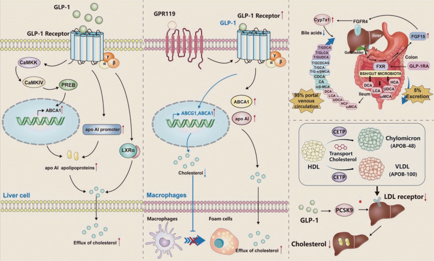
As the central organ of cholesterol metabolism, the liver directly affects the body’s cholesterol metabolism homeostasis. Hepatic cholesterol metabolism involves multiple parts, including cholesterol uptake, synthesis, biotransformation, and efflux. And it is related to cholesterol metabolism genes such as LXRα, ATP-binding cassette transporter A1 (ABCA1), ATP-binding cassette G1 (ABCG1), acyl-coenzyme A oxidase 1 (ACOX1), apolipoprotein AI (apo AI) (Fig. 2) [69]. Ben-Shlomo et al. [70] found in vivo and in vitro studies that GLP-1 and its analog exendin-4 were able to induce an increase in ABCA1 mRNA, increase the expression of apo AI mRNA and promoter, thereby increasing apo AI secretion, indicating that GLP-1 in hepatocytes may mediate the outflow of intracellular cholesterol to apo AI through the ABCA1 pathway. Recent studies have also demonstrated that exendin-4 is mediated by Ca2+/calmodulin (CaM)-dependent protein kinase kinase/CAM-dependent protein kinase IV/prolactin regulatory element binding (CaMKK/CaMKIV/PREB) pathway increases liver ABCA1 expression and reduces cholesterol accumulation [71]. Similarly, studies such as Ben-Shlomo et al. [70] have found that GLP-1 directly acts on LXRα in liver cells to mediate cholesterol outflow. These studies suggest that GLP-1 may mediate cholesterol outflow in hepatocytes through the LXRα-ABCA1 pathway, consequently avoiding large amounts of cholesterol accumulation in cells.
Recent studies have pointed out that GLP-1 can increase the expression of ABCA1 and ABCG1 in ApoE-/- mice, thereby reducing the area of aortic plaque in HF raised ApoE-/- mice and realizing the reverse transport of aortic plaque. At the cellular level, GLP-1 can significantly increase the expression levels of ABCA1 and ABCG1, and decrease the level of intracellular cholesterol, which inhibits the transformation of macrophages into foam cells [72]. Studies have also shown that GLP-1 can significantly increase the expression of GLP-1R through G protein-coupled receptor 119 (GPR119), enhance the activity of ABCA1, lead to the increase of apo AI, and thus promote intracellular cholesterol outflow [73]. The GLP-1 analog Lira has been shown to have similar effects on cholesterol metabolism with GLP-1 [74].
Meanwhile, in the latest animal studies, some researchers found that exendin-4 can reduce levels of serum VLDL cholesterol and low-density lipoprotein cholesterol (LDL-C) by reducing liver sterol regulatory element binding protein 2 (SREBP2) level and cholesterol absorption [75]. The hepato-enteral circulation theory of bile acids is well known; that is, cholesterol is converted to bile acids in the liver, 95% bile acids are absorbed by the distal ileum and returned to the liver, and 5% bile acids are excreted in the stool. In order to maintain the bile acid pool, therefore, the amount of bile acid excreted is almost equal to the amount newly synthesized by the liver. Therefore, a possible mechanism proposed by the authors is that the activation of the farnesoid X receptor (FXR) by exendin-4 stimulates the expression of FGF15 in the small intestine and inhibits the expression of cytochrome P450 family 7 subfamily A member 1 (Cyp7a1) in the liver through fibroblast growth factor receptor 4 (FGFR4), thereby reducing fecal bile acid excretion and liver bile acid synthesis [75]. Interestingly, Lira has also been shown to attenuate negative feedback inhibition of bile acids by inhibiting intestinal FXR activity and ultimately reduce bile acid synthesis in the liver [76]. It has also been reported that cholesteryl ester transfer protein (CETP) can transport cholesterol from high-density lipoprotein to lipoprotein containing apolipoprotein B (ApoB) and rich in TG, such as chylomicron remains (containing ApoB-48) and VLDL remains (containing ApoB-100). These cholesterol can then be absorbed into the liver via low-density lipoprotein receptors (LDLRs) or LDLR-associated proteins [77]. In theory, if the LDLR is inhibited, the reverse transport of cholesterol is also inhibited, and less cholesterol is absorbed into the liver, thus reducing the accumulation of cholesterol in the liver. There are varieties of studies that confirm this conclusion. For example, Yang et al. [78] found that Lira inhibited proprotein convertase subtilisin/kexin type 9 (PCSK9) expression in HepG2 cells and db/db mice through hepatocyte nuclear factor 1 (HNF1)-dependent mechanism, and may inhibit LDLR expression in db/db mice through PCSK9 independent pathway, and ultimately reduce liver cholesterol accumulation. Animal studies have also shown that portal vein GLP-1 weakens intestinal fat absorption and chylomicron production through the vagal enteric-brain-liver axis, and reduces postprandish and fasting blood lipids [79].
These studies show that GLP-1 is closely related to cholesterol metabolism. GLP-1 can promote intracellular cholesterol outflow, reduce intracellular cholesterol content, and maintain intracellular cholesterol homeostasis through the ABCA1 pathway. While GLP-1 has not been clearly studied and reported through other ways to regulate intracellular cholesterol outflow. In addition, GLP-1 can reduce liver cholesterol accumulation by inhibiting SREBP2 and PCSK9, and reduce liver bile acid synthesis by weakening the negative feedback inhibition of bile acid after inhibiting FXR.

GLP-1 modulates fat lipid metabolism

GLP-1 modulates adiposynthetic and differentiation

Fat synthesis includes three aspects: de novo synthesis of saturated fatty acids, extension of fatty acid carbon chains, and synthesis of unsaturated fatty acids. The differentiation of adipose precursor cells consists of two stages, the former stage is the differentiation of adipose precursor cells into immature adipocytes, and the latter stage is the transformation of immature adipocytes to mature adipocytes, which associated genes include PPARγ, C/EBPα, adipocyte fatty acid-binding protein (AP2), lipoprotein lipase (LPL), etc.
After treating 3T3-L1 cells with GLP-1, some scholars found that the number of small and medium-sized adipocytes increased. And oil red O staining showed that GLP-1 had no significant effect on lipid accumulation. Compared with the control group, the treated cells can enhance the phosphorylation of the Akt signal 24 hours before differentiation [80]. Overall, these results suggest that GLP-1 may regulate 3T3-L1 adipogenesis through the Akt signaling pathway.
GLP-1 has also been found to increase mRNA and protein expression levels of AP2 and PPARγ in a dose-dependent manner during pre-3T3-L1 adipocyte differentiation. The real-time reverse transcription polymerase chain reaction (RT-PCR) results showed that GLP-1 promoted the expression of C/EBPα and LPL [80]. Recent studies have also found that the promotion of GLP-1 on 3T3-L1 pre-adipocyte differentiation depends on the long non-coding RNA protein folding activity of the ribosome (lncRNA PFAR), which can target miR-138, remove the inhibition of miR-138 on Yes-associated protein 1 (YAP1) mRNA, and then upregulate the expression of YAP1 [81]. These data suggest that GLP-1 can promote the differentiation of pre-3T3-L1 adipocytes by promoting the expression of the specific markers LPL and AP2, as well as the transcription factors PPARγ and C/EBPα. Activation and phosphorylation of signaling pathways such as Akt, and yes-associated protein 1-TEA domain transcription factor (YAP1-TEADs) also play a key role in fat differentiation.

GLP-1 modulates fat browning

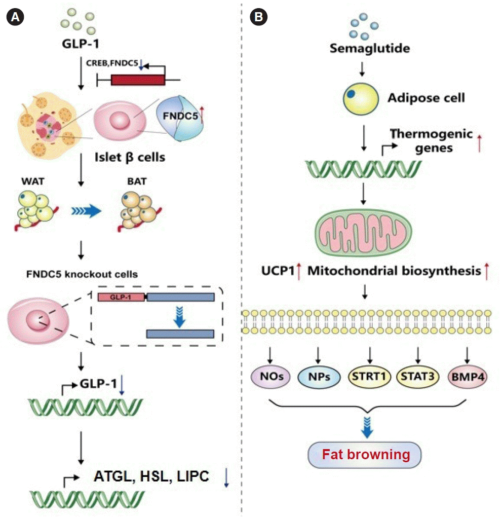
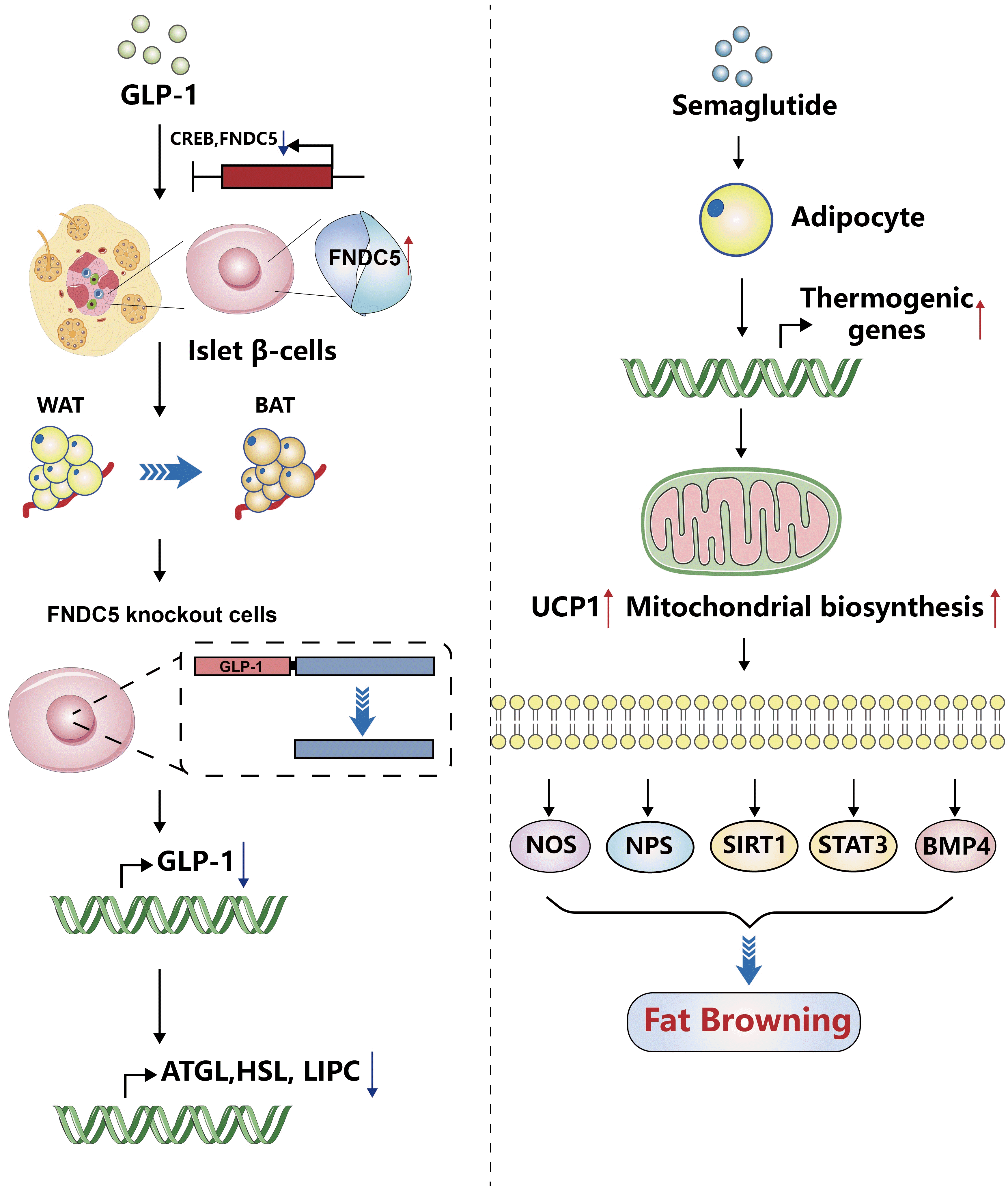
Adipose tissue is produced by the aggregation of a large number of fat cells. Adipose tissue is divided into WAT with a small number of mitochondria in the cell and BAT with a large number of mitochondria in the cell. WAT mainly plays the role of storing energy, while the main role of BAT is to decompose lipid droplets for energy. WAT can also be further divided into visceral adipose tissue (VAT) and subcutaneous adipose tissue [82]. Fat browning refers to the high plasticity of white fat cells, which can be transformed into beige fat cells with many similar morphological and functional characteristics to brown fat cells under the stimulation of exercise, cold exposure, adrenergic receptor stimulation, and other factors [83]. Since BAT has the function of thermogenesis and heat generation, promoting TG clearance and glucose metabolism. White fat browning is a good way to promote white fat consumption and accelerate human metabolism, which is associated with uncoupling protein 1 (UCP1), nitric oxide synthase (NOS), SIRT1, signal transducer and activator of transcription 3 (STAT3) (Fig. 3) [84].
The latest research pointed out that irisin is derived from its precursor protein fibronectin type III domain-containing protein 5 (FNDC5), which can be released from various tissues such as skeletal muscle, fat, and pancreas, mainly involved in the transformation of WAT to BAT, thereby increasing the body’s energy expenditure. GLP-1 can induce the expression of FNDC5 in pancreatic β-cells through the interaction of its downstream transcription factor cAMP-response element binding protein (CREB) with binding elements in the FNDC5 promoter, thereby promoting the conversion of WAT to BAT and enhancing energy metabolism [85]. In addition, GLP-1 expression was down-regulated in wild-type β Lox5 cells knocked out by FNDC5, and the induced expressions of lipolysis and autophagy-related genes and proteins were also inhibited, such as ATGL, HSL, and hepatic lipase deficiency gene (LIPC), suggesting that GLP-1 expression is also regulated by FNDC5, and may also provide therapeutic targets for lipid metabolism-related diseases.
Animal studies have shown that GLP-1RAs activate fat cell browning and may regulate BAT through the central nervous system. Martins et al. [86] found that semaglutide, as a GLP-1 analog, can directly act on fat cells, increase the expression of thermogenic genes for browning phenotype maintenance, improve the expression of UCP1, mitochondrial biosynthesis and thermogenic marker, thereby contributing to weight loss. So how does GLP-1 modulate BAT? Lockie et al. [44] observed the activity of BAT in the shoulder blade region of C57BL/6 mice by injecting GLP-1 (7–36) amide into the lateral ventricle of C57BL/6 mice. And they found that the mice lost weight, increased sympathetic activity, increased BAT temperature in the scapular region, and increased thermogenesis. However, the same dose of the above drug intervention was given by intraperitoneal injection, and there was no significant change in the temperature of BAT in the scapular area. This study suggests that GLP-1 can enhance the function of brown fat, increase heat production in BAT, and reduce body weight in mice by modulating the activity of the sympathetic system through binding with GLP-1R in the center. In recent years, extensive literature has reported that a variety of signaling pathways and transcription factors are involved in the fat differentiation process of WAT, such as the NOS pathway, neuropeptide S [87] pathway, SIRT1 [88] pathway, STAT3 [89], bone morphogenetic protein 4 (BMP4) [90] pathway, etc. These pathways and signaling pathways can promote BAT heat production. The above studies show that GLP-1 and GLP-1RAs can enhance BAT function or promote WAT browning through the center. However, the specific pathways or pathways involved are not clear enough and need further research and exploration.

CLINICAL APPLICATION OF GLP-1 IN LIPID METABOLISM AND ADIPOSE-RELATED DISORDERS

Clinical application of GLP-1 in obesity

Obesity is a major health problem worldwide. Studies in human and animal models suggest that GLP-1 may play a role in lipid metabolism and obesity. Similar to rodents, GLP-1 also plays a key role in human lipid metabolism and obesity [91]. Most studies have found a decrease in GLP-1 secretion in obese individuals [92], and some studies have found no difference in GLP-1 levels between obese and normal-weight controls [93]. The mechanism of action of GLP-1RA to promote weight loss mainly includes the reduction of fat deposition through regulation of adipose tissue (lipolysis, fatty acid oxidation, and adipocyte differentiation) and the reduction of food intake through the central and peripheral nervous system.
In obese patients, acipimox can inhibit adipose tissue lipolysis by activating protein upregulated in macrophage by INF-γ (PUMA-G) (also known as the hydroxycarboxylic acid receptor 2 [HCA2] and GPR109a) receptors. In addition, an inverse correlation between circulating free fatty acids and GLP-1 concentrations have been found in vivo and in vitro experiments, and systemic GLP-1 levels can be increased in obese patients after treatment with acipimox [94]. Studies demonstrated that the fat-damaging effects of GLP-1 are effective for weight loss and may also improve insulin resistance in obese patients [95]. There is also evidence that GLP-1RA (such as Lira, etc.) can significantly reduce food intake and promote fatty acid oxidation, thus promoting weight loss and fat mass reduction [16,96]. In addition, in vitro models of primary human adipose stem cells, researchers detected intracellular fat content and quantitative expression of early and mature adipocyte markers (PPARγ, FABP4, and HSL), demonstrating that glucagon can significantly reduce adipocyte differentiation, inhibit adipocyte proliferation and adipogenesis. Moreover, this effect can be reversed by specific GLP-1R (exendin-9) antagonists, which means that GLP-1RA may inhibit the proliferation and differentiation of human adipocyte precursors [97,98]. These studies suggest that GLP-1RA can achieve weight loss by interfering with the proliferation and differentiation of human fat precursors.
Besides, Baggio and Drucker [99] and Allegretti et al. [100] investigated the relationship between intestinal flora and obesity. They found that intestinal flora may mediate the release of the intestinal hormone GLP-1 through the short-chain fatty acid (SCFA)-GPCR pathway, thereby increasing satiety and slowing gastric emptying, ultimately reducing obesity. There is also evidence that GLP-1RA has positive effect on metabolic syndrome and abdominal obesity. In a recent randomized, double-blind, placebo-controlled trial, obese adults without diabetes were assigned to the exercise group, the GLP-1RA (Lira) group, and the exercise+Lira group. The results showed that abdominal fat percentage decreased by 2.6, 2.8, and 6.1 percentage points in the exercise, Lira, and combination groups compared to placebo. The combination of adherent exercise and Lira treatment reduced abdominal obesity and may therefore reduce obesity risk more than the individual treatments [101]. The difference between elevated or unchanged GLP-1 levels in obese patients may be related to differences in metabolic status and detection methods, but the significant correlation between GLP-1 and lipid metabolism suggests that it can control weight and regulate lipid metabolism in HF diet groups, and GLP-1 has the potential to treat obesity caused by HF diet [102].

Clinical application of GLP-1 in fatty liver

NAFLD is one of the most common metabolic syndromes in today’s society and has become the common chronic liver disease that seriously affects the health of adults and even children worldwide. There are currently no specific agents for NAFLD [103]. Previous studies have shown that GLP-1 can inhibit the formation and progression of NAFLD by enhancing lipid metabolism through oxidation and reducing the influx of fatty acids into hepatocytes [104]. The mechanisms of GLP-1 to improve NAFLD include improving lipid metabolism by regulating metabolism-related signaling pathways such as AMPK pathway [33] and JNK pathway [105]. And GLP-1 can inhibite hepatocyte apoptosis which induced by NLRP3 through autophagy in mitochondria, weakening the intrahepatic inflammatory response, thereby inhibiting the transition from simple fatty liver to nonalcoholic steatohepatitis (NASH). Finally, GLP-1 can achieve the purpose of preventing the deterioration of NAFLD [106,107]. Recent research data has shown that the expression of lipid metabolism genes adiponectin (APN), Adipo R, AMPK, PPARα mRNA and protein is significantly increased after 48 hours of treatment with different concentrations of GLP-1 compared with the model group of adipose hepatocytes [108]. GLP-1 alleviates hepatocyte steatosis and reduces intracellular lipid droplets by activating the expression of the above lipid metabolism genes. This also shows that GLP-1 can significantly improve clinical indexes, reduce inflammatory indicators, improve liver fat deposition, and be effective in the treatment of fatty liver.
In addition, Sharma et al. [64] studied GLP-1 and receptors on hepatocytes and found that the combination of them can reduce the lipid stress response in hepatocytes, reduce the lipid load of hepatocytes, delay steatosis, and ultimately prevent NAFLD. Several other studies have also provided evidence that GLP-1RA can reduce hepatic fat accumulation and reduce steatosis in liver tissue by regulating the expression of genes and proteins related to the fat synthesis pathway [109,110]. However, there are no targeted drugs specifically designed to treat NAFLD. According to this characteristic, GLP-1RA gradually entered the clinic. Hopkins et al. [111] treated T2DM patients with exenatide and found that the liver fat content (LFC) of patients decreased from 21.3%±19.3% to 12.7%±10.6%, a trend that suggested that GLP-1RA drugs could improve liver lipid metabolism. Similarly, Gastaldelli et al. [112] found a decrease in LFC in patients with moderate to severe NAFLD with T2DM using exenatide in combination with dapagliflozin. Additionally, a further follow-up study was conducted in patients with poor T2DM control and found a significant 31% reduction in LFC in patients treated with Lira. Recent studies suggest that treatment with semaglutide (GLP-1RA) in combination with firsocostat (ACC-a inhibitor) and/or cilofexor (nonsteroidal FXR agonist) can further improve hepatic steatosis and thus NAFLD compared with semaglutide alone in exploratory efficacy analyses [113]. Flint et al. [114] randomized subjects to semaglutide or placebo to observe the change of liver fat from baseline to week 48, and found that semaglutide significantly reduced hepatic steatosis, improved liver enzymes and metabolic parameters, and had a positive effect on the diagnosis and treatment of NAFLD compared with placebo. The above studies suggest that GLP-1 can significantly reduce LFC and inflammatory response in patients with T2DM and NAFLD, and even slow down the process of liver fibrosis [115]. And the emergence of GLP-1RA, such as semaglutide, exenatide, and efinopegdutide [116], and so on, can accelerate the drug development of NAFLD which has great therapeutic potential for the treatment of NAFLD [117].
Interestingly, there are studies on the safety and effectiveness of Lira in the treatment of NAFLD. It is found that in the clinical follow-up studies of NAFLD patients, compared with the control group, hepatic fat content and ALT levels were not significantly reduced in Lira group and were associated with an increased incidence of gastrointestinal adverse events [118]. These studies suggest that despite the potential benefits of Lira, current evidence does not advocate the use of Lira in patients with NAFLD.
In addition to NAFLD, NASH is also of concern, with limited treatment options. If NASH is left untreated, the liver can deteriorate over time. In a recent study, Newsome et al. [119] conducted a phase II clinical trial in patients with NASH, that is, a placebo-controlled trial of subcutaneous sommarutide in the treatment of NASH, and found that sommarutide treatment resulted in a significantly higher proportion of patients in remission of NASH compared with placebo. A number of studies have also validated the therapeutic potential of sommarutide in NASH [113,120,121]. While sommarutide is undoubtedly an exciting potential treatment option, there is still a lack of clear clinical data on the mechanism of action of GLP-1 agonists in NASH, such as whether GLP-1 agonists have a direct, independent role in improving NASH, or simply by improving body weight, insulin resistance, and so on.

Clinical application of GLP-1 in atherosclerosis

There is substantial evidence that GLP-1 is involved in the function of the cardiovascular system [9]. GLP-1 has multiple beneficial effects in cardiovascular diseases such as hypertension, myocardial ischemia, atherosclerosis, and heart failure. And it may prevent the occurrence of cardiovascular disease. This article mainly reviews the anti-atherosclerotic effect of GLP-1 in clinical applications. A clinical trial of sitagliptin in a small sample of patients with impaired glucose tolerance and mild T2DM found significant reductions in carotid intimal thickness in sitagliptin compared with diet control alone [122]. In addition, a multicenter, randomized controlled trial in patients with T2DM without cardiovascular disease also found that the mean carotid intimal thickness and left carotid intimal thickness after sitagliptin treatment were significantly reduced from baseline, with no significant difference on the right side. The results suggested that sitagliptin has a delayed effect on the progression of carotid atherosclerosis (CAS). This is consistent with the results of GLP-1 in animal experiments conducted by Burgmaier et al. [123] to reduce intraplaque inflammation and increase plaque stability. Also, data analysis showed that the serum GLP-1 level of patients with newly diagnosed T2DM combined with CAS was significantly reduced compared with the control group. And it was related to the severity of CAS. GLP-1 levels can predict CAS in patients with newly diagnosed T2DM to some extent, and the optimal GLP-1 level of it is 6.59 pmol/L. However, Koska et al. [124] conducted a double-blind, practical trial in which participants were randomly assigned to receive exenatide or placebo to measure changes in carotid plaque volume and composition at 9 and 18 months of treatment, and found no difference in mean change in plaque volume in the exenatide group compared with placebo. This suggests that the antiatherosclerotic effects of short-term GLP-1 may not play a central role in atherosclerosis. Therefore, studies that extend the follow-up time and increase the sample size to observe the role of GLP-1 in atherosclerosis can be further developed. It can also provide new ideas for the prevention and treatment of cardiovascular diseases.

GLP-1 AND DUAL AGONISTS

Effects of glp-/glucose-dependent insulinotropic peptide dual agonists on lipid metabolism

GLP-/glucose-dependent insulinotropic peptide (GIP) dual agonists are a new type of oral hypoglycemic drug, which can activate both GLP-1 and GIP incretin at the same time, thereby exerting a more comprehensive blood glucose control effect [125]. Recent studies have shown that GLP-/GIP dual agonists also have significant lipid-regulating effects. Frias et al. [126] demonstrated in a drug clinical study in 2018 that GLP-/GIP dual agonists could significantly reduce TG and TC levels in patients compared with placebo. In mouse models of NASH, GLP-/GIP dual agonists have also been found to significantly reduce TG and LDL-C and increase high-density lipoprotein cholesterol (HDL-C) [127]. Specifically, tezepatide can reduce TC, LDL-C and TG levels by 12.9%, 15.5%, and 24.9%, respectively, and increase HDL-C levels by 0.9% [128]. In addition, more and more studies have proved that such drugs can reduce TC, LDL-C and TG, and increase HDL-C, so as to effectively correct lipid metabolism disorders [129].
In rodent models, a number of studies have found that GLP-/GIP dual agonists group mice have reduced fat mass, decreased circulating cholesterol, and lost weight, which has excellent weight loss effect. Furthermore, the weight loss effect of GLP-/GIP dual agonists is achieved by combining an increase in energy expenditure with a decrease in food intake [130], and its potential in the treatment of obesity has been validated in clinical trials. In addition, a large number of clinical studies have shown that GLP-/GIP dual agonists can reduce LFC, VAT, and body weight, and have an improvement effect on both NAFLD and NASH [131,132]. These studies all showed that GLP-/GIP dual agonists had similar effects on lipid metabolism to GLP-1, and even had better effects on lipid reduction and weight loss.

Comparison of GLP-/GIP dual agonists and GLP-1

GLP-1 and GIP, both major members of the incretin family, play a physiological role by binding and activating corresponding receptors to promote postprandial insulin secretion [125]. GIP consists of 42 amino acids and is synthesized and secreted by endocrine K cells located mainly in the proximal duodenum and jejunum. GIP receptor (GIPR) is also a type B G-protein-coupled receptor, which exists in various tissues such as pancreas, adipose tissue, gastric mucosa, heart, adrenal cortex, bone and brain. Binding of GIP to its receptor also stimulates glucose-dependent insulin secretion and produces a greater proportion of the incretin effect than GLP-1 (about 45% for GIP and 29% for GLP-1) [133]. It is worth mentioning that, GLP-1 inhibits glucagon secretion in α-cells during hyperglycemia and normal glycemia states, but does not have this effect at low glycemia levels. Unlike GLP-1, GIP can promote glucagon secretion in normal and hypoglycemic states, and inhibit it in hyperglycemia, thus acting as a bifunctional hormone to maintain blood glucose homeostasis. In addition, GIP enhances the lipid buffering capacity of WAT in lipid metabolism and promotes suitable lipid allocation, thereby reducing ectopic fat deposition [134]. However, GLP-1 regulates lipid metabolism by enhancing the function of BAT or promoting WAT browning. GLP-/GIP dual agonist innovatively integrates the effects of two incretin into one molecule, which can simultaneously activate GLP-1R and GIPR. The main representative drug of it is tirzepatide [135], which is a representative dual agonist commonly studied. There are also many multi-target dual agonists under development, such as GLP-1R/GCGR, GLP-1R/fibroblast growth factor 21 receptor (FGF21R), etc. Compared with GLP-1, dual agonists can avoid the degradation of dipeptidyl peptidase-4, thereby prolongating drug administration. A number of studies have shown that GLP-/GIPR dual agonists can achieve better blood glucose control, insulin sensitivity, lipid metabolism and weight loss compared with GLP-1 alone [136,137]. GLP-/GIPR dual agonists have fewer side effects and greater safety, which means that the combination therapy has greater metabolic benefits than single use. Moreover, although GLP-1 has a significant effect on blood glucose control, its effect on lipid metabolism is relatively weak. In contrast, GLP-/GIP dual agonists, while controlling blood sugar, can more comprehensively regulate lipid metabolism and reduce the risk of cardiovascular diseases [138,139]. In addition, all currently available GLP-1R therapies are peptide agonists, most of which require subcutaneous administration [140]. However, GLP-/GIP dual agonists have been proved to be orally effective in animal experiments, and its state in plasma is more stable than GLP-1RA [141], and this oral administration method has gradually been applied to the clinic [142]. Therefore, GLP-/GIP dual agonists have advantages in terms of administration mode, and their oral administration mode is more convenient than GLP-1 injection administration. The development of double agonists and even multiple agonists has also improved the limitations of GLP-1R in the treatment of metabolic diseases and provided better treatment options.
In summary, GLP-/GIP dual agonists have shown superior potential over GLP-1 in improving lipid metabolism. With the deepening of clinical research, we expect that this kind of new drug can bring more comprehensive treatment benefits to the majority of diabetes patients. At the same time, GLP-/GIP dual agonists may also be an effective treatment option for non-diabetic patients with abnormal lipid metabolism.

CONCLUSIONS

In summary, the research progress of GLP-1 in lipid metabolism shows its potential in the treatment of metabolic disorders such as NAFLD, especially combined with its significant weight loss effect, making the application of GLP-1 in obese patients with NAFLD more worthy of in-depth exploration. However, the use time and effect of GLP-1RAs, and the adverse reactions after long-term use must also be fully paid attention to and studied before clinical application. In addition, attention should also be paid to the effect of GLP-1 at different sites of administration and blood concentrations. Therefore, basic and clinical trials further reveal the mechanism of GLP-1 on lipid metabolism, which is helpful to improve our understanding of GLP-1 function and is essential for advancing its clinical application. The development of GLP-/GIP dual agonists provides a more complete scheme for the treatment of metabolic diseases, but the safety of more dual agonists or multiple agonists in clinical trials is also worthy of attention.

Notes

CONFLICTS OF INTEREST

No potential conflict of interest relevant to this article was reported.

FUNDING

This work was supported by the National Natural Science Foundation of China (82300903), the Key R&D Plan Social Development Project of Jiangsu Province (SBE2023741128), the National Natural Science Foundationof Jiangsu Province (SBK20 23022865), the sixth phase 333 second level talent training project of Jiangsu Province (tackling bottleneck technology) (BRA2022008), the Projects from Social Development of Zhenjiang (SH2023029), the Youth Science and Technology Talent Recruitment Project of Zhenjiang city (ZJTJ2101), the Affiliated Hospital of Jiangsu University Beigu Talent Cultivation Plan Project (BGYCB202206), the Affiliated Hospital of Jiangsu University Doctoral Initiation Fund Project (jdfykc2021005).

ACKNOWLEDGMENTS

None

REFERENCES

1. Holst JJ. The physiology of glucagon-like peptide 1. Physiol Rev. 2007; 87:1409–39.
crossref
2. Kimball CP, Murlin JR. Aqueous extracts of pancreas. III. Some precipitation reactions of insulin. J Biol Chem. 1923; 58:337–46.
3. Bell GI, Sanchez-Pescador R, Laybourn PJ, Najarian RC. Exon duplication and divergence in the human preproglucagon gene. Nature. 1983; 304:368–71.
crossref
4. Kreymann B, Williams G, Ghatei MA, Bloom SR. Glucagon-like peptide-1 7-36: a physiological incretin in man. Lancet. 1987; 2:1300–4.
crossref
5. Mehdi SF, Pusapati S, Anwar MS, Lohana D, Kumar P, Nandula SA, et al. Glucagon-like peptide-1: a multi-faceted antiinflammatory agent. Front Immunol. 2023; 14:1148209.
crossref
6. Gentilella R, Pechtner V, Corcos A, Consoli A. Glucagon-like peptide-1 receptor agonists in type 2 diabetes treatment: are they all the same? Diabetes Metab Res Rev. 2019; 35:e3070.
crossref
7. Drucker DJ, Nauck MA. The incretin system: glucagon-like peptide-1 receptor agonists and dipeptidyl peptidase-4 inhibitors in type 2 diabetes. Lancet. 2006; 368:1696–705.
crossref
8. Rowlands J, Heng J, Newsholme P, Carlessi R. Pleiotropic effects of GLP-1 and analogs on cell signaling, metabolism, and function. Front Endocrinol (Lausanne). 2018; 9:672.
crossref
9. Campos RV, Lee YC, Drucker DJ. Divergent tissue-specific and developmental expression of receptors for glucagon and glucagon-like peptide-1 in the mouse. Endocrinology. 1994; 134:2156–64.
crossref
10. Shin S, Le Lay J, Everett LJ, Gupta R, Rafiq K, Kaestner KH. CREB mediates the insulinotropic and anti-apoptotic effects of GLP-1 signaling in adult mouse β-cells. Mol Metab. 2014; 3:803–12.
crossref
11. Madsbad S. Review of head-to-head comparisons of glucagon-like peptide-1 receptor agonists. Diabetes Obes Metab. 2016; 18:317–32.
crossref
12. Yang H, Li H, Wang Z, Shi Y, Jiang G, Zeng F. Exendin-4 ameliorates renal ischemia-reperfusion injury in the rat. J Surg Res. 2013; 185:825–32.
crossref
13. Jensen EP, Poulsen SS, Kissow H, Holstein-Rathlou NH, Deacon CF, Jensen BL, et al. Activation of GLP-1 receptors on vascular smooth muscle cells reduces the autoregulatory response in afferent arterioles and increases renal blood flow. Am J Physiol Renal Physiol. 2015; 308:F867–77.
crossref
14. Muskiet MH, Tonneijck L, Smits MM, van Baar MJ, Kramer MH, Hoorn EJ, et al. GLP-1 and the kidney: from physiology to pharmacology and outcomes in diabetes. Nat Rev Nephrol. 2017; 13:605–28.
crossref
15. Meng J, Ma X, Wang N, Jia M, Bi L, Wang Y, et al. Activation of GLP-1 receptor promotes bone marrow stromal cell osteogenic differentiation through β-catenin. Stem Cell Reports. 2016; 6:579–91.
crossref
16. Imbernon M, Saponaro C, Helms HC, Duquenne M, Fernandois D, Deligia E, et al. Tanycytes control hypothalamic liraglutide uptake and its anti-obesity actions. Cell Metab. 2022; 34:1054–63.
crossref
17. Yoon G, Kim YK, Song J. Glucagon-like peptide-1 suppresses neuroinflammation and improves neural structure. Pharmacol Res. 2020; 152:104615.
crossref
18. Lee SJ, Sanchez-Watts G, Krieger JP, Pignalosa A, Norell PN, Cortella A, et al. Loss of dorsomedial hypothalamic GLP-1 signaling reduces BAT thermogenesis and increases adiposity. Mol Metab. 2018; 11:33–46.
crossref
19. Zhang T, Ruan HZ, Wang YC, Shao YQ, Zhou W, Weng SJ, et al. Signaling mechanism for modulation by GLP-1 and exendin-4 of GABA receptors on rat retinal ganglion cells. Neurosci Bull. 2022; 38:622–36.
crossref
20. Kabahizi A, Wallace B, Lieu L, Chau D, Dong Y, Hwang ES, et al. Glucagon-like peptide-1 (GLP-1) signalling in the brain: from neural circuits and metabolism to therapeutics. Br J Pharmacol. 2022; 179:600–24.
crossref
21. Ussher JR, Baggio LL, Campbell JE, Mulvihill EE, Kim M, Kabir MG, et al. Inactivation of the cardiomyocyte glucagon-like peptide-1 receptor (GLP-1R) unmasks cardiomyocyte-independent GLP-1R-mediated cardioprotection. Mol Metab. 2014; 3:507–17.
crossref
22. Ban K, Kim KH, Cho CK, Sauve M, Diamandis EP, Backx PH, et al. Glucagon-like peptide (GLP)-1(9-36)amide-mediated cytoprotection is blocked by exendin(9-39) yet does not require the known GLP-1 receptor. Endocrinology. 2010; 151:1520–31.
crossref
23. Ban K, Noyan-Ashraf MH, Hoefer J, Bolz SS, Drucker DJ, Husain M. Cardioprotective and vasodilatory actions of glucagon-like peptide 1 receptor are mediated through both glucagon-like peptide 1 receptor-dependent and -independent pathways. Circulation. 2008; 117:2340–50.
crossref
24. Giblett JP, Clarke SJ, Dutka DP, Hoole SP. Glucagon-like peptide-1: a promising agent for cardioprotection during myocardial ischemia. JACC Basic Transl Sci. 2016; 1:267–76.
25. Meloni AR, DeYoung MB, Lowe C, Parkes DG. GLP-1 receptor activated insulin secretion from pancreatic β-cells: mechanism and glucose dependence. Diabetes Obes Metab. 2013; 15:15–27.
crossref
26. Ding WG, Gromada J. Protein kinase A-dependent stimulation of exocytosis in mouse pancreatic beta-cells by glucosedependent insulinotropic polypeptide. Diabetes. 1997; 46:615–21.
crossref
27. Skelin M, Rupnik M. cAMP increases the sensitivity of exocytosis to Ca2+ primarily through protein kinase A in mouse pancreatic beta cells. Cell Calcium. 2011; 49:89–99.
crossref
28. Wagner S, Brierley DI, Leeson-Payne A, Jiang W, Chianese R, Lam BY, et al. Obesity medication lorcaserin activates brainstem GLP-1 neurons to reduce food intake and augments GLP-1 receptor agonist induced appetite suppression. Mol Metab. 2023; 68:101665.
crossref
29. Vargas EJ, Rizk M, Gomez-Villa J, Edwards PK, Jaruvongvanich V, Storm AC, et al. Effect of endoscopic sleeve gastroplasty on gastric emptying, motility and hormones: a comparative prospective study. Gut. 2023; 72:1073–80.
crossref
30. Sannaa W, Dilmaghani S, BouSaba J, Maselli D, Atieh J, Eckert D, et al. Factors associated with successful weight loss after liraglutide treatment for obesity. Diabetes Obes Metab. 2023; 25:377–86.
crossref
31. Cantini G, Mannucci E, Luconi M. Perspectives in GLP-1 research: new targets, new receptors. Trends Endocrinol Metab. 2016; 27:427–38.
crossref
32. Perez-Garcia A, Hurtado-Carneiro V, Herrero-De-Dios C, Dongil P, Garcia-Maurino JE, Sanchez MD, et al. Storage and utilization of glycogen by mouse liver during adaptation to nutritional changes are GLP-1 and PASK dependent. Nutrients. 2021; 13:2552.
crossref
33. He Q, Sha S, Sun L, Zhang J, Dong M. GLP-1 analogue improves hepatic lipid accumulation by inducing autophagy via AMPK/mTOR pathway. Biochem Biophys Res Commun. 2016; 476:196–203.
crossref
34. Wang C, Li Q, Wang W, Guo L, Guo C, Sun Y, et al. GLP-1 contributes to increases in PGC-1α expression by downregulating miR-23a to reduce apoptosis. Biochem Biophys Res Commun. 2015; 466:33–9.
crossref
35. Mahalingam S, Bellamkonda R, Arumugam MK, Perumal SK, Yoon J, Casey C, et al. Glucagon-like peptide 1 receptor agonist, exendin-4, reduces alcohol-associated fatty liver disease. Biochem Pharmacol. 2023; 213:115613.
crossref
36. Ding X, Saxena NK, Lin S, Gupta NA, Anania FA. Exendin-4, a glucagon-like protein-1 (GLP-1) receptor agonist, reverses hepatic steatosis in ob/ob mice. Hepatology. 2006; 43:173–81.
crossref
37. Fang Y, Ji L, Zhu C, Xiao Y, Zhang J, Lu J, et al. Liraglutide alleviates hepatic steatosis by activating the TFEB-regulated autophagy-lysosomal pathway. Front Cell Dev Biol. 2020; 8:602574.
crossref
38. Omanovic Kolaric T, Kizivat T, Mihaljevic V, Zjalic M, BilicCurcic I, Kuna L, et al. Liraglutide exerts protective effects by downregulation of PPARγ, ACSL1 and SREBP-1c in Huh7 cell culture models of non-alcoholic steatosis and drug-induced steatosis. Curr Issues Mol Biol. 2022; 44:3465–80.
crossref
39. Czech TY, Wang Q, Seki E. A new mechanism of action of glucagon-like peptide-1 agonist in hepatic steatosis: promotion of hepatic insulin clearance through induction of carcinoembryonic antigen-related cell adhesion molecule 1. Hepatol Commun. 2018; 2:9–12.
crossref
40. Geloneze B, de Lima-Junior JC, Velloso LA. Glucagon-like peptide-1 receptor agonists (GLP-1RAs) in the brain-adipocyte axis. Drugs. 2017; 77:493–503.
crossref
41. Challa TD, Beaton N, Arnold M, Rudofsky G, Langhans W, Wolfrum C. Regulation of adipocyte formation by GLP-1/GLP-1R signaling. J Biol Chem. 2012; 287:6421–30.
crossref
42. Beiroa D, Imbernon M, Gallego R, Senra A, Herranz D, Villarroya F, et al. GLP-1 agonism stimulates brown adipose tissue thermogenesis and browning through hypothalamic AMPK. Diabetes. 2014; 63:3346–58.
crossref
43. Kooijman S, Wang Y, Parlevliet ET, Boon MR, Edelschaap D, Snaterse G, et al. Central GLP-1 receptor signalling accelerates plasma clearance of triacylglycerol and glucose by activating brown adipose tissue in mice. Diabetologia. 2015; 58:2637–46.
crossref
44. Lockie SH, Heppner KM, Chaudhary N, Chabenne JR, Morgan DA, Veyrat-Durebex C, et al. Direct control of brown adipose tissue thermogenesis by central nervous system glucagon-like peptide-1 receptor signaling. Diabetes. 2012; 61:2753–62.
crossref
45. Li X, Jiang L, Yang M, Wu Y, Sun S, Sun J. GLP-1 receptor agonist increases the expression of CTRP3, a novel adipokine, in 3T3-L1 adipocytes through PKA signal pathway. J Endocrinol Invest. 2015; 38:73–9.
crossref
46. Chen J, Zhao H, Ma X, Zhang Y, Lu S, Wang Y, et al. GLP-1/GLP-1R signaling in regulation of adipocyte differentiation and lipogenesis. Cell Physiol Biochem. 2017; 42:1165–76.
crossref
47. El Bekay R, Coin-Araguez L, Fernandez-Garcia D, Oliva-Olivera W, Bernal-Lopez R, Clemente-Postigo M, et al. Effects of glucagon-like peptide-1 on the differentiation and metabolism of human adipocytes. Br J Pharmacol. 2016; 173:1820–34.
crossref
48. Jin ES, Malloy CR, Sharma G, Finn E, Fuller KN, Reyes YG, et al. Glycerol as a precursor for hepatic de novo glutathione synthesis in human liver. Redox Biol. 2023; 63:102749.
crossref
49. Huby T, Gautier EL. Immune cell-mediated features of nonalcoholic steatohepatitis. Nat Rev Immunol. 2022; 22:429–43.
crossref
50. Bu H, Li L, Li X, Guo J. Long-term intake of a high protein diet modulates liver steatosis and fat accumulation in OVX mice. Chin J Comp Med. 2021; 31:69–75.
51. Hou H, Qiu Y, Zhang J, Hu Y, Su S, Wang Y. GLP-1 down-regulates mRNA expression of SOCS-3 and SREBP-1c in rats with nonalcoholic fatty liver disease. Chin J Pathophysiol. 2016; 32:1312–6.
52. Boland ML, Laker RC, Mather K, Nawrocki A, Oldham S, Boland BB, et al. Resolution of NASH and hepatic fibrosis by the GLP-1R/GcgR dual-agonist cotadutide via modulating mitochondrial function and lipogenesis. Nat Metab. 2020; 2:413–31.
crossref
53. Burmeister MA, Ferre T, Ayala JE, King EM, Holt RM, Ayala JE. Acute activation of central GLP-1 receptors enhances hepatic insulin action and insulin secretion in high-fat-fed, insulin resistant mice. Am J Physiol Endocrinol Metab. 2012; 302:E334–43.
crossref
54. Lu J, Chen L, Liu W, Guo W. Effects of liraglutide on lipid metabolism and expression of PI3K protein in KKAy mice. Chin J Drug Appl Monit. 2021; 18:363–6.
55. Zhu H, Deng X, Gao J, Li X, Wang W, Wang M. Effects of liraglutide on expressions of lipid metabolism-related genes in rats with T2DM and fatty liver. Chin J Clin Res. 2017; 30:7–10.
56. Lee J, Hong SW, Kim MJ, Moon SJ, Kwon H, Park SE, et al. Dulaglutide ameliorates palmitic acid-induced hepatic steatosis by activating FAM3A signaling pathway. Endocrinol Metab (Seoul). 2022; 37:74–83.
crossref
57. Wang H, Wang L, Li Y, Luo S, Ye J, Lu Z, et al. The HIF-2α/PPARα pathway is essential for liraglutide-alleviated, lipid-induced hepatic steatosis. Biomed Pharmacother. 2021; 140:111778.
crossref
58. Zhu CG, Luo Y, Wang H, Li JY, Yang J, Liu YX, et al. Liraglutide ameliorates lipotoxicity-induced oxidative stress by activating the NRF2 pathway in HepG2 cells. Horm Metab Res. 2020; 52:532–9.
crossref
59. Zhou D, Chen YW, Zhao ZH, Yang RX, Xin FZ, Liu XL, et al. Sodium butyrate reduces high-fat diet-induced non-alcoholic steatohepatitis through upregulation of hepatic GLP-1R expression. Exp Mol Med. 2018; 50:1–12.
crossref
60. Yu P, Xu X, Zhang J, Xia X, Xu F, Weng J, et al. Liraglutide attenuates nonalcoholic fatty liver disease through adjusting lipid metabolism via SHP1/AMPK signaling pathway. Int J Endocrinol. 2019; 2019:1567095.
crossref
61. Zheng X, Xu F, Liang H, Cao H, Cai M, Xu W, et al. SIRT1/ HSF1/HSP pathway is essential for exenatide-alleviated, lipidinduced hepatic endoplasmic reticulum stress. Hepatology. 2017; 66:809–24.
crossref
62. Zhu W, Feng PP, He K, Li SW, Gong JP. Liraglutide protects non-alcoholic fatty liver disease via inhibiting NLRP3 inflammasome activation in a mouse model induced by high-fat diet. Biochem Biophys Res Commun. 2018; 505:523–9.
crossref
63. Gupta NA, Mells J, Dunham RM, Grakoui A, Handy J, Saxena NK, et al. Glucagon-like peptide-1 receptor is present on human hepatocytes and has a direct role in decreasing hepatic steatosis in vitro by modulating elements of the insulin signaling pathway. Hepatology. 2010; 51:1584–92.
crossref
64. Sharma S, Mells JE, Fu PP, Saxena NK, Anania FA. GLP-1 analogs reduce hepatocyte steatosis and improve survival by enhancing the unfolded protein response and promoting macroautophagy. PLoS One. 2011; 6:e25269.
crossref
65. Liu D, Pang J, Shao W, Gu J, Zeng Y, He HH, et al. Hepatic fibroblast growth factor 21 is involved in mediating functions of liraglutide in mice with dietary challenge. Hepatology. 2021; 74:2154–69.
crossref
66. Zhang G, Han J, Wang L, Yang X, Yan Z, Qu M, et al. The vesicular transporter STX11 governs ATGL-mediated hepatic lipolysis and lipophagy. iScience. 2022; 25:104085.
crossref
67. Huang Y. Study on the mechanism of liraglutide to improve hepatic lipid metabolism in mice [dissertation]. Yangzhou: Yangzhou University;2022.
68. Matikainen N, Soderlund S, Bjornson E, Pietilainen K, Hakkarainen A, Lundbom N, et al. Liraglutide treatment improves postprandial lipid metabolism and cardiometabolic risk factors in humans with adequately controlled type 2 diabetes: a single-centre randomized controlled study. Diabetes Obes Metab. 2019; 21:84–94.
crossref
69. Luo J, Yang H, Song BL. Mechanisms and regulation of cholesterol homeostasis. Nat Rev Mol Cell Biol. 2020; 21:225–45.
crossref
70. Ben-Shlomo S, Zvibel I, Rabinowich L, Goldiner I, Shlomai A, Santo EM, et al. Dipeptidyl peptidase 4-deficient rats have improved bile secretory function in high fat diet-induced steatosis. Dig Dis Sci. 2013; 58:172–8.
crossref
71. Lyu J, Imachi H, Fukunaga K, Sato S, Kobayashi T, Dong T, et al. Role of ATP-binding cassette transporter A1 in suppressing lipid accumulation by glucagon-like peptide-1 agonist in hepatocytes. Mol Metab. 2020; 34:16–26.
crossref
72. Han Y, Yue Y, Zhang K, Shen X. GLP-1(7-36) inhibits atherosclerosis progression in ApoE-/- mouse aorta by lowering expressions of CD36 and ACAT1 in foam cells. J Amry Med Univ. 2019; 41:1947–53.
73. Hu YW, Yang JY, Ma X, Chen ZP, Hu YR, Zhao JY, et al. A lincRNA-DYNLRB2-2/GPR119/GLP-1R/ABCA1-dependent signal transduction pathway is essential for the regulation of cholesterol homeostasis. J Lipid Res. 2014; 55:681–97.
74. Wang YG, Yang TL. Liraglutide reduces oxidized LDL-induced oxidative stress and fatty degeneration in Raw 264.7 cells involving the AMPK/SREBP1 pathway. J Geriatr Cardiol. 2015; 12:410–6.
75. Hori M, Hasegawa Y, Hayashi Y, Nakagami T, Harada-Shiba M. Acute cholesterol-lowering effect of exendin-4 in Ldlr-/- and C57BL/6J mice. J Atheroscler Thromb. 2023; 30:74–86.
76. Zhang Z, Du Z, Liu Q, Wu T, Tang Q, Zhang J, et al. Glucagonlike peptide 1 analogue prevents cholesterol gallstone formation by modulating intestinal farnesoid X receptor activity. Metabolism. 2021; 118:154728.
crossref
77. Fielding CJ, Fielding PE. Molecular physiology of reverse cholesterol transport. J Lipid Res. 1995; 36:211–28.
crossref
78. Yang SH, Xu RX, Cui CJ, Wang Y, Du Y, Chen ZG, et al. Liraglutide downregulates hepatic LDL receptor and PCSK9 expression in HepG2 cells and db/db mice through a HNF-1a dependent mechanism. Cardiovasc Diabetol. 2018; 17:48.
crossref
79. Hoffman S, Alvares D, Adeli K. GLP-1 attenuates intestinal fat absorption and chylomicron production via vagal afferent nerves originating in the portal vein. Mol Metab. 2022; 65:101590.
crossref
80. Yang J, Ren J, Song J, Liu F, Wu C, Wang X, et al. Glucagon-like peptide 1 regulates adipogenesis in 3T3-L1 preadipocytes. Int J Mol Med. 2013; 31:1429–35.
crossref
81. Cui J. Study on mechanisms of 3T3-L1 adipocyte differentiation promoted by GLP-1 mediated upregulation of LncRNA PFAR and its effect on obesity in mice [dissertation]. Taiyuan: Shanxi Medical University;2020.
82. Rodriguez-Barrueco R, Latorre J, Devis-Jauregui L, Lluch A, Bonifaci N, Llobet FJ, et al. A microRNA cluster controls fat cell differentiation and adipose tissue expansion by regulating SNCG. Adv Sci (Weinh). 2022; 9:e2104759.
crossref
83. Tabuchi C, Sul HS. Corrigendum: signaling pathways regulating thermogenesis. Front Endocrinol (Lausanne). 2021; 12:698619.
crossref
84. Kaisanlahti A, Glumoff T. Browning of white fat: agents and implications for beige adipose tissue to type 2 diabetes. J Physiol Biochem. 2019; 75:1–10.
crossref
85. Li H. GLP-1 induces the expression of FNDC5 derivatives that execute lipolytic actions [dissertation]. Jinan: Shandong University;2022.
86. Martins FF, Marinho TS, Cardoso LE, Barbosa-da-Silva S, Souza-Mello V, Aguila MB, et al. Semaglutide (GLP-1 receptor agonist) stimulates browning on subcutaneous fat adipocytes and mitigates inflammation and endoplasmic reticulum stress in visceral fat adipocytes of obese mice. Cell Biochem Funct. 2022; 40:903–13.
crossref
87. Maiseyeu A, Di L, Ravodina A, Barajas-Espinosa A, Sakamoto A, Chaplin A, et al. Plaque-targeted, proteolysis-resistant, activatable and MRI-visible nano-GLP-1 receptor agonist targets smooth muscle cell differentiation in atherosclerosis. Theranostics. 2022; 12:2741–57.
crossref
88. Boutant M, Joffraud M, Kulkarni SS, Garcia-Casarrubios E, Garcia-Roves PM, Ratajczak J, et al. SIRT1 enhances glucose tolerance by potentiating brown adipose tissue function. Mol Metab. 2014; 4:118–31.
crossref
89. Gutierrez AD, Gao Z, Hamidi V, Zhu L, Saint Andre KB, Riggs K, et al. Anti-diabetic effects of GLP1 analogs are mediated by thermogenic interleukin-6 signaling in adipocytes. Cell Rep Med. 2022; 3:100813.
crossref
90. Wang X, Ma B, Chen J, You H, Sheng C, Yang P, et al. Glucagon-like peptide-1 improves fatty liver and enhances thermogenesis in brown adipose tissue via inhibiting BMP4-related signaling pathway in high-fat-diet-induced obese mice. Int J Endocrinol. 2021; 2021:6620289.
crossref
91. Yoon HS, Cho CH, Yun MS, Jang SJ, You HJ, Kim JH, et al. Akkermansia muciniphila secretes a glucagon-like peptide1-inducing protein that improves glucose homeostasis and ameliorates metabolic disease in mice. Nat Microbiol. 2021; 6:563–73.
crossref
92. Muscelli E, Mari A, Casolaro A, Camastra S, Seghieri G, Gastaldelli A, et al. Separate impact of obesity and glucose tolerance on the incretin effect in normal subjects and type 2 diabetic patients. Diabetes. 2008; 57:1340–8.
crossref
93. Vilsboll T, Krarup T, Sonne J, Madsbad S, Volund A, Juul AG, et al. Incretin secretion in relation to meal size and body weight in healthy subjects and people with type 1 and type 2 diabetes mellitus. J Clin Endocrinol Metab. 2003; 88:2706–13.
crossref
94. Vilsboll T, Krarup T, Deacon CF, Madsbad S, Holst JJ. Reduced postprandial concentrations of intact biologically active glucagon-like peptide 1 in type 2 diabetic patients. Diabetes. 2001; 50:609–13.
crossref
95. Vendrell J, El Bekay R, Peral B, Garcia-Fuentes E, Megia A, Macias-Gonzalez M, et al. Study of the potential association of adipose tissue GLP-1 receptor with obesity and insulin resistance. Endocrinology. 2011; 152:4072–9.
crossref
96. Wang L, Chen T, Wang H, Wu X, Cao Q, Wen K, et al. Engineered bacteria of MG1363-pMG36e-GLP-1 attenuated obesity-induced by high fat diet in mice. Front Cell Infect Microbiol. 2021; 11:595575.
crossref
97. Cantini G, Trabucco M, Di Franco A, Mannucci E, Luconi M. Glucagon modulates proliferation and differentiation of human adipose precursors. J Mol Endocrinol. 2019; 63:249–60.
crossref
98. Cantini G, Di Franco A, Samavat J, Forti G, Mannucci E, Luconi M. Effect of liraglutide on proliferation and differentiation of human adipose stem cells. Mol Cell Endocrinol. 2015; 402:43–50.
crossref
99. Baggio LL, Drucker DJ. Glucagon-like peptide-1 receptors in the brain: controlling food intake and body weight. J Clin Invest. 2014; 124:4223–6.
crossref
100. Allegretti JR, Kassam Z, Mullish BH, Chiang A, Carrellas M, Hurtado J, et al. Effects of fecal microbiota transplantation with oral capsules in obese patients. Clin Gastroenterol Hepatol. 2020; 18:855–63.
crossref
101. Sandsdal RM, Juhl CR, Jensen SB, Lundgren JR, Janus C, Blond MB, et al. Combination of exercise and GLP-1 receptor agonist treatment reduces severity of metabolic syndrome, abdominal obesity, and inflammation: a randomized controlled trial. Cardiovasc Diabetol. 2023; 22:41.
crossref
102. Zhang Y, Zhao X, Dong X, Zhang Y, Zou H, Jin Y, et al. Activity-balanced GLP-1/GDF15 dual agonist reduces body weight and metabolic disorder in mice and non-human primates. Cell Metab. 2023; 35:287–98.
crossref
103. El-Agroudy NN, Kurzbach A, Rodionov RN, O’Sullivan J, Roden M, Birkenfeld AL, et al. Are lifestyle therapies effective for NAFLD treatment? Trends Endocrinol Metab. 2019; 30:701–9.
crossref
104. Li S, Wang X, Zhang J, Li J, Liu X, Ma Y, et al. Exenatide ameliorates hepatic steatosis and attenuates fat mass and FTO gene expression through PI3K signaling pathway in nonalcoholic fatty liver disease. Braz J Med Biol Res. 2018; 51:e7299.
crossref
105. Czaja MJ. JNK regulation of hepatic manifestations of the metabolic syndrome. Trends Endocrinol Metab. 2010; 21:707–13.
crossref
106. Yu X, Hao M, Liu Y, Ma X, Lin W, Xu Q, et al. Liraglutide ameliorates non-alcoholic steatohepatitis by inhibiting NLRP3 inflammasome and pyroptosis activation via mitophagy. Eur J Pharmacol. 2019; 864:172715.
crossref
107. Carvalho T. Efruxifermin combined with a GLP-1 receptor agonist reduces liver fat in NASH. Nat Med. 2023; 29:1881.
crossref
108. Xie J, Yang M, Xing Y. Effects of liraglutide on glucose and lipid metabolism and insulin resistance in type 2 diabetes mellitus patients with non-alcoholic fatty liver disease. Chin J Liver Dis. 2021; 13:46–53.
109. Patel Chavez C, Cusi K, Kadiyala S. The emerging role of glucagon-like peptide-1 receptor agonists for the management of NAFLD. J Clin Endocrinol Metab. 2022; 107:29–38.
crossref
110. Jianping W, Xuelian Z, Anjiang W, Haiying X. Efficacy and safety of glucagon-like peptide-1 receptor agonists in the treatment of metabolic associated fatty liver disease: a systematic review and meta-analysis. J Clin Gastroenterol. 2021; 55:586–93.
111. Hopkins ND, Cuthbertson DJ, Kemp GJ, Pugh C, Green DJ, Cable NT, et al. Effects of 6 months glucagon-like peptide-1 receptor agonist treatment on endothelial function in type 2 diabetes mellitus patients. Diabetes Obes Metab. 2013; 15:770–3.
112. Gastaldelli A, Repetto E, Guja C, Hardy E, Han J, Jabbour SA, et al. Exenatide and dapagliflozin combination improves markers of liver steatosis and fibrosis in patients with type 2 diabetes. Diabetes Obes Metab. 2020; 22:393–403.
crossref
113. Alkhouri N, Herring R, Kabler H, Kayali Z, Hassanein T, Kohli A, et al. Safety and efficacy of combination therapy with semaglutide, cilofexor and firsocostat in patients with non-alcoholic steatohepatitis: a randomised, open-label phase II trial. J Hepatol. 2022; 77:607–18.
crossref
114. Flint A, Andersen G, Hockings P, Johansson L, Morsing A, Sundby Palle M, et al. Randomised clinical trial: semaglutide versus placebo reduced liver steatosis but not liver stiffness in subjects with non-alcoholic fatty liver disease assessed by magnetic resonance imaging. Aliment Pharmacol Ther. 2021; 54:1150–61.
115. Song N, Xu H, Liu J, Zhao Q, Chen H, Yan Z, et al. Design of a highly potent GLP-1R and GCGR dual-agonist for recovering hepatic fibrosis. Acta Pharm Sin B. 2022; 12:2443–61.
116. Romero-Gomez M, Lawitz E, Shankar RR, Chaudhri E, Liu J, Lam RL, et al. A phase IIa active-comparator-controlled study to evaluate the efficacy and safety of efinopegdutide in patients with non-alcoholic fatty liver disease. J Hepatol. 2023; 79:888–97.
117. Yabut JM, Drucker DJ. Glucagon-like peptide-1 receptor-based therapeutics for metabolic liver disease. Endocr Rev. 2023; 44:14–32.
118. Kalogirou MS, Patoulias D, Haidich AB, Akriviadis E, Sinakos E. Liraglutide in patients with non-alcoholic fatty liver disease: a systematic review and meta-analysis of randomized controlled trials. Clin Res Hepatol Gastroenterol. 2021; 45:101568.
crossref
119. Newsome PN, Buchholtz K, Cusi K, Linder M, Okanoue T, Ratziu V, et al. A placebo-controlled trial of subcutaneous semaglutide in nonalcoholic steatohepatitis. N Engl J Med. 2021; 384:1113–24.
crossref
120. Romero-Gomez M, Armstrong MJ, Funuyet-Salas J, Mangla KK, Ladelund S, Sejling AS, et al. Improved health-related quality of life with semaglutide in people with non-alcoholic steatohepatitis: a randomised trial. Aliment Pharmacol Ther. 2023; 58:395–403.
crossref
121. Dichtel LE. The glucagon-like peptide-1 receptor agonist, semaglutide, for the treatment of nonalcoholic steatohepatitis. Hepatology. 2021; 74:2290–2.
crossref
122. Ishikawa S, Shimano M, Watarai M, Koyasu M, Uchikawa T, Ishii H, et al. Impact of sitagliptin on carotid intima-media thickness in patients with coronary artery disease and impaired glucose tolerance or mild diabetes mellitus. Am J Cardiol. 2014; 114:384–8.
crossref
123. Burgmaier M, Liberman A, Mollmann J, Kahles F, Reith S, Lebherz C, et al. Glucagon-like peptide-1 (GLP-1) and its split products GLP-1(9-37) and GLP-1(28-37) stabilize atherosclerotic lesions in apoe-/- mice. Atherosclerosis. 2013; 231:427–35.
crossref
124. Koska J, Migrino RQ, Chan KC, Cooper-Cox K, Reaven PD. The effect of exenatide once weekly on carotid atherosclerosis in individuals with type 2 diabetes: an 18-month randomized placebo-controlled study. Diabetes Care. 2021; 44:1385–92.
crossref
125. Min T, Bain SC. The role of tirzepatide, dual GIP and GLP-1 receptor agonist, in the management of type 2 diabetes: the SURPASS clinical trials. Diabetes Ther. 2021; 12:143–57.
crossref
126. Frias JP, Nauck MA, Van J, Kutner ME, Cui X, Benson C, et al. Efficacy and safety of LY3298176, a novel dual GIP and GLP-1 receptor agonist, in patients with type 2 diabetes: a randomised, placebo-controlled and active comparator-controlled phase 2 trial. Lancet. 2018; 392:2180–93.
crossref
127. Liu C, Li C, Cai X, Zou Y, Mo J, Chen B, et al. Discovery of a novel GLP-1/GIP dual receptor agonist CY-5 as long-acting hypoglycemic, anti-obesity agent. Bioorg Chem. 2021; 106:104492.
crossref
128. Dahl D, Onishi Y, Norwood P, Huh R, Bray R, Patel H, et al. Effect of subcutaneous tirzepatide vs placebo added to titrated insulin glargine on glycemic control in patients with type 2 diabetes: the SURPASS-5 randomized clinical trial. JAMA. 2022; 327:534–45.
crossref
129. Yu D, Shen S, Zhang J, Wang Q. Effect of the dual glucose‐dependent insulinotropic peptide/gulcagon‐like peptide 1 receptor agonist tirzepatide on lipid profile and waist circumference: a systematic review and meta‐analysis. Clin Ther. 2023; 45:787–96.
crossref
130. Zhao F, Zhou Q, Cong Z, Hang K, Zou X, Zhang C, et al. Structural insights into multiplexed pharmacological actions of tirzepatide and peptide 20 at the GIP, GLP-1 or glucagon receptors. Nat Commun. 2022; 13:1057.
crossref
131. Gastaldelli A, Cusi K, Fernandez Lando L, Bray R, Brouwers B, Rodriguez A. Effect of tirzepatide versus insulin degludec on liver fat content and abdominal adipose tissue in people with type 2 diabetes (SURPASS-3 MRI): a substudy of the randomised, open-label, parallel-group, phase 3 SURPASS-3 trial. Lancet Diabetes Endocrinol. 2022; 10:393–406.
crossref
132. Syed YY. Tirzepatide: first approval. Drugs. 2022; 82:1213–20.
crossref
133. Pedrosa MR, Franco DR, Gieremek HW, Vidal CM, Bronzeri F, de Cassia Rocha A, et al. GLP-1 agonist to treat obesity and prevent cardiovascular disease: what have we achieved so far? Curr Atheroscler Rep. 2022; 24:867–84.
crossref
134. Samms RJ, Coghlan MP, Sloop KW. How may GIP enhance the therapeutic efficacy of GLP-1? Trends Endocrinol Metab. 2020; 31:410–21.
crossref
135. Wang L. Designing a dual GLP-1R/GIPR agonist from tirzepatide: comparing residues between tirzepatide, GLP-1, and GIP. Drug Des Devel Ther. 2022; 16:1547–59.
crossref
136. Jastreboff AM, Aronne LJ, Ahmad NN, Wharton S, Connery L, Alves B, et al. Tirzepatide once weekly for the treatment of obesity. N Engl J Med. 2022; 387:205–16.
crossref
137. Garvey WT, Frias JP, Jastreboff AM, le Roux CW, Sattar N, Aizenberg D, et al. Tirzepatide once weekly for the treatment of obesity in people with type 2 diabetes (SURMOUNT-2): a double-blind, randomised, multicentre, placebo-controlled, phase 3 trial. Lancet. 2023; 402:613–26.
138. Rizvi AA, Rizzo M. The emerging role of dual GLP-1 and GIP receptor agonists in glycemic management and cardiovascular risk reduction. Diabetes Metab Syndr Obes. 2022; 15:1023–30.
crossref
139. Fisman EZ, Tenenbaum A. The dual glucose-dependent insulinotropic polypeptide (GIP) and glucagon-like peptide-1 (GLP-1) receptor agonist tirzepatide: a novel cardiometabolic therapeutic prospect. Cardiovasc Diabetol. 2021; 20:225.
crossref
140. Buse JB, Wexler DJ, Tsapas A, Rossing P, Mingrone G, Mathieu C, et al. 2019 Update to: management of hyperglycemia in type 2 diabetes, 2018: a consensus report by the American Diabetes Association (ADA) and the European Association for the Study of Diabetes (EASD). Diabetes Care. 2020; 43:487–93.
crossref
141. Ma T, Lu W, Wang Y, Qian P, Tian H, Gao X, et al. An oral GLP1 and GIP dual receptor agonist improves metabolic disorders in high fat-fed mice. Eur J Pharmacol. 2022; 914:174635.
crossref
142. Nogueiras R, Nauck MA, Tschop MH. Gut hormone co-agonists for the treatment of obesity: from bench to bedside. Nat Metab. 2023; 5:933–44.
crossref

Fig. 1.
Physiological function of glucagon-like peptide-1 (GLP-1). AMPK, AMP-activated protein kinase; mTOR, mammalian target of rapamycin; TFEB, transcription factor EB; ACSL1, acyl-CoA synthetase long chain family member 1; C/EBPα, CCAAT/enhancer binding protein α; PPARγ, peroxisome proliferator-activated receptor-γ; SREBP-1c, sterol regulatory element binding protein-1c.
dmj-2023-0277f1.tif
Fig. 2.
Possible mechanisms by which glucagon-like peptide-1 (GLP-1) modulates liver cholesterol metabolism. (Left) In liver cells, GLP-1 can mediate cholesterol efflux by acting directly on LXRα, or by inducing an increase in ATP-binding cassette transporter A1 (ABCA1) mRNA through Ca2+/calmodulin (CaM)-dependent protein kinase kinase/CAM-dependent protein kinase IV/Prolactin regulatory element binding (CaMKK/CaMKIV/PREB), increasing apolipoprotein AI (apo AI) mRNA and promoter expression, and increasing apo AI secretion, thereby mediating intracellular cholesterol efflux. (Center) In macrophages, GLP-1 can significantly increase the expression of GLP-1 receptor through G protein-coupled receptor 119 (GPR119), enhance the activity of ABCA1, lead to the increase of apo AI, and thus promote intracellular cholesterol outflow. The expression levels of ABCA1 and ATP-binding cassette G1 (ABCG1) can also be increased by GLP-1, which inhibits the transformation process of macrophages into foam cells and reduces the level of intracellular cholesterol. (Right) GLP-1 receptor agonist (GLP-1RA) reduces cholesterol accumulation by affecting the expression of genes in the hepato-enteral circulation and cholesterol reverse transport pathway, such as farnesoid X receptor (FXR), fibroblast growth factor-15 (FGF15), fibroblast growth factor receptor 4 (FGFR4), cytochrome P450 family 7 subfamily A member 1 (Cyp7a1), proprotein convertase subtilisin/kexin type 9 (PCSK9), low-density lipoprotein receptor (LDLR), etc. T/GDCA, T/glycodeoxycholic acid; T/GLCA, T/glucoronic acid; T/GUDCA, T/glycoursodexoycholic acid; T/ GCA, T/glycocholic acid; MCA, muricholic acid; CDCA, chenodeoxycholic acid; CA, cholic acid; DCA, deoxycholic acid; LCA, lithocholicacid; UDCA, ursodeoxycholic acid; HCA, hydroxy carboxylic acid; BSH, bile salt hydrolase; CETP, cholesteryl ester transfer protein; HDL, high density lipoprotein; VLDL, very low density lipoprotein.
dmj-2023-0277f2.tif
Fig. 3.
Possible mechanisms by which glucagon-like peptide-1 (GLP-1) modulates the browning of white fat. (A) GLP-1 can induce fibronectin type III domain-containing protein 5 (FNDC5) expression in pancreatic β-cells through the interaction of its downstream transcription factor cAMP-response element binding protein (CREB) with binding elements in the FNDC5 promoter, thereby promoting the conversion of white adipose tissue (WAT) to brown adipose tissue (BAT). GLP-1 expression was downregulated in wild-type β Lox5 cells knocked out by FNDC5, and the induced expression of lipolysis and autophagy-related genes and proteins were also inhibited, such as adipose triacylglyceride lipase (ATGL), hormone-sensitive lipase (HSL), and hepatic lipase deficiency gene (LIPC). (B) Semaglutide, as a GLP-1 analog, can directly act on fat cells, increase the expression of thermogenic genes for browning phenotype maintenance, improve the expression of uncoupling protein 1 (UCP1), mitochondrial biosynthesis, and thermogenic marker, or promote fatty browning through nitric oxide synthase (NOS), neuropeptides (NPs), sirtuin 1 (STRT1), signal transducer and activator of transcription 3 (STAT3), and bone morphogenetic protein 4 (BMP4) signaling.
dmj-2023-0277f3.tif
dmj-2023-0277f4.tif
TOOLS
Similar articles