On October 4, 2023, the Nobel Prize in Chemistry was awarded to Moungi Bawendi, Louis Brus, and Aleksey Yekimov for the discovery and synthesis of quantum dots (QDs). The Nobel Foundation credited QDs as being the “seeds of nanoscience”. As suggested by its name, QDs are extremely small matter resembling a dot. A QD is a photoluminescent nanoparticle composed of semiconductor materials with diameters ranging between 2 and 10 nm. It often consists of only a few thousand atoms and are smaller than human cells and viruses [1,2].
Quantum confinement effect
When objects become extremely small, quantum confinement effect begin to occur. When we consider bulk materials, trillions of free electrons move around unrestrictedly, forming electron clouds. Their movement is not confined, resulting in a continuous energy level that forms a band. When an electron absorbs energy, it transitions to an excited state. Conversely, when energy is released, the electron moves to the ground state. However, as the semiconductor size decreases to the nanoscale, free electrons have less space to move. Essentially, they become confined in all directions owing to size reduction. This confinement results in fewer available states, causing the energy band to become narrower and more discrete. When the size of the QD is reduced, the energy gap increases. This means smaller QD can emit light with higher energy and shorter wavelength. Therefore, the color of the emitted light can be controlled by manipulating the QD size [2]. Moreover, because of their discrete energy states, their brightness can be measured and calibrated. Which means that QDs can be used for quantitative measurements [3].
Go to : 
Structure and properties of QD
QDs have a unique structure composed of a core and a shell. The choice of materials for the core is important because it determines their fluorescent properties. The core of a QD is typically composed of metallic elements, primarily from the second to sixth groups of the periodic table. Common materials used include cadmium selenide and cadmium sulfide. The shell plays a protective role and serves to enhance the quantum yield of the QD [4].
QDs exhibit exceptional optical properties, which makes them invaluable for various applications [5]. These properties include high brightness, broad excitation spectra, a narrow emission band, multiplexing capacity, resistance to photobleaching, and quantifiable fluorescence intensity. QDs are highly luminous, up to 20 times brighter than conventional organic dyes. This intense brightness makes them particularly useful in imaging applications, in which sensitivity is key. The precise and narrow emission of QDs prevents interference from autofluorescence. QDs of different sizes emit different colors. A single light source can simultaneously excite multiple QDs of different sizes, enabling the detection of multiple targets. Their brightness can be measured and calibrated, offering a standardized and consistent output.
Because of their unique optical and electrical properties, QDs are highly valuable for diverse applications. QD technology has brought about notable advances in the display sector including quantum-dot LED TVs, and has been investigated for use in cameras, solar energy harvesting, and informatics [6]. QDs are also being actively researched in the fields of biology and medicine since the 2000s. When QDs are conjugated with various ligands, such as antibodies, peptides, small-molecule drugs, and inhibitors, they can be used for intracellular tracking, in vivo imaging, and diagnostics for detecting viruses, proteins, nucleic acids, and drugs [7,8].
Go to : 
Limitations of lateral flow immunoassays and application of QD
Lateral flow immunoassays (LFIAs) are widely used for point-of-care testing because of their rapid turnaround times and simplicity. However, these methods have several limitations, including low sensitivity and specificity, dependence on visual readout, imprecise results (especially when the viral or bacterial load is low), difficulty in multiplexing, and lack of digital connectivity for data collection. Despite these challenges, LFIAs still play a crucial role in diagnostics, owing to their convenience and rapid results.
Various strategies are being explored to overcome the limitations of LFIAs, especially in terms of sensitivity. These strategies encompass sample enrichment, optimization of assay kinetics, and signal amplification. Techniques for sample enrichment, such as preconcentration or pre-amplification, are being utilized, although they tend to increase the overall assay time. Optimizing assay kinetics involves the identification and utilization of reagents that are both high in affinity and specificity. For signal amplification, the adoption of newly developed labels and readers is being considered, which includes the use of QDs as labels.
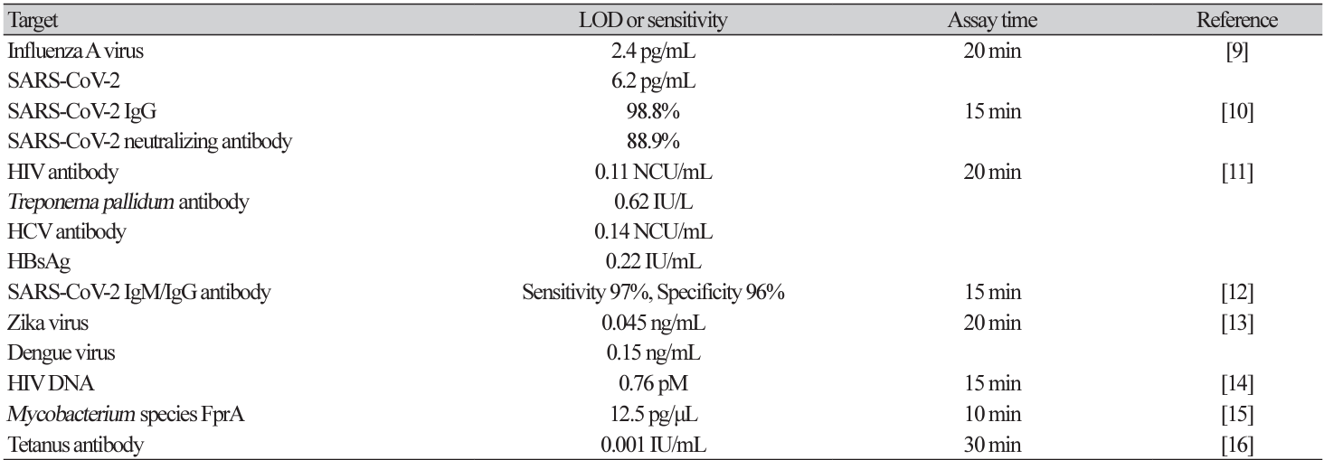
To enhance the sensitivity of LFIA, the implementation of QDs as labels is being explored for the detection of various pathogens. These include SARS-CoV-2, hepatitis C virus, hepatitis B virus, HIV, influenza virus, zika virus, dengue virus, Treponema pallidum, Mycobacterium species FprA, and tetanus antibodies (Table 1). These assays are ultrasensitive with very low detection limits and can be completed in 20 min [9-16].
Go to : 
Challenges of QDs
Although QDs are one of the most promising fluorescent labels in LFIAs, they also present several challenges. For uniform size and consistent characteristics, QDs require complex synthesis. Modification of the surface to attach antibodies or proteins can be intricate and can also affect the optical properties, posing challenges when transitioning from laboratory-scale to large-scale manufacturing. The materials and techniques required to synthesize high-quality QDs are more expensive than those for conventional dyes, which increases the cost. Some QDs are composed of toxic heavy metals such as cadmium, which introduces potential environmental and health hazards. Traditional LFIAs are often read visually, whereas quantum-dot LFIAs require specialized equipment for fluorescence detection.
Various strategies are being explored to address the limitations of QD-based LFIA. These include the introduction of less toxic, cadmium-free QDs, such as those based on zinc or silver. Additionally, new synthesis methods aimed at reducing environmental impact are under investigation. To minimize the leaching of toxic elements, biocompatible coatings like silica or organic polymers are being utilized. Efforts are also being made to enhance the stability and functionality of these QDs to improve their interaction with biological molecules. Furthermore, research is focusing on multiplexing capabilities and the development of equipment-free reading methods.
The future of lateral flow assays promises quicker and more sensitive pathogen detection. Although QDs currently have many limitations, ongoing research and technological breakthroughs suggest that diagnostic revolution might be just around the corner.
Go to : 


 PDF
PDF Citation
 Print
Print


 XML Download