Abstract
A 22-year-old male patient presented to the clinic with severe pain in the preauricular area with an inability to completely occlude the jaw. Facial computed tomography and magnetic resonance imaging revealed a well-defined lesion that was tentatively diagnosed as a benign tumor or cystic mass. Surgical approach of a lesion in the condyle is delicate and problematic as many vulnerable anatomical structures are present. There are several methods for surgery in this area. Typically, an extraoral approach is dangerous because of potential injuries to nerves and arteries. The intraoral approach also presents difficulties due to the lack of visibility and accessibility. On occasion, coronoidectomy may be performed. The goal here was to determine an easier and safer new surgical approach to the condyle. We reached the anterior part of the pterygoid plate in the same method as in Le Fort I surgery. From this point, through the external pterygoid muscle, approaching the anterior aspect of the condyle is relatively easy and safe, with minimal damage to the surrounding tissues. Pus was drained at the site, and the lesion was diagnosed as an abscess. Pain and inability to close the mouth resolved without recurrence.
Abscess caused by infection in facial areas occurs mainly in buccal, sub-mandibular, sub-lingual, and infraorbital spaces. Although abscesses can occur in all the spaces in the head and neck areas, abscesses occurring in the external pterygoid muscle space are rare.
In this case, abscess occurred without any previous history of infection. Most abscess cases have a previous history and sometimes involve transmission to other regions to create secondary abscess. There have been no reports of abscesses that are generated primarily within the external pterygoid muscle.
For removal of tumors or other lesions that are generated on the mandibular condyle or the surrounding tissues, many surgical techniques have been introduced. Several methods approach the temporomandibular joint area extraorally or intraorally. Most surgeons historically chose the extraoral access method due to visibility. However, in 1972, Sear1 introduced an intraoral approach method to treat temporomandibular joints. In addition, in 1977, Eller et al.2 reported an intraoral approach method to remove osteochondromas in the condyle.
Since then, the intraoral approach has been the preferred method. In cases using the intraoral approach, only a narrow field of vision can be obtained, making the operation difficult and requiring extra care from surgeons to not damage anatomical structures such as the maxillary artery.
However, in cases using the extraoral approach, there is potential for damage to facial nerves, the auriculotemporal nerve, the superficial temporal artery, and the maxillary artery. Due to these potentially dangerous situations with both methods of approach, limitations regarding temporomandibular joint operations are unavoidable.
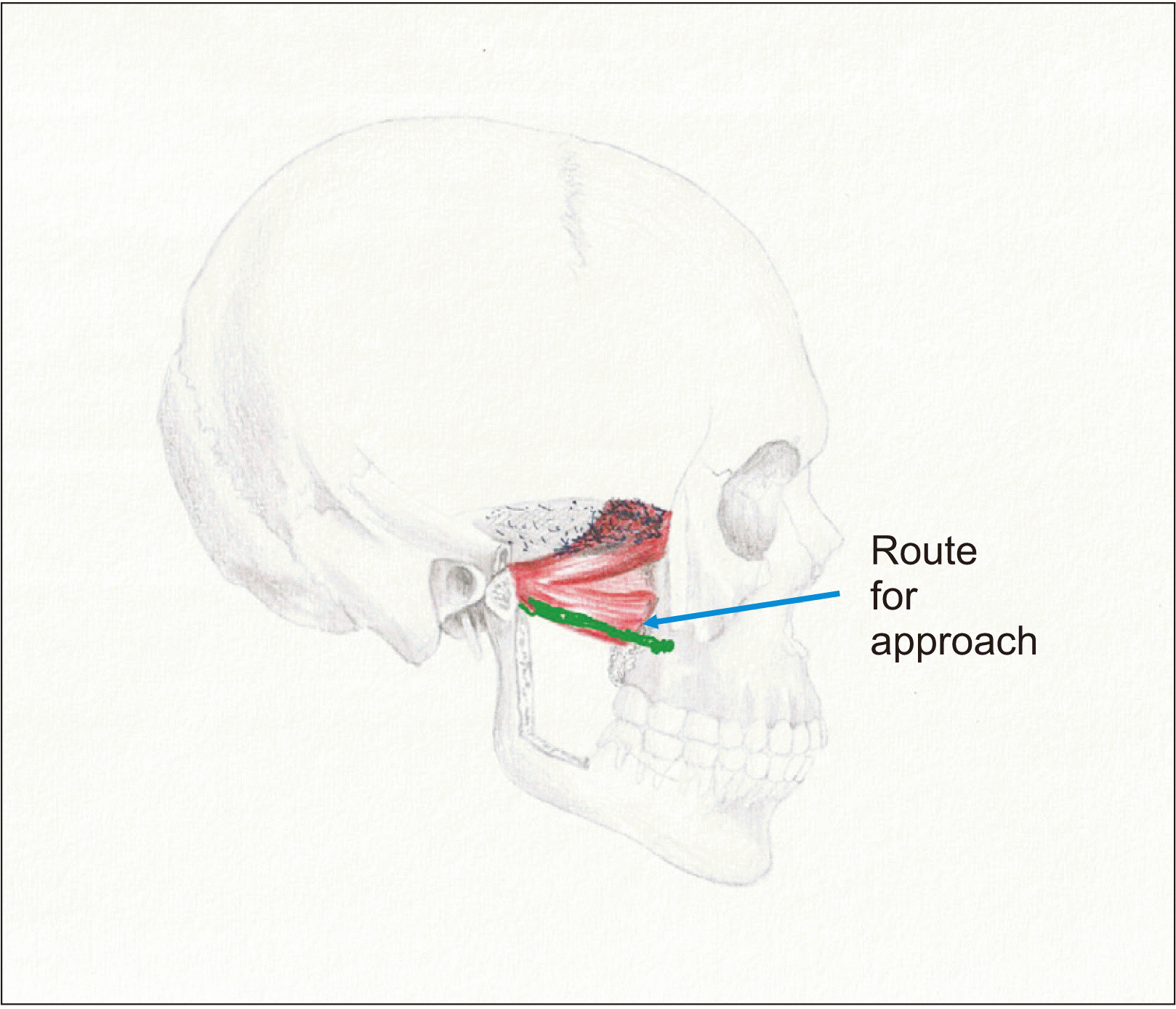
Therefore, we intend to determine safe and convenient surgical routes for gradually approaching the tissues surrounding the temporomandibular joint, including the mandibular condyle itself. Approaching the maxillary tuberosity and the external pterygoid plate, we can reach the external pterygoid muscle and contact the anterior portion of the condyle through the muscle. Abscesses can be easily removed from the external pterygoid muscle without complications. Thus, we are introducing a new method of approach to the anterior condylar area.
A 22-year-old male patient visited my clinic with extreme pain in the anterior portion of the left ear and left condyle area, especially when closing the mouth. The pain was unable to be suppressed by medication, and the teeth on the left side of the mouth did not occlude well. Although the cause of the pathogenesis was unclear, due to the medical history of pain in the vicinity of the left ear, a local clinic had diagnosed referred pain from a wisdom tooth in the lower jaw. The patient was subsequently referred for extraction of the wisdom tooth in the left lower jaw.
In the panoramic radiograph, no lesion was found surrounding the wisdom tooth. Considering the symptoms of the patient, lesions surrounding the tissues of the mandibular condyle were deemed suspicious. Computed tomography showed a radiolucent focus in the front portion of the mandibular condyle. To confirm the diagnosis, magnetic resonance imaging was performed. Based on the images, the lesion was tentatively diagnosed as an abscess or a liquid-containing benign tumor within the external pterygoid muscle.(Fig. 1-4)
Most tumors or diseased dental masses must be removed. Therefore, safe methods of approach are needed. Extraoral approaches present many difficulties and have a high probability of complications. Thus, caution is needed regarding the anatomy of the area including the maxillary artery and facial nerve. Even when the condyle is successfully approached, care must be taken to access a lesion in the external pterygoid muscle in the anterior-mesial portion of the condyle. As a result, the intraoral approach has been preferentially considered. However, even with these methods, the coronoid process must be removed to secure an adequate view and to approach the lesion. Therefore, to determine a more effective and safer route of approach, we devised a new method of operation.
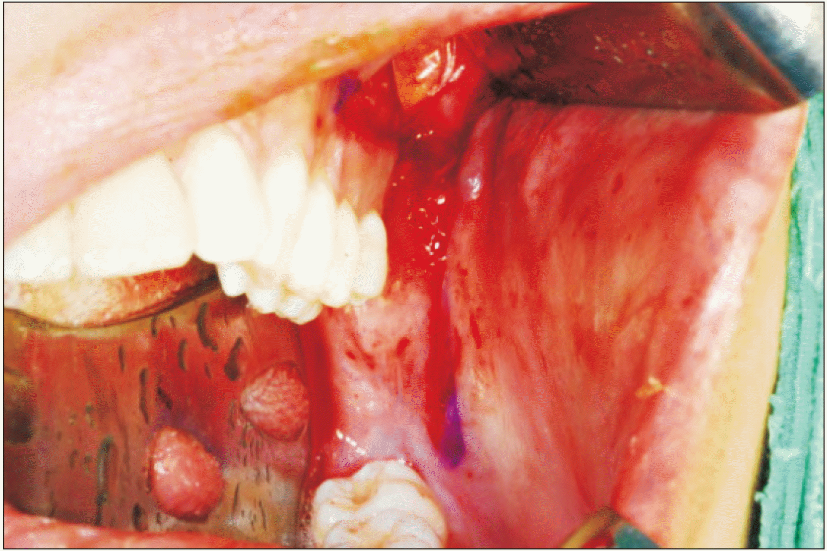
Under general anesthesia through naso-endotracheal intubation, the patient was positioned supine on the surgical table. The incision line was extended from the first premolar to the second molar on the left side of the upper jaw and extended to the anterior border of the ramus through the external oblique ridge. When the incision line passes over the maxillary tuberosity and down through the external oblique ridge, care must be taken to not harm any underlying vessels, nerves, fat pads, etc. As a result, incisions must be created within submucosal level in these areas. The mucous membrane was dissected to obtain wide exposure of the area of surgery for good visibility and accessibility. Exposure of the fat pad in the upper aspect of the coronoid process must be avoided if possible. If exposure of the fat pad occurs, it should be set aside laterally. Dissection of the mucosa continues through the anterior aspect of the ramus gradually approaching the lower part of the external oblique ridge.(Fig. 5, 6)
In the anterior border aspect of the ramus, the fascia of the temporal muscle is observed. Further dissection is carried out in the upper aspect. In the upper jaw, the periosteum is reflected to create an approach to the underlying bone and to allow exposure of the maxillary tuberosity.
Additional approach to the posterior aspect of the upper jaw following the maxillary bone approaches a border point between the maxillary tuberosity and the pterygoid plate. In this region, caution is needed in the direction of the condyle through the muscle fascia of the lateral pterygoid muscle.(Fig. 7)
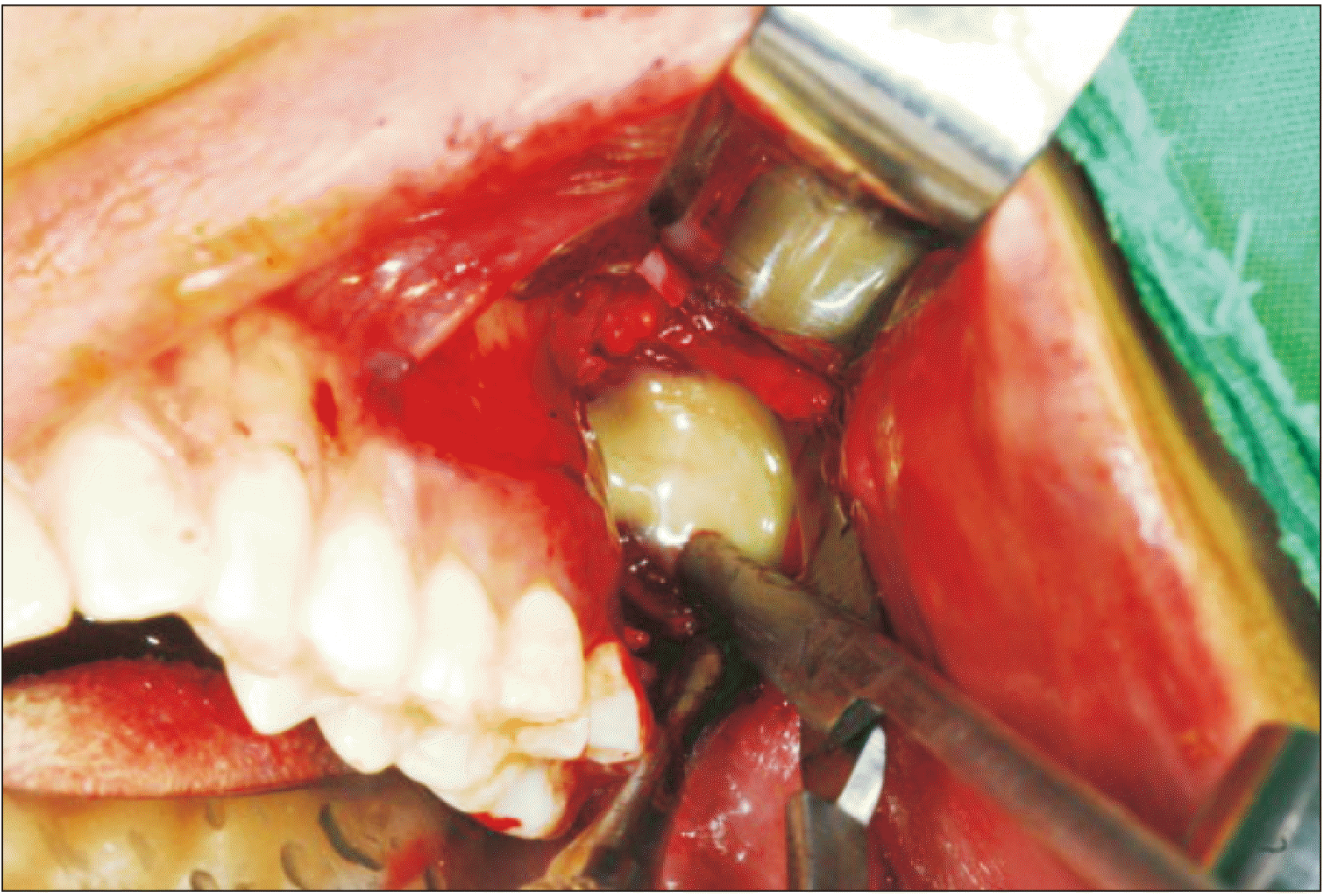
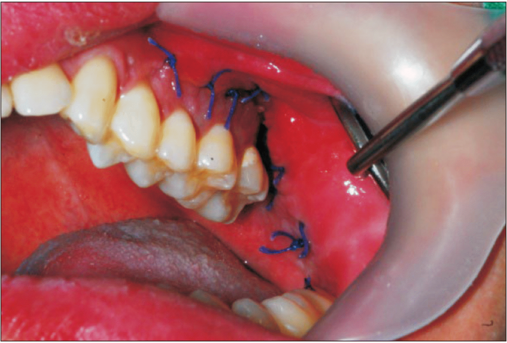
After verifying the external pterygoid muscle, the muscle was dissected using a Metzenbaum scissor, and the abscess within the muscle was drained.(Fig. 8) After eliminating the pus from the lesion through sufficient saline irrigation, a rubber drain was inserted and a suture was placed for retention and additional drainage.(Fig. 9)
After the surgery, the patient could occlude normally and the pain resolved. The healing progressed without pain or edema in the region. The drain was retained intraorally for 2 days after the surgery. The patient did not exhibit any signs of recurrence or symptoms.
The temporo-mandibular joint is located deep in the facial area and is surrounded by many tissues. Due to this location, abscesses in this region are very rare. In this clinical report, the patient had no history of trauma or abscess in the head and neck area. However, he visited the clinic with the chief complaint of limited mouth closure.
Since the lesion was located in the front portion of the mandibular condyle, a surgical method approaching the anterior and mesial parts of the condyle can be devised. With such an intraoral approach, surgery must be preceded by removal of the coronoid process. This removal also is needed with an extraoral approach to access the distal aspect of coronoid process.
The various methods of approach for surgery on the temporomandibular joint have been well organized by Kreutziger3. As representative methods of incisions, Kreutziger3 introduced the preauricular incision, the endaural incision, the postauricular approach, and the Risdon incision. Great caution must be taken with the facial nerves during these surgeries. In addition, the auriculotemporal nerve, the superficial temporal artery, and the maxillary artery must be avoided.
Regarding the complications of procedures on the mandibular condyle, Vallerand and Dolwick4 highlighted cases with complications, together with the criteria for successful surgeries. In addition, in 2003, Keith5 explained complications of the surgeries. In both studies, damage to the facial nerves was the most common. Other complications include damage to blood vessels, infections, otologic complications, parotid gland injuries, cranial fossa perforations, failure of implanted materials, malocclusions, degenerative arthritis, adhesions, and ankylosis.
Generally, when performing surgery on the mandibular condyle or the front area of the condylar head, an extra-oral approach is used. The issue of potential damage to the facial nerves remains with this surgical method. In 2011, Knepil et al.6 presented the many surgical approaches to the mandibular condyle and showed that the anatomical locations of the facial nerves were the most important to recognize.
Various methods for open reduction of condyle fractures have been reported. Vesnaver and colleagues7,8 stated that such surgical intervention for condyle fractures produces more positive outcomes in jaw function in comparison with the conservative approach. However, reported side effects include temporary facial nerves paralysis, parotid gland fistulas, loss of hearing due to damage to the greater auricular nerve, fracture of the miniplate, bleeding during the operation, and edema or infection after the operation.
Various methods for reduction of bone fractures of the condyle have been reported. Trost et al.9 introduced an approach to the mandibular condyle involving cutting and entering the masseter muscle using the space occupied by the anterior parotid gland. Currently, this approach is performed between the upper and lower buccal branches of the facial nerves.
In addition, Vesnaver7 used a preauricular approach method entering through the articular capsule based on the location of the zygoma arch. That method is one of the easier ways to avoid the branches of the facial nerves.
Narayanan et al.10 published a retromandibular approach, and Choi et al.11 reported endoscope-assisted open reduction. Trost et al.12, Shi et al.13, and Guo and Guo14 also have introduced surgical methods, though most are no longer used. In addition, for treatment of chondroma in the condyle of the lower jawbone, there have been reports using a modified temporal incision15, a preauricular incision16, and a hemicoronal incision17.
In our presented case, a diseased mass was present within the external pterygoid muscle rather than in the mandibular condyle itself. Therefore, no extraoral method of approach allowed direct access to the mass. To expose such a mass or diseased tissue, the mandibular condyle must be removed.
The modified transoral approach proposed by Deng et al.18 can avoid damage to the facial nerves without leaving any extraoral scars. The method approaches the condyle without damaging the blood vessels widely distributed in the facial areas. However, the coronoid process must be removed to approach the condyle. In addition, the external pterygoid muscle lays in the internal direction and not in the direction of the mandibular condyle. Also, there is danger of damaging the maxillary artery and its branches between the mandibular condyle and the diseased tissue. For these reasons, the method proposed by Deng et al.18 is not suitable for approach to the external pterygoid muscle.
We designed an approach path by modifying the incision line used in Le Fort I surgeries, moving it more posteriorly and extending it to the second molar. When the mucosa and periosteum are reflected, the maxillary tuberosity and pterygoid plate are exposed. The approach can be created posteriorly following the fascia of the external pterygoid muscle to the condyle head. A mass designated for removal will be located between these two reference points.
In our case, drainage was allowed when the external pterygoid muscle was dissected and was supported by insertion of a surgical drain. The drain was removed the following day after the chief complaints of the patient were resolved.
Immediately after the procedure, normal occlusion was achieved with pain resolution. Lower jaw movements subsequently were normalized. The patient completely recovered without any complications of edema or damage to nerves or vessels. At the 18-month follow-up, the patient exhibited no signs of recurrence.
To remove lesions in the external pterygoid muscle, a new intraoral approach was presented. With this method, damage to nerves and blood vessels can be avoided. Based on this result, it is expected that this new method will effectively remove lesions in this of or in the external pterygoid muscle. After the present surgery, all symptoms resolved. The patient exhibited rapid recovery without any complications.
Notes
References
1. Sear AJ. 1972; Intra-oral condylectomy applied to unilateral condylar hyperplasia. Br J Oral Surg. 10:143–53. https://doi.org/10.1016/s0007-117x(72)80030-1. DOI: 10.1016/S0007-117X(72)80030-1. PMID: 4509977.

2. Eller DJ, Blakemore JR, Stein M, Byers S. 1977; Transoral resection of a condylar osteochondroma: report of case. J Oral Surg. 35:409–13.
3. Kreutziger KL. 1984; Surgery of the temporomandibular joint. I. Surgical anatomy and surgical incisions. Oral Surg Oral Med Oral Pathol. 58:637–46. https://doi.org/10.1016/0030-4220(84)90027-6. DOI: 10.1016/0030-4220(84)90027-6. PMID: 6594653.

4. Vallerand WP, Dolwick MF. 1990; Complications of temporomandibular joint surgery. Oral Maxillofac Surg Clin North Am. 2:481–8.
5. Keith DA. 2003; Complications of temporomandibular joint surgery. Oral Maxillofac Surg Clin North Am. 15:187–94. https://doi.org/10.1016/s1042-3699(03)00016-5. DOI: 10.1016/S1042-3699(03)00016-5. PMID: 18088674.

6. Knepil GJ, Kanatas AN, Loukota RJ. 2011; Classification of surgical approaches to the mandibular condyle. Br J Oral Maxillofac Surg. 49:664–5. https://doi.org/10.1016/j.bjoms.2011.02.009. DOI: 10.1016/j.bjoms.2011.02.009. PMID: 21453998.

7. Vesnaver A. 2008; Open reduction and internal fixation of intra-articular fractures of the mandibular condyle: our first experiences. J Oral Maxillofac Surg. 66:2123–9. https://doi.org/10.1016/j.joms.2008.06.010. DOI: 10.1016/j.joms.2008.06.010. PMID: 18848112.

8. Vesnaver A, Ahčan U, Rozman J. 2012; Evaluation of surgical treatment in mandibular condyle fractures. J Craniomaxillofac Surg. 40:647–53. https://doi.org/10.1016/j.jcms.2011.10.029. DOI: 10.1016/j.jcms.2011.10.029. PMID: 22079126.

9. Trost O, Trouilloud P, Malka G. 2009; Open reduction and internal fixation of low subcondylar fractures of mandible through high cervical transmasseteric anteroparotid approach. J Oral Maxillofac Surg. 67:2446–51. https://doi.org/10.1016/j.joms.2009.04.109. DOI: 10.1016/j.joms.2009.04.109. PMID: 19837315.

10. Narayanan V, Kannan R, Sreekumar K. 2009; Retromandibular approach for reduction and fixation of mandibular condylar fractures: a clinical experience. Int J Oral Maxillofac Surg. 38:835–9. https://doi.org/10.1016/j.ijom.2009.04.008. DOI: 10.1016/j.ijom.2009.04.008. PMID: 19467846.

11. Choi EJ, Cha IH, Nam W. 2011; The result of endoscope-assisted open reduction and internal fixation (EAORIF) of lateral overridden subcondyle fracture. J Korean Assoc Oral Maxillofac Surg. 37:62–6. https://doi.org/10.5125/jkaoms.2011.37.1.62. DOI: 10.5125/jkaoms.2011.37.1.62.

12. Trost O, Abu El-Naaj I, Trouilloud P, Danino A, Malka G. 2008; High cervical transmasseteric anteroparotid approach for open reduction and internal fixation of condylar fracture. J Oral Maxillofac Surg. 66:201–4. https://doi.org/10.1016/j.joms.2006.09.031. DOI: 10.1016/j.joms.2006.09.031. PMID: 18083441.

13. Shi J, Yuan H, Xu B. 2013; Treatment of mandibular condyle fractures using a modified transparotid approach via the parotid mini-incision: experience with 31 cases. PLoS One. 8:e83525. https://doi.org/10.1371/journal.pone.0083525. DOI: 10.1371/journal.pone.0083525. PMID: 24386221. PMCID: PMC3873388.

14. Guo Y, Guo C. 2014; Maxillary-fronto-temporal approach for removal of recurrent malignant infratemporal fossa tumors: anatomical and clinical study. J Craniomaxillofac Surg. 42:206–12. https://doi.org/10.1016/j.jcms.2013.05.001. DOI: 10.1016/j.jcms.2013.05.001. PMID: 23932542.

15. Utumi ER, Pedron IG, Perrella A, Zambon CE, Ceccheti MM, Cavalcanti MG. 2010; Osteochondroma of the temporomandibular joint: a case report. Braz Dent J. 21:253–8. https://doi.org/10.1590/s0103-64402010000300014. DOI: 10.1590/S0103-64402010000300014. PMID: 21203710.

16. Yun KI, Park MK, Kim CH, Park JU. 2008; Chondrosarcoma in the mandibular condyle: case report. J Korean Oral Maxillofac Surg. 34:95–8.
17. Ryu DM, Kim HJ, Lee SC, Kim YG, Lee BS. 2002; Osteochonrdoma of the mandibular condyle: a case report. J Korean Oral Maxillofac Surg. 28:132–5.
18. Deng M, Long X, Cheng AH, Cheng Y, Cai H. 2009; Modified trans-oral approach for mandibular condylectomy. Int J Oral Maxillofac Surg. 38:374–7. https://doi.org/10.1016/j.ijom.2009.01.020. DOI: 10.1016/j.ijom.2009.01.020. PMID: 19282151.

Fig. 1
Panoramic view. The patient was referred from a local clinic for extraction of #38. His chief complaint was severe pain around the left facial area with difficulty in mouth closing. He was diagnosed with referred pain from pericoronitis of #38. However, no lesion was observed in the #38 area and the patient had no previous history of pain.
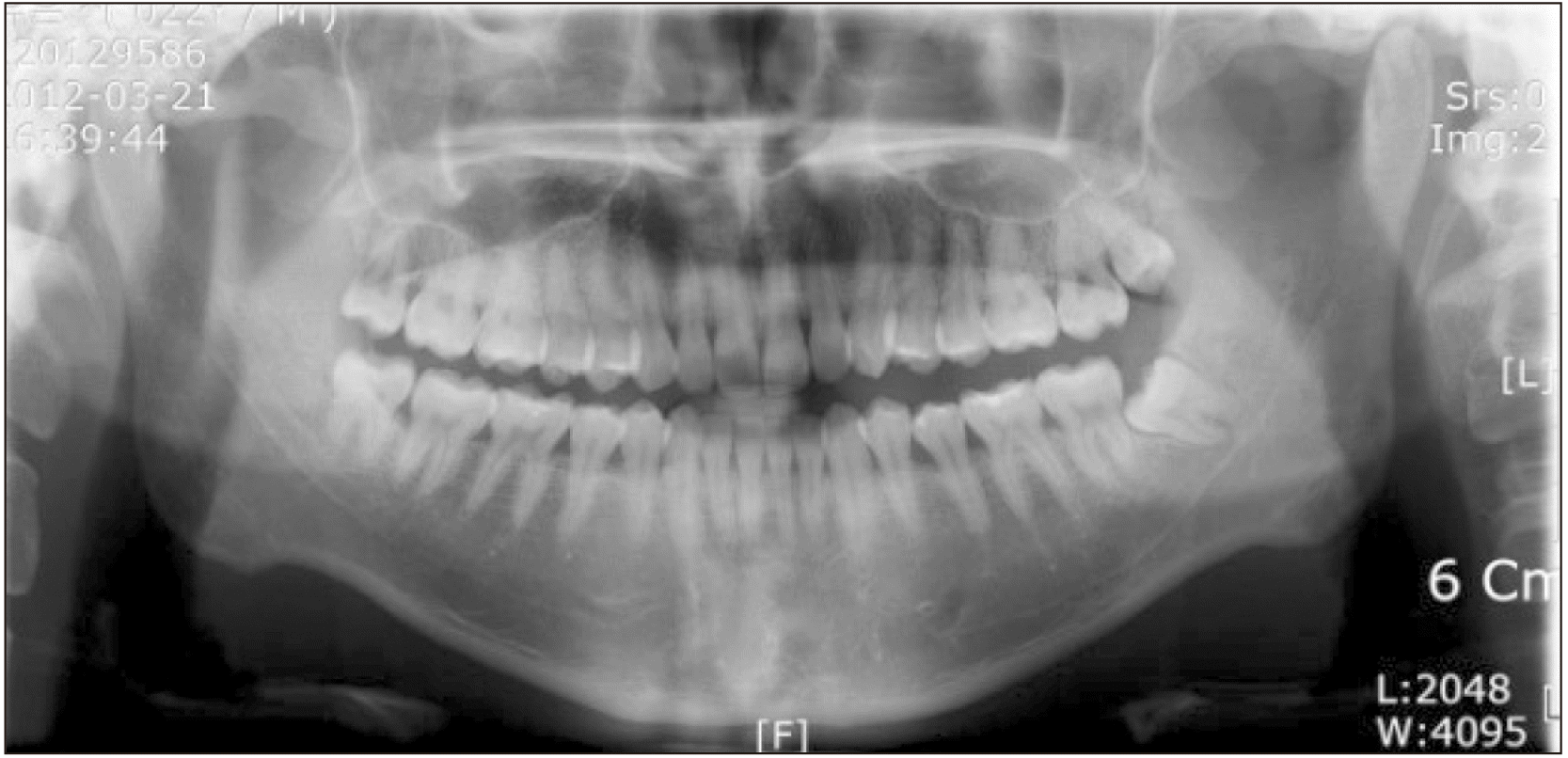
Fig. 2
A computed tomography view showing the tumorous lesion, located from the anterior portion of the condyle to the posterior portion of the pterygoid plate and from the condylar head to the sigmoid notch.
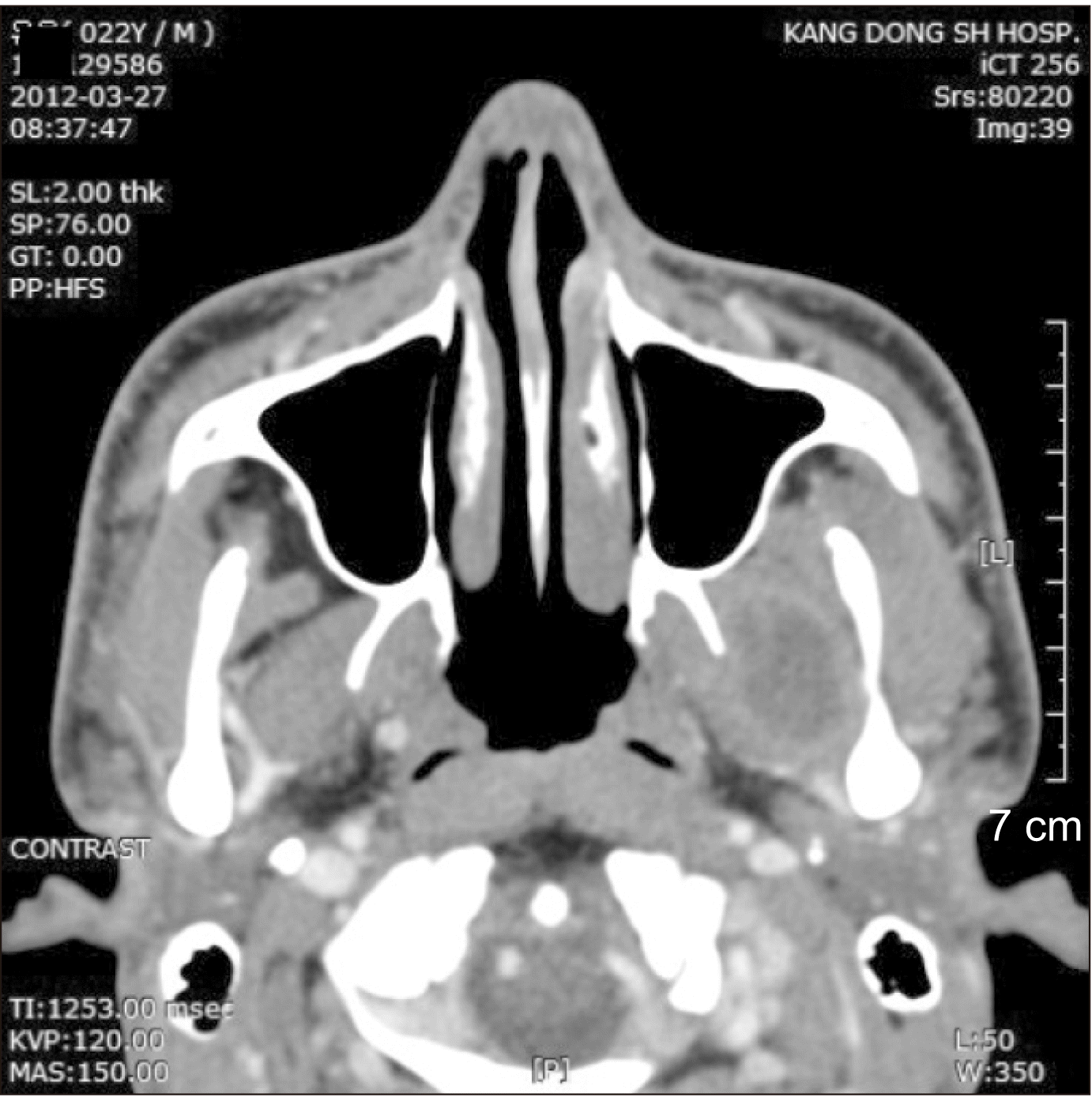
Fig. 3
Coronal view of computed tomography showing a liquid-like mass in the upper and lower areas of the pterygoid muscle.
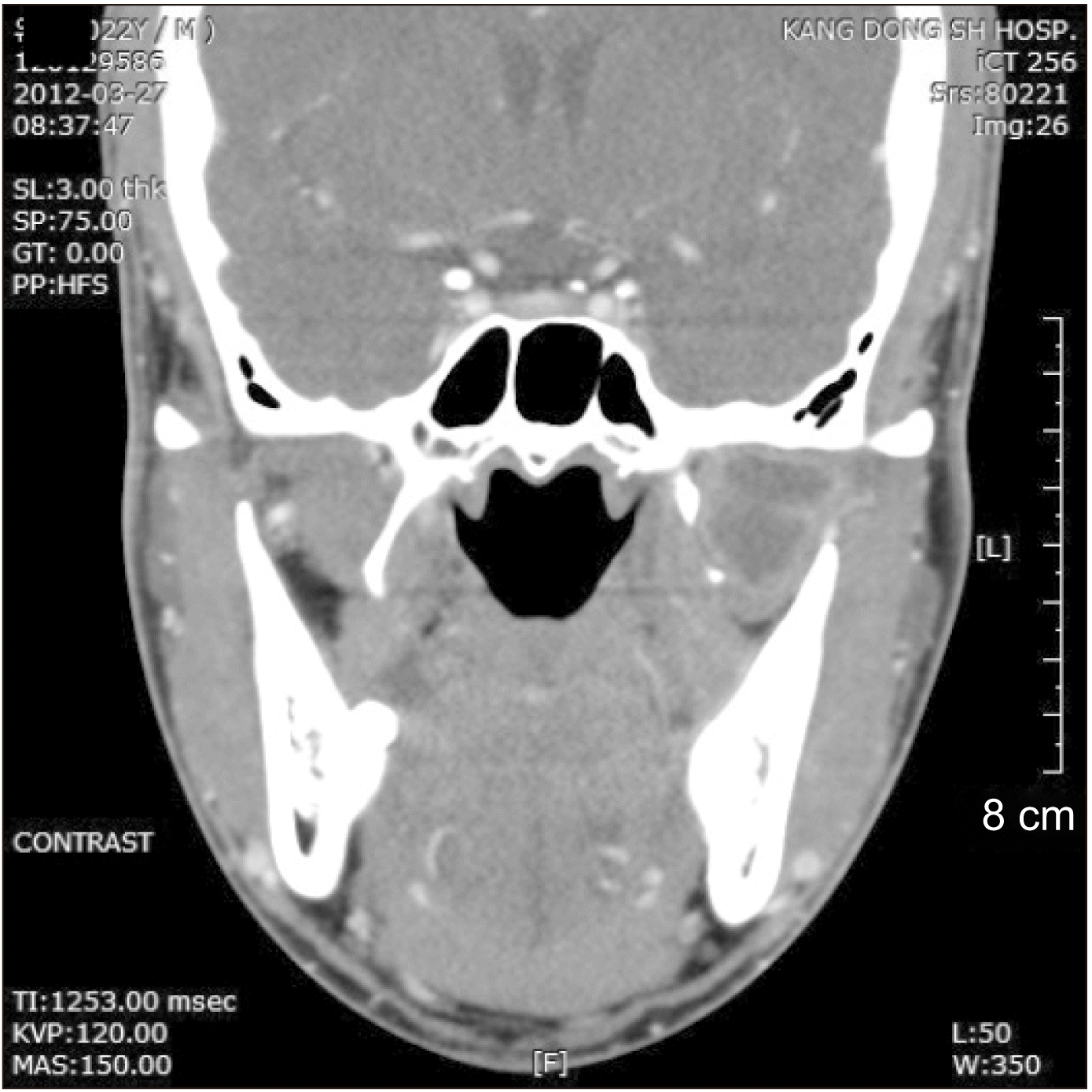
Fig. 4
Magnetic resonance imaging showing a liquified lesion indicating an abscess and ruling out a liquid-containing benign tumor.
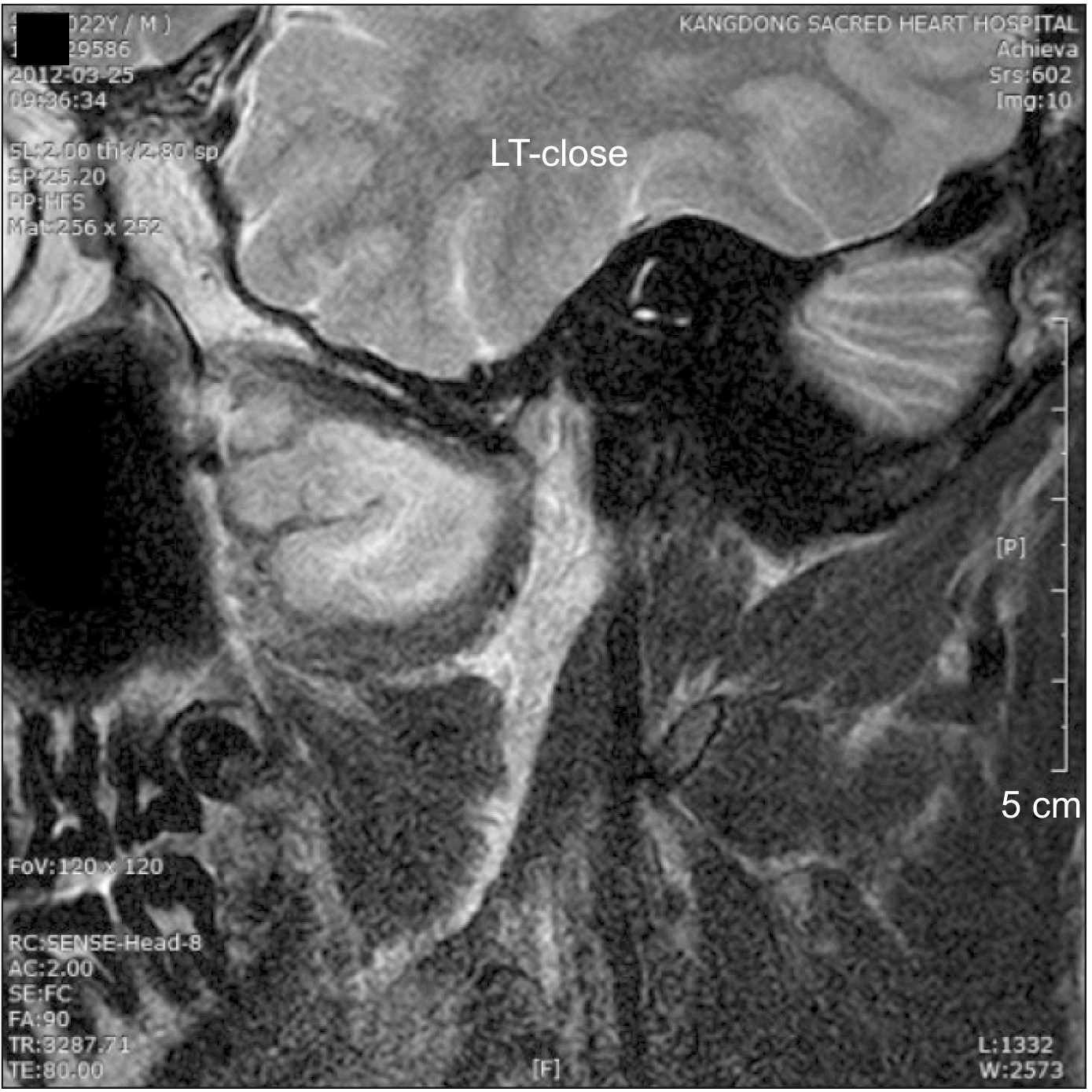
Fig. 5
An incision line was created from the premolar region and extended through the anterior border of the mandible. The maxillary area was dissected fully to expose bone, while the mandibular portion was dissected with partial thickness.
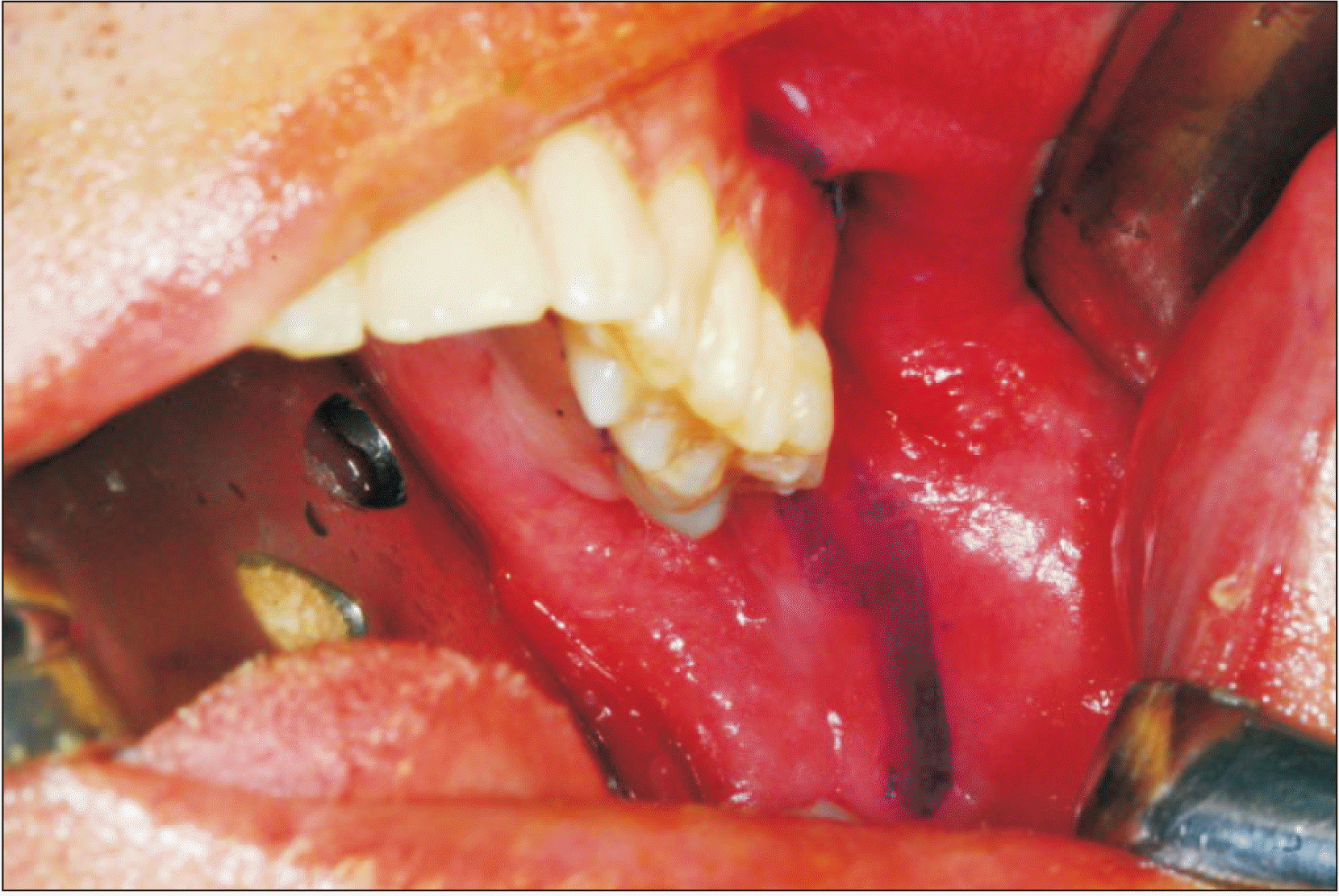
Fig. 6
The incision was completed at the submucosal level. The fat pad was observed at the antero-superior aspect of the coronoid process and was moved laterally to not disturb the surgical field.


 PDF
PDF Citation
 Print
Print


 XML Download