Abstract
The occurrence of an abdominal wall hematoma caused by abdominal paracentesis in patients with liver cirrhosis is rare. This paper presents a case of an abdominal wall hematoma caused by abdominal paracentesis in a 67-year-old woman with liver cirrhosis with a review of the relevant literature. Two days prior, the patient underwent abdominal paracentesis for symptom relief for refractory ascites at a local clinic. Upon admission, a physical examination revealed purpuric patches with swelling and mild tenderness in the left lower quadrant of the abdominal wall. Abdominal computed tomography revealed advanced liver cirrhosis with splenomegaly, tortuous dilatation of the para-umbilical vein, a large volume of ascites, and a large acute hematoma at the left lower quadrant of the abdominal wall. An external iliac artery angiogram showed the extravasation of contrast media from the left deep circumflex iliac artery. Embolization of the target arterial branches using N-butyl-2-cyanoacrylate was then performed, and the bleeding was stopped. The final diagnosis was an abdominal wall hematoma from the left deep circumflex iliac artery after abdominal paracentesis in a patient with liver cirrhosis.
Liver cirrhosis is a progressive and diffuse liver disease characterized by collagen deposition in the extracellular matrix of the hepatic parenchyma and fibrosis. The disease can lead to many complications, including ascites, varices, hepatic encephalopathy, hepatocellular carcinoma, hepatorenal syndrome, hepatopulmonary syndrome, and coagulation disorders.1,2
Among its complications, ascites is associated with an impaired quality of life, frequent hospitalization, and chronic treatment costs. In addition, it causes problems, such as dilutional hyponatremia, respiratory dysfunction, spontaneous bacterial peritonitis, umbilical hernia formation, and hepatorenal syndrome. Important treatment approaches for the management of ascites include sodium and fluid restriction, diuretic treatment, abdominal paracentesis, portosystemic shunting, and, ultimately, liver transplantation.3-5 In particular, although there are some potential complications, such as ascitic fluid leakage, infection, circulatory failure, bleeding, and bowel perforation, abdominal paracentesis is generally a safe procedure commonly recommended for managing excessive ascites.6-9 Furthermore, the occurrence of an abdominal wall hematoma caused by abdominal paracentesis in patients with liver cirrhosis is rare.
This paper presents a case of an abdominal wall hematoma caused by abdominal paracentesis in a 67-year-old woman with liver cirrhosis with a review of the relevant literature.
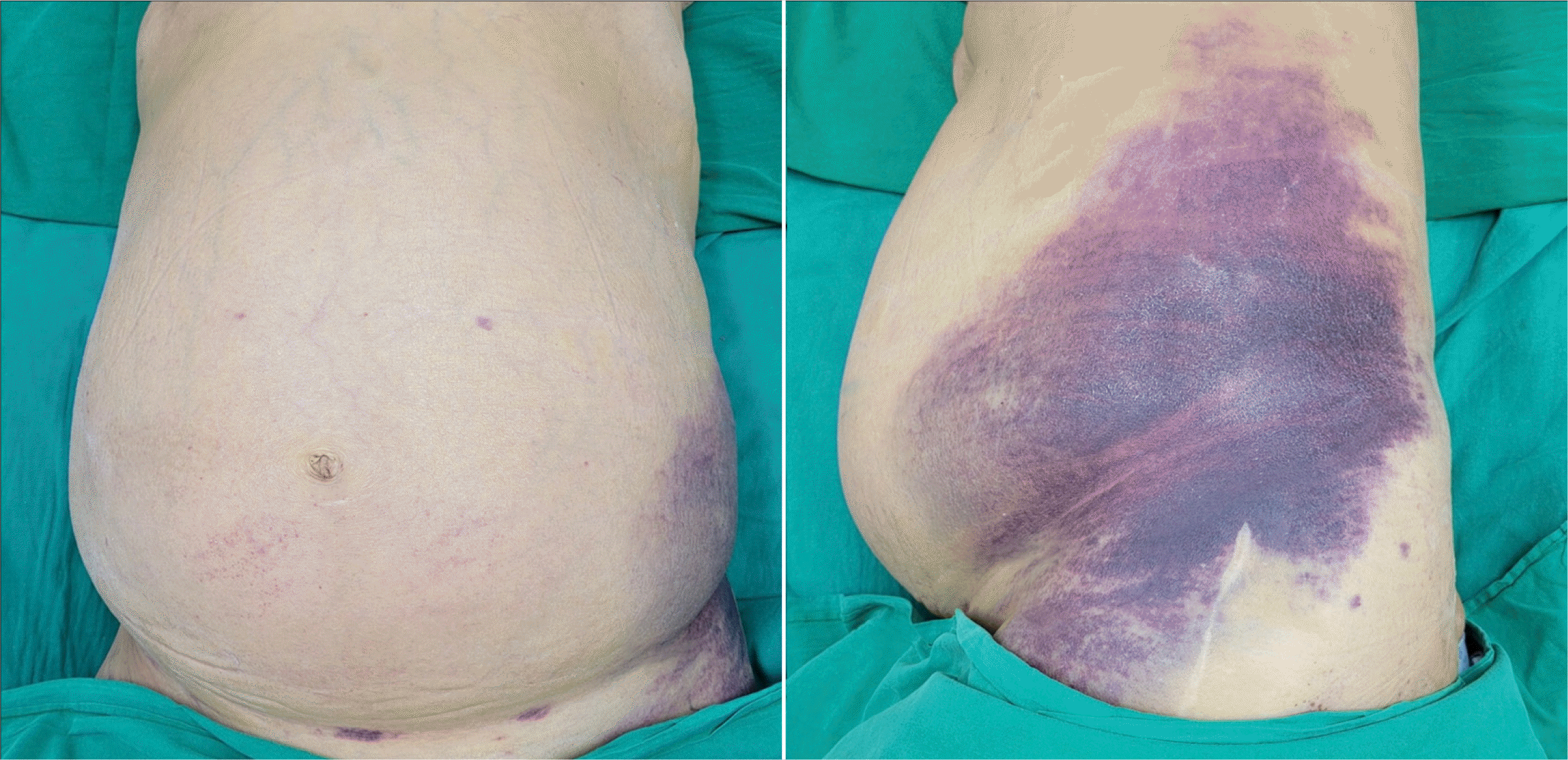
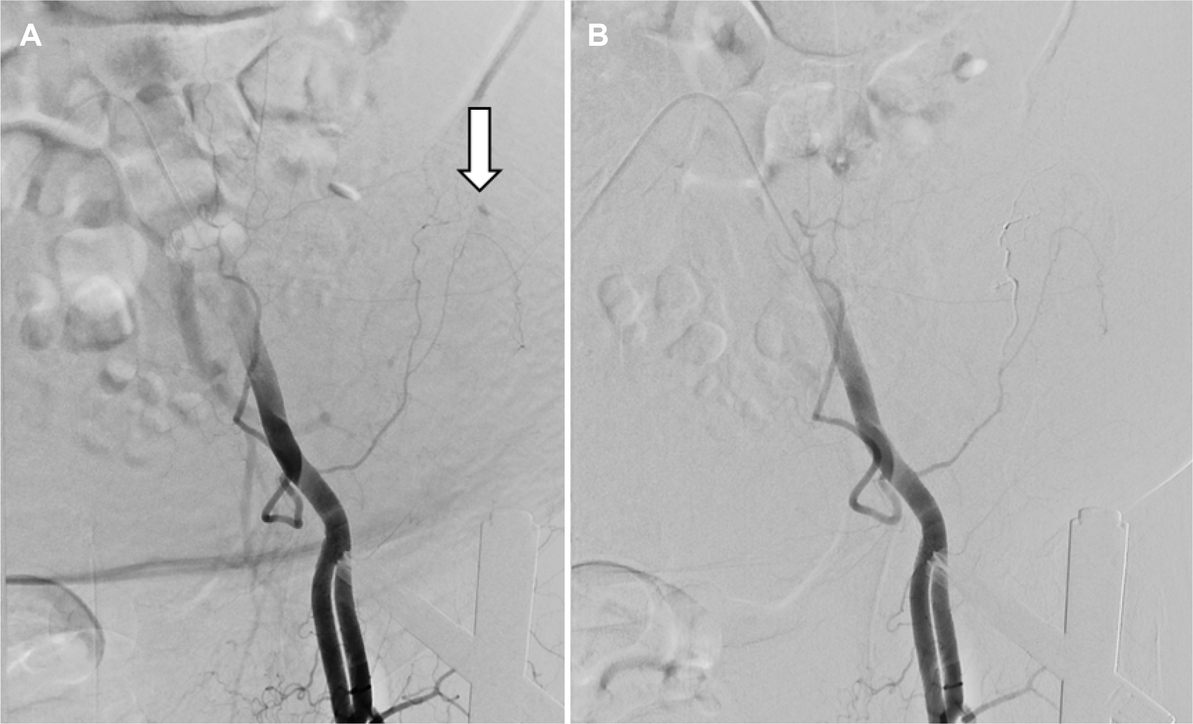
A 67-year-old woman was admitted to the authors’ hospital with a two-day history of left lower quadrant abdominal pain. She had suffered from liver cirrhosis secondary to chronic alcohol abuse for 10 years. Two days earlier, she had undergone ultrasonography-guided abdominal paracentesis for symptom relief for refractory ascites at a local clinic. On admission, she was found to have a blood pressure of 100/60 mmHg, a heart rate of 102 beats/min, a pulse rate of 15 breaths/min, and a temperature of 36.6°C. A physical examination revealed purpuric patches with swelling and mild tenderness in the left lower quadrant of the abdominal wall (Fig. 1). A laboratory examination revealed the following: a white blood cell count of 9,500/mm3 (reference range, 6,000–10,000/mm3), a hemoglobin level of 8.1 g/dL (reference range, 12–16 g/dL), a platelet count of 20,000/mm3 (reference range, 130,000– 450,000/mm3), a serum albumin level of 2.9 g/dL (reference range, 3.0–5.0 g/dL), an aspartate aminotransferase level of 49 U/L (reference range, 5–37 U/L), an alanine aminotransferase level of 20 U/L (reference range, 5–40 U/L), an alkaline phosphatase level of 73 U/L (reference range, 39–117 U/L), and a γ-glutamyl transpeptidase level of 42 U/L (reference range, 7–49 U/L). The total bilirubin level was 9.73 mg/dL (reference range, 0.2–1.2 mg/dL) with a direct fraction of 6.73 mg/dL (reference range, 0.05–0.3 mg/dL). The alpha-fetoprotein (AFP) level was 10.27 IU/mL (reference range, 0.74–7.29 IU/mL), and the protein induced by the vitamin K absence-2 (PIVKA-II) level was 81 mAU/mL (reference range, 0–40 mAU/mL). The coagulation profile results were as follows: a prothrombin time (PT) of 22.6 seconds (control 11.8 seconds), an activated partial thromboplastin time (aPTT) of 50.3 seconds (control, 28 to 40 seconds), and a fibrinogen of 106 mg/dL (reference range 180–300 mg/dL). The follow-up blood test revealed an improved coagulation profile: a platelet count of 116,000/mm3, a PT of 19.9 seconds, an aPTT of 49.0 seconds, and a fibrinogen of 163 mg/dL. These results suggested the possibility of accompanying disseminated intravascular coagulation caused by bleeding. The serum hepatitis B surface antigen, hepatitis B e-antigen, and hepatitis B surface antibody tests were negative; the anti-HBc and anti-HBe were positive, and the hepatitis C antibody testing was negative. Enhanced axial computed tomography (CT) images of the abdomen revealed advanced liver cirrhosis with splenomegaly, tortuous dilatation of the para-umbilical vein, and a large volume of ascites (Fig. 2A, B). A coronal CT image showed a large acute hematoma with suspicious contrast media extravasation at the left lower quadrant of the abdominal wall (Fig. 2C). Despite the AFP and PIVKA-II elevation, there was no definite evidence of hepatocellular carcinoma in the CT scan. Angiography was performed due to continued bleeding and an increasing hematoma size despite medical therapy. An external iliac artery angiogram revealed the extravasation of contrast media from the left deep circumflex iliac artery (Fig. 3A). Embolization of the target arterial branches using N-butyl-2-cyanoacrylate was then performed, and the bleeding was stopped (Fig. 3B). The final diagnosis was an abdominal wall hematoma from the left deep circumflex iliac artery after abdominal paracentesis in a patient with liver cirrhosis with coagulopathy and thrombocytopenia. Informed consent for publication was obtained from the patient.
Ascites develop in more than 50% of patients with liver cirrhosis within 10 years of a diagnosis of compensated liver cirrhosis. Treatment approaches for ascites include sodium restriction, albumin treatment, aldosterone antagonists, loop diuretics, large-volume paracentesis, peritoneovenous shunting, transjugular intrahepatic portosystemic shunting, and liver transplantation.3-5
Abdominal paracentesis is a commonly performed diagnostic or therapeutic procedure for patients with liver cirrhosis with ascites. It is usually performed in the left lower quadrant of the abdomen and is considered a safe procedure with a 1% overall complication rate. The known complications include ascitic fluid leakage, infection, circulatory failure, bleeding, and bowel perforation. Hemorrhagic complications are rare, and the presentations of hemorrhage are classified as an abdominal wall hematoma, a pseudoaneurysm, and hemoperitoneum. An abdominal wall hematoma is the main presentation of abdominal paracentesis-related hemorrhage.6-9 The symptoms and signs of hemorrhage become evident from hours to one week after abdominal paracentesis.9 In the present case, the hemorrhagic complication presented as an abdominal wall hematoma and became evident two days after the abdominal paracentesis.
The superficial and deep arcades supply the anterior abdominal wall. The superficial arcade consists of the superficial inferior epigastric artery, superficial superior epigastric artery, superficial circumflex iliac artery, and lateral superficial branches of the subcostal and intercostal arteries. The deep arcade consists of the deep inferior epigastric artery, the deep superior epigastric artery, a branch of the external iliac artery, and a terminal branch of the internal thoracic artery. These arteries anastomose laterally with the subcostal, intercostal, and deep circumflex iliac artery, a branch of the external iliac artery.10-12 In patients with tense ascites, these arteries and the abdominal musculature are stretched and displaced laterally. Therefore, these vessels are more susceptible to injury during abdominal paracentesis.7,13
The most frequent site of hemorrhagic complications was previously reported to be the inferior epigastric arteries and their branches originating from the external iliac artery, followed by mesenteric varix.7 One study showed that eight out of 16 patients had a deep circumflex iliac artery injury on angiography for the management of hemorrhagic complications occurring after paracentesis. Although this study was a single-center study, they concluded that the true incidence of injury to the deep circumflex iliac artery was significantly underreported.14 In the present case, angiography revealed the extravasation of contrast media from the left deep circumflex iliac artery. Embolization using N-butyl-2-cyanoacrylate was then performed, and the bleeding stopped.
If the puncture site is too close to the midline, it can injure the inferior epigastric artery. If the puncture site is too close to the iliac crest, it may injure the deep circumflex iliac artery. To prevent these bleeding complications, targeting the left lower abdominal quadrant is recommended based on the anatomic landmarks 2–4 cm cephalad and medial to the anterior superior iliac spine, which is sufficiently lateral to the rectus sheath to avoid the risk of injury to the inferior epigastric artery.15 Injuries to the inferior epigastric artery can be detected early compared to injuries of other abdominal wall vessels, resulting in hematoma formation within the rectus muscle sheath rather than the lateral abdominal wall and the peritoneal cavity.16 On the other hand, deep circumflex iliac artery bleeding may be more difficult to identify compared to inferior epigastric bleeding because it is less likely to form a visible abdominal wall hematoma.17 When deep circumflex iliac artery bleeding occurs, occult hemoperitoneum rather than visible abdominal wall hematoma is the main hemorrhagic feature, and delayed presentation of clinically significant bleeding up to four days later may occur.18 Avoiding an internal iliac artery injury requires practitioners to choose a puncture site that is lateral along the iliac crest where the deep circumflex iliac artery courses.7,13 In addition, coagulopathy and thrombocytopenia are risk factors for hemorrhagic complications occurring after abdominal paracentesis, particularly in patients with liver cirrhosis.6-9
Abdominal wall hematomas are generally managed conservatively, and they rarely cause morbidity and mortality. On the other hand, if an abdominal wall hematoma is not managed conservatively, as in the present case, the possible treatment options are transcatheter arterial embolization and open or laparoscopic surgery.6-9 Previous studies have shown that transcatheter arterial embolization is superior to conservative management and surgery regarding treatment outcomes.6-9,19-22
In summary, this paper reported the successful transcatheter arterial embolization of an abdominal wall hematoma from the left deep circumflex iliac artery after abdominal paracentesis in a patient with alcoholic liver cirrhosis with coagulopathy and thrombocytopenia. First, despite its rarity, injury to the deep circumflex iliac artery should be considered as a causative vascular injury in patients with hemorrhagic complications occurring after abdominal paracentesis. Second, basic knowledge of the vessel anatomy of the anterior abdominal wall is vital to avoiding any major hemorrhagic complications, particularly in patients with liver cirrhosis with coagulopathy and thrombocytopenia. Third, transcatheter arterial embolization can treat an abdominal wall hematoma after abdominal paracentesis.
REFERENCES
1. GBD 2017 Cirrhosis Collaborators. The global, regional, and national burden of cirrhosis by cause in 195 countries and territories, 1990-2017: a systematic analysis for the Global Burden of Disease Study 2017. Lancet Gastroenterol Hepatol. 2020; 5:245–266. DOI: 10.1016/S2468-1253(19)30349-8. PMID: 31981519.
2. Huang DQ, Terrault NA, Tacke F, et al. 2023; Global epidemiology of cirrhosis- aetiology, trends and predictions. Nat Rev Gastroenterol Hepatol. 20:388–398. DOI: 10.1038/s41575-023-00759-2. PMID: 36977794. PMCID: PMC10043867.

3. Jagdish RK, Roy A, Kumar K, et al. 2023; Pathophysiology and management of liver cirrhosis: from portal hypertension to acute-onchronic liver failure. Front Med (Lausanne). 10:1060073. DOI: 10.3389/fmed.2023.1060073. PMID: 37396918. PMCID: PMC10311004.

4. Tapper EB, Parikh ND. 2023; Diagnosis and management of cirrhosis and its complications: A review. JAMA. 329:1589–1602. DOI: 10.1001/jama.2023.5997. PMID: 37159031.

5. Tonon M, Piano S. 2023; Cirrhosis and portal hypertension: How do we deal with ascites and its consequences. Med Clin North Am. 107:505–516. DOI: 10.1016/j.mcna.2022.12.004. PMID: 37001950.
6. Chandel K, Rana S, Patel RK, Tripathy TP, Mukund A. 2022; Bedside USG-guided paracentesis - A technical note for beginners. J Med Ultrasound. 30:215–216. DOI: 10.4103/jmu.jmu_141_21. PMID: 36484053. PMCID: PMC9724473.

7. Sharzehi K, Jain V, Naveed A, Schreibman I. 2014; Hemorrhagic complications of paracentesis: a systematic review of the literature. Gastroenterol Res Pract. 2014:985141. DOI: 10.1155/2014/985141. PMID: 25580114. PMCID: PMC4280650.

8. Lin S, Wang M, Zhu Y, et al. 2015; Hemorrhagic complications following abdominal paracentesis in acute on chronic liver failure: A propensity score analysis. Medicine (Baltimore). 94:e2225. DOI: 10.1097/MD.0000000000002225. PMID: 26656363. PMCID: PMC5008508.
9. Webster ST, Brown KL, Lucey MR, Nostrant TT. 1996; Hemorrhagic complications of large volume abdominal paracentesis. Am J Gastroenterol. 91:366–368.
10. Rozen WM, Ashton MW, Taylor GI. 2008; Reviewing the vascular supply of the anterior abdominal wall: redefining anatomy for increasingly refined surgery. Clin Anat. 21:89–98. DOI: 10.1002/ca.20585. PMID: 18189276.

11. Konerding MA, Gaumann A, Shumsky A, Schlenger K, Hockel M. 1997; The vascular anatomy of the inner anterior abdominal wall with special reference to the transversus and rectus abdominis musculoperitoneal (TRAMP) composite flap for vaginal reconstruction. Plast Reconstr Surg. 99:705–710. discussion 711–712. DOI: 10.1097/00006534-199703000-00016. PMID: 9047190.

12. Thein T, Kreidler J, Stocker E, Herrmann M. 1997; Morphology and blood supply of the iliac crest applied to jaw reconstruction. Surg Radiol Anat. 19:217–225. DOI: 10.1007/BF01627860. PMID: 9381326.

13. Murthy SV, Hussain ST, Gupta S, Thulkar S, Seenu V. 2002; Pseudoaneurysm of inferior epigastric artery following abdominal paracentesis. Indian J Gastroenterol. 21:197–198.
14. Kalantari J, Nashed MH, Smith JC. 2019; Post paracentesis deep circumflex iliac artery injury identified at angiography, an underreported complication. CVIR Endovasc. 2:24. DOI: 10.1186/s42155-019-0068-y. PMID: 32026994. PMCID: PMC6966403.

15. Sakai H, Sheer TA, Mendler MH, Runyon BA. 2005; Choosing the location for non-image guided abdominal paracentesis. Liver Int. 25:984–986. DOI: 10.1111/j.1478-3231.2005.01149.x. PMID: 16162157.

16. Rimola J, Perendreu J, Falcó J, Fortuño JR, Massuet A, Branera J. 2007; Percutaneous arterial embolization in the management of rectus sheath hematoma. AJR Am J Roentgenol. 188:W497–502. DOI: 10.2214/AJR.06.0861. PMID: 17515337.

17. Day RW, Huettl EA, Naidu SG, Eversman WG, Douglas DD, O'Donnell ME. 2014; Successful coil embolization of circumflex iliac artery pseudoaneurysms following paracentesis. Vasc Endovascular Surg. 48:262–266. DOI: 10.1177/1538574413518115. PMID: 24399129.

18. Arnold C, Haag K, Blum HE, Rössle M. 1997; Acute hemoperitoneum after large-volume paracentesis. Gastroenterology. 113:978–982. DOI: 10.1016/S0016-5085(97)70210-5. PMID: 9287992.

19. Moon SN. 2019; Transarterial embolization for incorrectable abdominal wall hematoma after abdominal paracentesis. Korean J Intern Med. 34:938–939. DOI: 10.3904/kjim.2017.339. PMID: 29294599. PMCID: PMC6610186.

20. Kang JW, Kim YD, Hong JS, et al. 2012; [A case of lateral abdominal wall hematoma treated with transcatheter arterial embolization]. Korean J Gastroenterol. 59:185–188. Korean. DOI: 10.4166/kjg.2012.59.2.185. PMID: 22387839.

21. Park YJ, Lee SY, Kim SH, Kim IH, Kim SW, Lee SO. 2011; Transcatheter coil embolization of the inferior epigastric artery in a huge abdominal wall hematoma caused by paracentesis in a patient with liver cirrhosis. Korean J Hepatol. 17:233–237. DOI: 10.3350/kjhep.2011.17.3.233. PMID: 22102392. PMCID: PMC3304649.

22. Morita S, Tsuji T, Yamagiwa T, Otsuka H, Inokuchi S. 2009; Arterial embolization for traumatic lethal lateral abdominal wall hemorrhage in a liver cirrhosis patient. Chin J Traumatol. 12:250–251.
Fig. 1
Physical examination revealed purpuric patches with swelling, mild tenderness, and skin color changes in the left lower quadrant of the abdominal wall.

Fig. 2
(A, B) Enhanced axial CT images of the abdomen showing advanced liver cirrhosis with splenomegaly, tortuous dilatation of the para-umbilical vein (arrows in A), and a large volume of ascites. (C) Coronal CT image of the abdomen showing a large acute hematoma with suspicious contrast media extravasation (arrowheads) at the left lower quadrant of the abdominal wall.


 PDF
PDF Citation
 Print
Print


 XML Download