Abstract
Background
Unintended subdural anesthesia accompanied by air bubbles compressing the cauda after attempting epidural anesthesia is rare.
Case
A 41-year-old pregnant woman was scheduled to undergo epidural anesthesia for cesarean section. After attempting epidural anesthesia, she experienced prolonged hypotension and recovery time, especially in the right extremity. Through magnetic resonance imaging we found subdural air bubbles compressing the right side of the cauda equina in the L3 region. Thus, we considered unintended subdural anesthesia and performed conservative management with close observation. Her symptoms completely resolved within 24 h.
Several cases of unintended subdural anesthesia during attempted epidural anesthesia have been reported [1-3]. Subdural anesthesia is not rare, but is often poorly recognized because of confusion and various features of neuraxial blockade. Therefore, diagnosis can be delayed and sometimes entirely missed. Understanding subdural anesthesia is important because extensive blockade can spread to the cervical or cranial nerves, leading to significant cardiovascular depression due to an impaired sympathetic nervous system [1-3]. When performing the loss-of-resistance technique for epidural anesthesia, the use of air has the advantage of facilitating the detection of dural puncture [4,5]. However, there is an accompanying higher incidence of spinal cord and nerve root compression owing to the mass effect of air bubbles [6]. Additionally, there is a higher risk of pneumocephalus and venous air embolism [7]. Cases of unintended subdural anesthesia accompanied by subdural air bubbles compressing the cauda, as revealed via magnetic resonance imaging (MRI) is rare. Herein, we report a case of unintended subdural anesthesia accompanied by subdural air bubbles compressing the cauda after epidural anesthesia was attempted.
A 41-year-old pregnant woman at 38 weeks of gestation, weighing 71 kg and 162 cm in height, was admitted to the hospital for a cesarean section. The patient was diagnosed with gestational diabetes mellitus at six weeks of pregnancy. She underwent myomectomy in 2010 and cesarean section in 2017 without complications.
Upon admission, laboratory tests were conducted, and the results were as follows: hemoglobin level, 13.0 g/dl and fasting glucose level, 94 mg/dl; and no other coagulation abnormalities. Considering the severe hypotension experienced by the patient during a previous cesarean section under spinal anesthesia, we decided to proceed with epidural anesthesia. No premedications were administered. In the preoperative area, 500 ml colloid (6% volume) was administered.
Upon arrival at the operating room, the patient’s initial vital signs were measured as follows: blood pressure, 120/70 mmHg; heart rate, 92 beats per min; respiratory rate, 18 breaths per min; and body temperature, 36.3°C. With the patient in the lateral decubitus position, the L3-4 interspace was identified, and the puncture site was marked. The surrounding skin was thoroughly disinfected with chlorhexidine and betadine. Local anesthesia was achieved by administering 3 ml of 2% lidocaine solution, followed by an initial puncture using an 18-G Tuohy needle. For the loss-of-resistance technique, 3 ml of air was applied. After confirming the loss of air, we verified that there was no regurgitation of cerebrospinal fluid (CSF) or blood. A 20-G epidural catheter was inserted 4 cm upward into the epidural space and anchored. Three milliliters of 0.75% ropivacaine were injected via an epidural catheter. When no signs of subarachnoid or intravascular injection were apparent, a mixture of 50 μg of fentanyl and 16 ml of 0.75% ropivacaine was administered. Sensory block assessment using a pinprick confirmed the level of T10 6 min after injection. Subsequently, 10 min after the injection, the sensory block level was determined to be T6 via a pinprick, and the patient did not experience any specific symptoms or complications. Surgery was initiated, and the patient remained at ease without experiencing any discomfort. The sensory block level was reached at T5 using a pinprick, 20 min after injection. The delivery of the fetus and surgical procedure progressed without complications. Forty minutes after the injection, the patient’s vital signs were measured as follows: blood pressure, 88/60 mmHg; and heart rate, 70 beats per min. Therefore, 30 mcg of phenylephrine was injected three times to address the hypotension. During this period, it was observed via pinprick that the sensory level reached a blockade at T3. The surgery lasted for 60 min. A total of 1,900 ml of fluid, including 1,200 ml of 0.9% normal saline and 500 ml of 6% Volulyte, was administered, and the estimated blood loss was 700 ml. The surgical procedure was completed without any major issues.
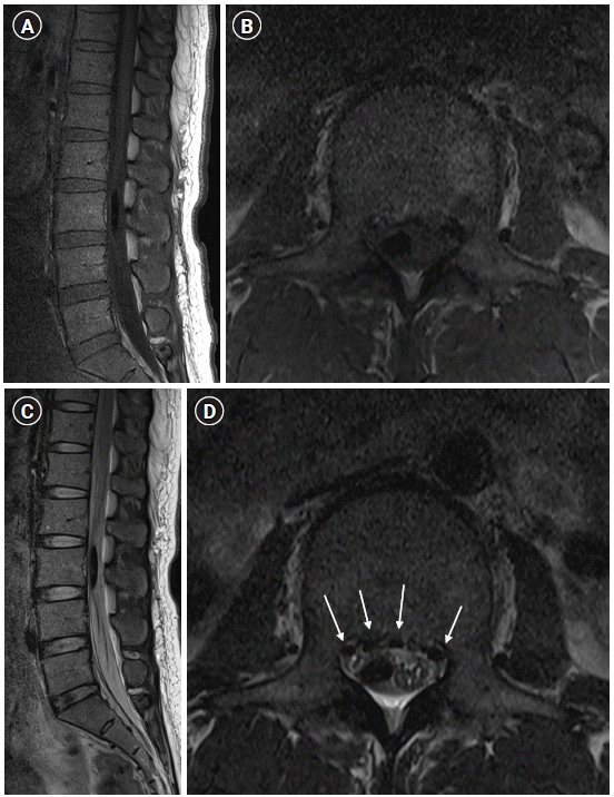
In the recovery room, the spinal level was confirmed to be T3 using a pinprick and the blood pressure was 96/65 mmHg. To manage the patient’s blood pressure, 30 μg of phenylephrine was administered six times to maintain systolic blood pressure above 90 mmHg. Due to persistently low blood pressure, a mixture of 10 mg of phenylephrine and 100 ml of 0.9% normal saline was infused at a rate of 10 ml/h. Five hours after the epidural injection, the sensory block level was confirmed to be T5, and the modified Bromage scale for motor block was 3 in both lower extremities. Considering the patient’s symptoms and condition, we suspected spinal cord compression. After providing a detailed explanation to the patient and obtaining informed consent, MRI of the thoracic and lumbar spine (Fig. 1) was performed, and the patient was subsequently transferred to the intensive care unit (ICU). The initial vital signs measured in the ICU were as follows: blood pressure, 102/71 mmHg; heart rate, 81 beats/min; respiratory rate, 16 breaths/min; and body temperature, 36.1°C.
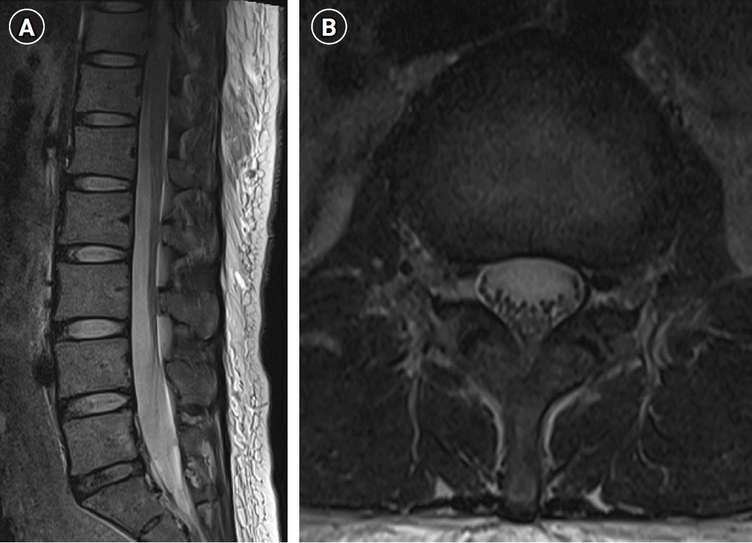
Spinal MRI revealed findings suggestive of an acute subdural hematoma or subdural air bubbles located on the right side of L3, with leftward deviation. Based on a comprehensive assessment of the MRI findings and physical examination, the initial opinion of the neurosurgeon was to consider surgery if the pain was severe. However, if there is a trend toward recovery without neurological deficits, observation and conservative treatment are recommended. The patient received dexamethasone (5 mg) every 4 h. Seven hours after epidural injection, full recovery of motor and sensory functions was observed on the left side. However, on the right side, there was no sensation below the L3 level, and the Bromage scale score for motor block was 2. Nine hours after the epidural injection, the sensory block level on the right side descended to L5, and the Bromage scale score for motor block was 0. Therefore, we decided to continue monitoring the patient while administering the dexamethasone. On postoperative day 1, no neurological deficits were observed and the patient exhibited stable vital signs in the morning. Consequently, the patient was transferred to a general ward and started on a tapering dose of 5 mg of dexamethasone every 6 h. On postoperative day 2, re-evaluation of the MRI findings suggested a higher possibility of an acute subdural air bubble rather than a subdural hematoma. On postoperative day 6, the patient had no complications, and follow-up MRI (Fig. 2) showed improved findings compared to those of the previous examination. The neurosurgery department confirmed that the patient had recovered without neurological deficits and was discharged without the need for further treatment. The patient was then discharged. Subsequent outpatient follow-ups confirmed the absence of abnormalities.
Epidural anesthesia during cesarean section offers several benefits. First, the epidural catheter can extend the duration of anesthesia and facilitate effective postoperative pain management. Second, it demonstrates minimal alterations in vital signs, thereby exhibiting a relatively low occurrence of hypotension, which is a significant concern during cesarean section. Third, the successful implementation of epidural anesthesia eliminates the risk of dural puncture, mitigating the potential development of postdural puncture headache, which is often associated with other anesthesia methods [8].
However, it is important to note that epidural anesthesia rarely leads to unintended subdural injections, as evidenced by the reported case [1-3]. Subdural spaces seem to be created by traumatic meningeal dissection between the dural and arachnoid layers, rather than a fixed potential space [9]. Thus, according to the subdural dissection plane, the neural blockade presented unexpected and variable features. Several risk factors have been associated with subdural injections, including difficult epidural catheter placement, combined spinal-epidural anesthesia, dural puncture, leakage of local anesthesia due to aggressive needle manipulation, and altered anatomy resulting from previous back surgeries [10,11]. In the present case, the patient, a parturient with a body mass index > 27, had a slightly challenging epidural placement, although she did not exhibit any other identifiable risk factors.
In cases of subdural injection, the onset of neuraxial blockade is variable (between 7 and 30 min) [7,10,11]. The extension of the blockade could be restricted or widespread to the cervical or cranial nerves, which may lead to significant cardiovascular depression due to impairment of the sympathetic nervous system [1]. Regarding the specific case of this patient, the onset of anesthesia was observed to be 5 min, indicating a relatively rapid onset. No segmental blocks were detected during the procedure. However, a greater sensory block up to T3 and hypotension, attributed to an impaired sympathetic nervous system, were observed.
When performing the loss-of-resistance technique for epidural anesthesia, there is no significant difference in the onset and analgesic quality between air and saline [4,5]. Air facilitated the detection of dural punctures. However, there is a higher incidence of spinal cord and nerve root compression due to the mass effect of air bubbles [6]. Additionally, there is a higher risk of pneumocephalus and venous air embolism [7].
In our patient, the cauda equina deviation occurred because of the mass effect caused by air bubbles in the subdural space at the L3 level, resulting in a prolonged sensory and motor block on the right side. Dural lacerations or punctures can cause severe complications such as unintended widespread block, lumbar pain, radiculopathy, and even cardiac arrest. Therefore, this procedure must be performed with awareness [7,12].
To prevent complications, care should be taken when rotating the Tuohy needle once it enters the epidural space. In addition, every top-up should be administered in a fractionated manner, as per usual safe practice. Using single-orifice catheters may be preferable to using orifice catheters [11]. Finally, it is crucial to administer the minimum amount of air during the loss of resistance for epidural procedures. In this case, the sensation of resistance loss after the initial air injection was unclear. Consequently, we attempted multiple times to confirm the epidural space with a total volume of 3 ml and an air bubble of size 2.8 cm × 0.4 cm was identified on MRI images. Therefore, we recommend a cautious approach involving confirmation of the epidural space with volumes less than 3 ml. Therefore, ultrasonography may also be beneficial. Although it cannot directly detect air bubbles, it can be used to confirm appropriate needle position during the procedure. However, their use in parturients is limited by positional restrictions.
Two diagnostic methods are available for identifying subdural air injections: an algorithmic approach and a radiological method. In 1988, Lubenow et al. [13] proposed the diagnostic criteria for subdural injection. The major criteria included a negative aspiration test and unintended extensive sensory block, while the minor criteria included a delayed onset by 10 min or more of sensory or motor block, a variable motor block, and sympatholysis out of proportion to the administered dose of local anesthetic. A subdural injection was considered to have occurred if both of the major criteria and at least one minor criterion were present. More recently, another diagnostic algorithm was proposed in 2009 by Hoftman and Ferrante [14] who expanded on the previously proposed algorithm and introduced a four-step diagnostic algorithm. The initial step relies on the operator’s judgment, considering the tactile sensation during catheter insertion and the presence or absence of CSF. The second step involved assessing the extent of the dermatomal distribution and categorizing it as excessive, restricted, or inconclusive. However, radiological analysis could clearly prove subdural injection from the heterogeneous features of the clinical symptoms. In radiological observations, air bubbles can be identified using X-rays when present in substantial quantities. This was further confirmed by CT scans. However, there are some limitations to the use of radiography and CT in pregnant women. MRI findings play a crucial role in differentiating between air and hematomas. Acute hematomas occurring within 12 h typically exhibit isointensity on T1-weighted images and hyperintensity on T2-weighted images. Additionally, it is important to consider the possibility of a hematoma in patients presenting with back pain, a history of clotting disorders, or those undergoing anticoagulant therapy [15]. Our patient’s postoperative MRI findings showed low signal intensity on both T1- and T2-weighted images. Thus, we ruled out the possibility of a hematoma by using MRI.
In an epidural block, the administration of 0.5–1.0% ropivacaine typically results in a duration with a two-dermatome regression time of 90–180 min, and a complete resolution time of 240–420 min. In this case, the maximal level reached was at T3, and despite 5 h passing after injection, the sensory block level remained at T5. Therefore, MRI was performed to suspect a mass effect due to an air bubble or hematoma, in addition to the local anesthetic-induced epidural block. While there are no predefined indications for imaging and the consequences of a missed diagnosis are serious, it may be necessary to consider an imaging study if the sensory block level shows less change compared to the prediction made by the physician.
Conservative treatment is generally effective in cases where spinal cord compression arises from air bubbles. If the symptoms persist, intravenous dexamethasone administration can be considered to alleviate them. Swift removal of the epidural catheter is crucial, and patients should undergo bed rest with meticulous monitoring of their vital signs. In the presence of sympathetic blockade symptoms and signs, vasopressors such as phenylephrine may be necessary to regulate blood pressure. Moreover, special attention must be paid to the potential occurrence of apnea, profound bradycardia, and loss of consciousness, which may result from higher neural blockade [1,2]. Generally, without specific interventions, symptoms in the majority of cases tend to resolve spontaneously over time. However, complications such as pneumocephalus, venous air embolism, and subcutaneous emphysema can occur, which may lead to poor prognosis and require invasive treatment. Subdural air collection usually causes minor complications, such as mild motor or sensory blocks or mild headaches due to increased intracranial pressure. Conservative treatments, including bed rest, Fowler’s position, and oxygen administration, are usually sufficient, and in 85% of the cases, air bubbles are absorbed within 2–3 weeks [16]. However, if symptoms persist or are severe, surgical decompression may be necessary [7].
Clinicians should be aware of the potential signs and symptoms of subdural anesthesia to enable its early diagnosis and effective management. Additionally, to mitigate the potential side effects of subdural air bubbles in the clinical field, it is crucial to minimize the injection of air during confirmation of the loss of resistance during epidural anesthesia.
Notes
DATA AVAILABILITY STATEMENT
The datasets generated during and/or analyzed during the current study are available from the corresponding author on reasonable request.
AUTHOR CONTRIBUTIONS
Writing - original draft: Eun Mi Choi. Writing - review & editing: Jung Eun Kim, Jinse Lee, Giyear Lee, Mi Hwa Chung, Young Ryong Choi. Conceptualization: Jinse Lee, Eun Mi Choi. Investigation: Jinse Lee. Resources: Eun Mi Choi. Software: Eun Mi Choi. Supervision: Mi Hwa Chung, Eun Mi Choi.
REFERENCES
1. Stevens RA, Stanton-Hicks MD. Subdural injection of local anesthetic: a complication of epidural anesthesia. Anesthesiology. 1985; 63:323–6.
2. Kalil A. Unintended subdural injection: a complication of epidural anesthesia--a case report. AANA J. 2006; 74:207–11.
3. Yun JS, Kang SY, Cho JS, Choi JB, Lee YW. Accidental intradural injection during attempted epidural block - a case report-. Korean J Anesthesiol. 2011; 60:205–8.

4. Sanford C 2nd, Rodriguez RE, Schmidt J, Austin PN. Evidence for using air or fluid when identifying the epidural space. AANA J. 2013; 81:23–8.
5. Norman D, Winkelman C, Hanrahan E, Hood R, Nance B. Labor epidural anesthetics comparing loss of resistance with air versus saline: does the choice matter? AANA J. 2006; 74:301–18.
6. Lee MH, Han CS, Lee SH, Lee JH, Choi EM, Choi YR, et al. Motor weakness after caudal epidural injection using the air-acceptance test. Korean J Pain. 2013; 26:286–90.

7. Overdiek N, Grisales DA, Gravenstein D, Bosek V, Nishman R, Modell JH. Subdural air collection: a likely source of radicular pain after lumbar epidural. J Clin Anesth. 2001; 13:392–7.

9. Reina MA, De Leon Casasola O, López A, De Andrés JA, Mora M, Fernández A. The origin of the spinal subdural space: ultrastructure findings. Anesth Analg. 2002; 94:991–5.

10. Alshoubi A, Newhide D. Inadvertent subdural catheter placement: a rare complication in obstetric anesthesia. Cureus. 2022; 14:e27252.

11. Agarwal D, Mohtaf M, Tyagi A, Sethi AK. Subdural block and the anaesthetist. Anaesth Intensive Care. 2010; 38:20–5.

12. Shin H, Choi HJ, Kim C, Lee I, Oh J, Ko BS. Cardiac arrest associated with pneumorrhachis and pneumocephalus after epidural analgesia: two case reports. J Med Case Rep. 2018; 12:387.
13. Lubenow T, Keh-Wong E, Kristof K, Ivankovich O, Ivankovich AD. Inadvertent subdural injection: a complication of an epidural block. Anesth Analg. 1988; 67:175–9.
14. Hoftman NN, Ferrante FM. Diagnosis of unintentional subdural anesthesia/analgesia: analyzing radiographically proven cases to define the clinical entity and to develop a diagnostic algorithm. Reg Anesth Pain Med. 2009; 34:12–6.
Fig. 1.
T1-weighted magnetic resonance imaging (A, B) shows a low signal intensity measuring approximately 2.8 × 0.4 cm accompanied by left deviation of the cauda equine in the L3 region. On T2-weighted MRI (C, D), multiple low-signal-intensity dots were observed in the anterior aspect of the spinal cord (indicated by arrows), suggesting the presence of air bubbles in the L3 region.


 PDF
PDF Citation
 Print
Print


 XML Download