

 PDF
PDF Citation
 Print
Print


Facial nerve schwannoma (FNS) is a rare tumor that arises along the course of the facial nerve.1 It typically has an insidious course. Rapid-onset FNS mimics Bell’s palsy, resulting in diagnostic difficulties.
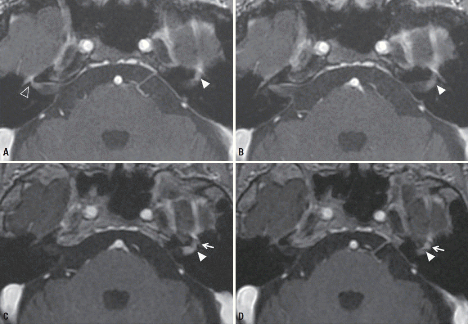
A 66-year-old male presented with sudden-onset right facial weakness with postauricular pain with a 2-day history. A neurologic examination revealed right peripheral facial weakness, classified as House-Brackmann (HB) grade-5 palsy. T1-weighted contrast-enhanced magnetic resonance imaging (MRI) demonstrated abnormal enhancement of the labyrinthine segment and genu of the right facial nerve without nerve enlargement. These findings were consistent with Bell’s palsy (Fig. 1A). MRI also showed abnormal small nodular enhancement of the labyrinthine segment of the left facial nerve, which was too small to characterize (Fig. 1A, B). His facial weakness resolved completely after treatment with an antiviral agent and steroid. Five years later the patient presented with left facial weakness with a 1-week history. A neurologic examination revealed HB grade-3 facial palsy. T1-weighted contrast-enhanced MRI revealed a fusiform mass. Compared with the previously identified lesion, the current mass was larger and exhibited more-intense enhancement. The mass was located in the distal cisternal, labyrinthine segment, and genu of the left facial nerve. These characteristics were suggestive of FNS (Fig. 1C, D).
FNS is often misdiagnosed as idiopathic facial nerve palsy or Bell’s palsy. It is difficult to detect FNS using MRI, especially when the lesion is small. On MRI, FNS presents as an enhanced tubular mass within an enlarged facial nerve canal,2 while uniformly linear facial nerve enhancement is characteristic of Bell’s palsy.3 Neurologists need to be able to recognize the different characteristic MRI findings of Bell’s palsy and FNS.
 XML Download