This article has been
cited by other articles in ScienceCentral.
Abstract
Acquired nasolacrimal duct obstruction may result from chronic infection, lacrimal stones, anatomical variations such as aberrant ethmoid cells, facial fractures, or complications following nasal surgery. In Korea, there has been no reported case of secondary nasolacrimal duct obstruction due to a retention cyst in the lacrimal sac. Recently, the authors encountered a 65-year-old female patient who presented with epiphora, was diagnosed with a lacrimal sac retention cyst, and was successfully treated with endoscopic marsupialization.
Go to :

Keywords: Lacrimal sac retention cyst, Nasolacrimal duct obstruction, Endoscopic marsupialization
INTRODUCTION
Acquired nasolacrimal duct obstruction can result from facial trauma, anatomical changes in the ethmoid cells, other nasal or paranasal sinus diseases, recurrent dacryocystitis, dacryolithiasis, complications arising from nasal surgery, and, on rare occasions, neoplasms or cysts of the nasolacrimal duct or adjacent structures [
1-
3]. The incidence of nasolacrimal duct obstruction has been reported as 20.24 per 100,000 [
4].
Although multiple case reports have stated that mucoceles can result from nasolacrimal duct obstruction [
5,
6], there have been no reported instances in Korea of secondary nasolacrimal duct obstruction caused by a retention cyst in the lacrimal sac. Furthermore, a retention cyst within the nasolacrimal duct may lead to stenosis of the lacrimal sac, culminating in epiphora.
Recently, we encountered a patient who presented to the hospital with epiphora and was diagnosed with a lacrimal retention cyst. In this report, we share our experience with the case, which resulted in a successful surgical outcome.
Go to :

CASE REPORT
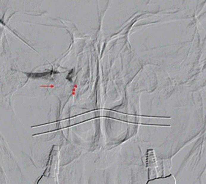
A 65-year-old woman presented to our hospital with a 5-year history of right-sided epiphora. The patient had no significant medical or trauma history. During the physical examination, a 1-cm cystic mass was palpated in the medial canthal area of the right side, and mucoid discharge was noted leaking from the inferior lacrimal punctum. Sinus endoscopic examination revealed no mass on the inferior turbinate, no mucosal edema, and no signs of sinusitis, such as purulent rhinorrhea. Dacryocystography (DCG) findings showed dilation in the proximal part of the right lacrimal sac and an illdefined defect on the inferolateral side of the sac, leading to stenosis of the nasolacrimal duct (
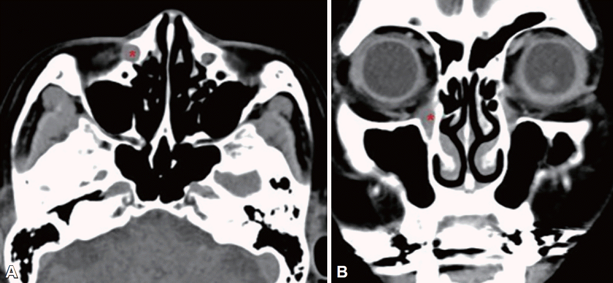
Fig. 1). A paranasal sinus computed tomography (CT) scan revealed a cystic mass measuring 6 mm×10 mm in the lacrimal sac (
Fig. 2).
 | Fig. 1.Dacryocystography shows proximal dilatation in the right lacrimal sac and complete obstruction at the duct-sac junction (arrowheads). There is a filling defect with a smooth margin on the inferolateral aspect of the lacrimal sac (arrow). 
|
 | Fig. 2.Paranasal sinus computed tomography (CT) scans. A: Axial CT shows a round cystic mass (*) in the right lacrimal sac. B: Coronal CT shows a mass (*) at the right nasolacrimal sac junction. 
|
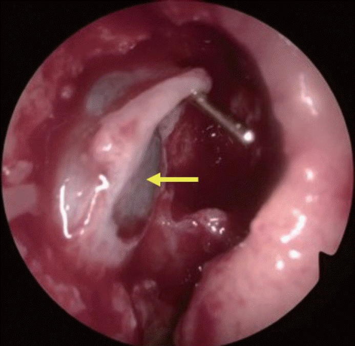
Under general anesthesia, we performed endoscopic dacryocystorhinostomy as a routine procedure. In the surgical field, we identified a 1-cm cyst on the inferolateral side of the lacrimal sac, which resulted in the obstruction of the lacrimal sac on the medial side (
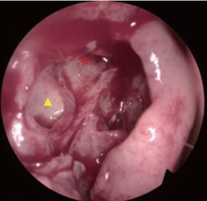
Fig. 3). After confirming that the mass was a retention cyst, not a mucocele, following the aspiration of 1 mL of mucoid discharge with a 10 mL syringe, we proceeded with endoscopic marsupialization with a sickle knife and seeker (
Fig. 4). The patient’s symptom of epiphora improved immediately after surgery.
 | Fig. 3.Intraoperative endoscopic finding shows a 1-cm cystic mass (arrow) in the lacrimal sac near the duct-sac junction. 
|
 | Fig. 4.Immediate postoperative endoscopic view shows a wide, patent opening (arrow) and a marsupialized mucous retention cyst (arrowhead). 
|
Go to :

DISCUSSION
Chronic obstruction of the distal portion of the nasolacrimal duct may lead to persistent inflammation and mucosal swelling. These conditions can subsequently cause the formation of a dacryocystocele, stemming from impaired nasal drainage and displacement of the nasolacrimal duct [
5,
6]. Nevertheless, there have been no reported cases of secondary stenosis of the nasolacrimal duct attributable to a retention cyst of the lacrimal sac.
There are various indications for performing DCG in patients with epiphora. The primary goal of DCG is to map the lacrimal drainage system, identifying areas of obstruction and any associated pathological conditions [
7]. When the duct dilates over an obstructed area during DCG, it is considered a positive sign, underscoring the significance of ductal patency in cases of stenosis. Neoplasms and dacryolithiasis within the lacrimal sac often present with a range of radiological abnormalities. In contrast-enhanced DCG, these may manifest as stable, dark, and poorly defined masses. Although air bubbles can be present, they typically exhibit a well-defined, round appearance, but variations in size and shape can occur. DCG is also instrumental in guiding surgical and therapeutic strategies by pinpointing the exact location of the blockage [
8].
When identifying obstructions and defects in the drainage system, it is important to examine the preoperative CT scan to identify any risk factors that may cause secondary obstruction of the nasolacrimal duct. Secondary obstruction of the nasolacrimal duct can arise from a variety of causes, which typically fall into five main categories: infectious, inflammatory, neoplastic, traumatic, and mechanical [
9]. The CT scan can be used for the differential diagnosis of masses around the lacrimal sac, such as carcinoma, mucoepidermoid carcinoma, dermoid cysts, and inflammatory diseases [
9]. Findings such as suspected malignancy, bony erosion, or invasion into adjacent structures on the CT scan may warrant further evaluation with magnetic resonance imaging. In the present case, a round cystic mass was detected in the lacrimal sac on the CT scan, which differed from the previously described lacrimal sac mucocele. A secondary obstruction due to a retention cyst was suspected, and this diagnosis was confirmed during surgery.
The treatment of a lacrimal sac retention cyst does not significantly differ from that of a mucocele of the nasolacrimal duct. If the cyst does not extend over the maxilla or is located in the proximal part of the nasolacrimal duct, surgeons typically perform dacryocystorhinostomy. When the distal part of the nasolacrimal duct is covered only by soft tissue, marsupialization using an endoscopic approach is required.
According to a meta-analysis [
10], silicone tube insertion does not significantly affect the success rate or the postoperative complication rate of the endoscopic dacryocystorhinostomy procedure. Silicone tube insertion should be considered if there is an injury to the intraductal mucosa or to the mucosa surrounding the nasolacrimal duct opening on the lateral wall of the nasal cavity. Historically, dacryocystorhinostomy was the primary procedure; however, with advancements in endoscopic techniques, endoscopic marsupialization has become more commonly performed. Stent insertion may not be necessary, especially in cases of a wide opening in the distal part of the nasolacrimal duct [
6].
In this case, the authors successfully treated a patient who had a 5-year history of epiphora. The patient was diagnosed with nasolacrimal duct obstruction, which was attributed to a lacrimal sac retention cyst. The treatment involved endoscopic marsupialization combined with dacryocystorhinostomy, and it was completed without any complications.
Go to :


 PDF
PDF Citation
 Print
Print


 XML Download