Journal List > Korean J Pain > v.37(2) > 1516086830

Suhaini, Pauzi, Juhari, Bakar, and Moon: Evaluation of the antinociceptive activities of natural propolis extract derived from stingless bee Trigona thoracica in mice

Abstract

Background

Stingless bee propolis is a popular traditional folk medicine and has been employed since ancient times. This study aimed to evaluate the antinociceptive activities of the chemical constituents of aqueous propolis extract (APE) collected by Trigona thoracica in a nociceptive model in mice.

Methods

The identification of chemical constituents of APE was performed using high-performance liquid chromatography (HPLC). Ninety-six male Swiss mice were administered APE (400 mg/kg, 1,000 mg/kg, and 2,000 mg/kg) before developing nociceptive pain models. Then, the antinociceptive properties of each APE dose were evaluated in acetic acid-induced abdominal constriction, hot plate test, and formalin-induced paw licking test. Administration of normal saline, acetylsalicylic acid (ASA, 100 mg/kg, orally), and morphine (5 mg/kg, intraperitoneally) were used for the experiments.

Results

HPLC revealed that the APE from Trigona thoracica contained p-coumaric acid (R2 = 0.999) and caffeic acid (R2 = 0.998). Although all APE dosages showed inhibition of acetic acid-induced abdominal constriction, only 2,000 mg/kg was comparable to the result of ASA (68.7% vs. 73.3%, respectively). In the hot plate test, only 2,000 mg/kg of APE increased the latency time significantly compared to the control. In the formalin test, the durations of paw licking were significantly reduced at early and late phases in all APE groups with a decrease from 45.1% to 53.3%.

Conclusions

APE from Trigona thoracica, containing p-coumaric acid and caffeic acid, exhibited antinociceptive effects, which supports its potential use in targeting the prevention or reversal of central and peripheral sensitization that may produce clinical pain conditions.

INTRODUCTION

Stingless bees are a monophyletic group of bees hibernating and pollinating in the tropical and subtropical regions [1,2]. They constitute about 500 species, known taxonomically as the family Apidae, and their most common genera are Trigona, including Trigona (Geniotrigona) thoracica [3]. Among products derived from stingless bees, propolis has been broadly used since ancient times as dietary supplementation, oral care products, creams, and ointments to enhance anti-inflammatory, anti-microbial, antioxidant, and immunomodulatory activities [48]. In particular, previous studies have focused on pharmacological properties of the phenolic compounds predominantly present in propolis from the stingless bees [811]. Although non-steroidal anti-inflammatory drugs (NSAIDs) and opioids have been the mainstream for anti-nociceptive analgesics in clinical practice [12,13], their use links to mild to severe adverse events from gastrointestinal disorder to psychologic addiction. Given that the naturally derived compounds from stingless bee’s propolis could be useful in various pharmacological activities, they have potential as new modern medicines, which could overcome adverse events from conventional analgesics. To date, the antinociceptive effects of propolis extracts from different species of bees have been studied in matters pertaining to accessible plants and cumulative evidence has reported analgesic effects of propolis from various regions [1416]. Chinese propolis extracts from Poplar sp. (populus sp.) enriched with polyphenolic constituents presented antinociceptive activities, which could be attributed to their antioxidant effects [16]. Brazilian organic propolis from Apis mellifera suppressed the p38mitogen-activated protein kinase (MAPK) and p-JNK phosphorylation, as well as nuclear factor NF-κB activation in the murine macrophages cell line RAW264.7, suggesting its anti-inflammatory potential [17]. Furthermore, the antinociceptive activity of such propolis has been reported in animal models in the acetic acid-induced abdominal constriction test, infrared hyperalgesia, and formalin-induced paw licking test [14,15,18].
Stingless bees are geographically specified, becoming essential factors in explaining the variety of chemical compositions of propolis. However, to the best of the authors’ knowledge, no study has been carried out on the possible antinociceptive effects of propolis collected by Geniotrigona thoracica, since this species is an endemic species of stingless bee in Malaysia. Recent studies have revealed the pancreatoprotective and antimicrobial properties of propolis from Geniotrigona thoracica [19,20]. Therefore, in this study, the authors aimed to investigate the antinociceptive effects of aqueous propolis extracts (APE) from Geniotrigona thoracica in chemical and thermal nociceptive animal models.

MATERIALS AND METHODS

1. Propolis samples collection

The fresh propolis samples produced by Trigona thoracica were collected in November 2021 from Kuala Besut, Terengganu Darul Iman, Malaysia (N 05°45’16” E 102°37’ 38”). The authentication of propolis from Geniotrigona thoracica stingless bee species was obtained from Forecast Research and Institute Malaysia (FRIM), Kepong, Malaysia, with the reference number ENTO/2022/03. The propolis was stored at –20°C after being washed with tap water to remove dust and foreign particles.

2. Preparation of APE

The frozen propolis was ground into powder in a commercial blender (Waring Commercial). Distilled water was used to extract the water compounds in propolis. It was prepared using distilled water from a Milli-Q Plus system (Millipore). Samples were repetitively changed for three days and filtered through Whatmann® No. 41 filter paper before lyophilization using a freeze dryer (Christ, GmbH). The dried brown APE was kept cool at –4°C. High-performance liquid chromatography UV spectrophotometer detector (HPLC-UV) (Shimadzu Corp.) analysis was carried out on a sample of this batch of APE and their retention time was considered with both caffeic acid (Lot no [#]: BCCF4731) and p-coumaric acid (Lot no [#]: SLCJ9012) reference standards. One percent dimethyl sulfoxide (DMSO) in distilled water was used to dissolve the APE and served as the vehicle used in the control group. The concentration of DMSO used is relatively low and has minimal impact on nociceptive or anti-nociceptive responses [21].

3. Identification of phenolic acids

The identification of phenolic acids in the APE was performed using the HPLC-UV, which was described in a previous study [22]. A chromatographic system comprises an LC unit with a UV-Vis spectrophotometer detector (SPD-10A), a degassing unit (DGU-20A5R), and a solvent delivery pump (LC-20AT). The reversed-phase separation was performed on an Infinity Poroshell 120 EC-C18 4.6 mm × 150 mm column, particle size 4 µm (Agilent Technologies). The UV detector was set at a wavelength of 300 nm for detection of targeted standards. The column temperature was operated at 40°C at 0.4 mL/min. The injection volume was 10 µL. A combination of mobile phase was composed of a mixture of solvents A (methanol:acetonitrile:deionized water) (Fisher Scientific) (40:5:55, v/v) containing 0.1% formic acid (v/v) and solvents B (methanol:acetonitrile:deionized water) (Fisher Scientific) (80:5:15, v/v) containing 0.1% formic acid (v/v). A gradient method of the solvent B was used as follows: 0 to 2 minutes, 8%; 2 to 4 minutes, 9%; 4 to 12 minutes, 10%; 12 to 18 minutes, 19%; and 18 to 20 minutes, 100%. The mobile phase was filtered using a filtration system followed by 15 minutes of sonication to degas the solvents in an ultrasonic bath before analysis. LC Solutions Software (Shimadzu Corp.) was used for peak integration, data acquisition, and calibrations. The calibration curves were performed in the range of 10–50 parts per million (ppm).

4. Drugs and chemicals

DMSO, all standards with purity > 98% of p-coumaric acid and caffeic acid, and acetylsalicylic acid (ASA, purity > 99.0%) were purchased from Sigma-Aldrich, Germany. Glacial acetic acid was procured from GmbH, Germany. Morphine sulphate was manufactured by Hameln, GmbH, Germany.

5. Experimental animals

The Animal and Plant Research Ethics Committee (UAPREC) Universiti Sultan Zainal Abidin evaluated and approved all animal experiments with permit number UAPREC/07/005. After obtaining the ethical approval, six male Albino Swiss mice (n = 6) aged four to seven weeks old (25–30 g) were randomly housed per cage for each treatment group at room temperature (24°C ± 2°C). The animals were maintained under standard environmental conditions (12-hr light/dark cycle) for seven days before the experiments began, and they were provided with free access to food and water supplied ad libitum. During ad libitum feeding, the mice were given drugs added to the water for a longer period whenever possible for ease of oral administration.

6. Acetic acid-induced abdominal constriction test

The acetic acid-induced abdominal constriction test was used to assess the antinociceptive potential of APE from Trigona thoracica, as described in detail in previous studies [23,24]. Briefly, mice (n = 6) were treated orally (p.o) with 1% DMSO in normal saline (10 mL/kg) as a control, ASA (100 mg/kg) as a positive control, or APE (400 mg/kg, 1,000 mg/kg, and 2,000 mg/kg) in the test groups for 60 minutes before the administration of the phlogistic agent (0.6%, acetic acid; intraperitoneal, i.p). The doses range for nociceptive study was determined from the reduction of the doses used in an acute toxicity study by Muhamad Suhaini et al. [25]. The cumulative number of abdominal constrictions observed was counted over 25 minutes, starting 5 minutes after the phlogistic agent injection. The percentage of inhibition of abdominal constrictions indicated the antinociceptive activity using the following formula as the mean of [(control group – test group) / control group × 100%].

7. Hot plate test

The central antinociceptive potential of APE extract from Trigona thoracica was assessed using the hot plate test as previously described [23,24]. The untreated mice were placed on the metal hot plate (Bio-CHP; Bioseb) heated to 52.5°C ± 0.5°C to select animals with suitable latency of response (5–7 seconds) to the thermal-induced nociceptive stimuli. The cut-off time of 20 seconds was chosen to avoid tissue injury. The selected mice (n = 6) were pre-treated p.o with 1% DMSO in normal saline (10 mL/kg), APE (400, 1,000, and 2,000 mg/kg) as test groups, or morphine sulfate (5 mg/kg, i.p) as a positive control for 60 minutes prior to being subjected to the test. The latency to a discomfort reaction (licking hind paws and jumping) for all treated and control groups was recorded before and at 0, 60, 90, 120, 150, 180, and 210 minutes after the oral administration of the respective test solutions.

8. Formalin test

The peripheral and central antinociceptive potential of APE from Trigona thoracica was assessed using the formalin test following the previous description [23,24]. The mice were treated p.o with 1% DMSO in normal saline (10 mL/kg) as a control, ASA (100 mg/kg) as a positive control, APE (400, 1,000, and 2,000 mg/kg) as test groups, or morphine (5 mg/kg, i.p) as a positive control for a peripherally- and centrally acting-analgesic for 60 minutes prior to the intraplantar injection of 5.0% (v/v) formalin (25 µL) into the region of the right hind paw. Immediately after the formalin injection, mice were individually placed in a Perspex cage for observation. The time the mice spent licking the injected paw was recorded in two distinct phases: the early (0–5 minutes) and late (15–30 minutes) phases. The percentage of reduction in paw licking and biting time using the following formula as the mean of [(control group–test group) / control group × 100%].

9. Statistical analysis

Results were expressed as mean ± standard error as descriptive statistics. For the acetic acid-induced abdominal constriction test and formalin-induced paw licking test, the mean differences between the APE (400, 1,000, and 2,000 mg/kg) and normal saline control group were compared and analyzed by 1-way ANOVA with Dunnett’s multiple comparisons tests. In contrast, the hot plate test used 2-way ANOVA with Dunnett’s multiple comparisons tests. Meanwhile, P values less than 0.05, 0.0001, and 0.00001 (P < 0.05, P < 0.0001, and P < 0.00001) were considered significant. GraphPad Prism 9.0 for Windows (GraphPad Software) was employed for data analysis.

RESULTS

1. HPLC analysis

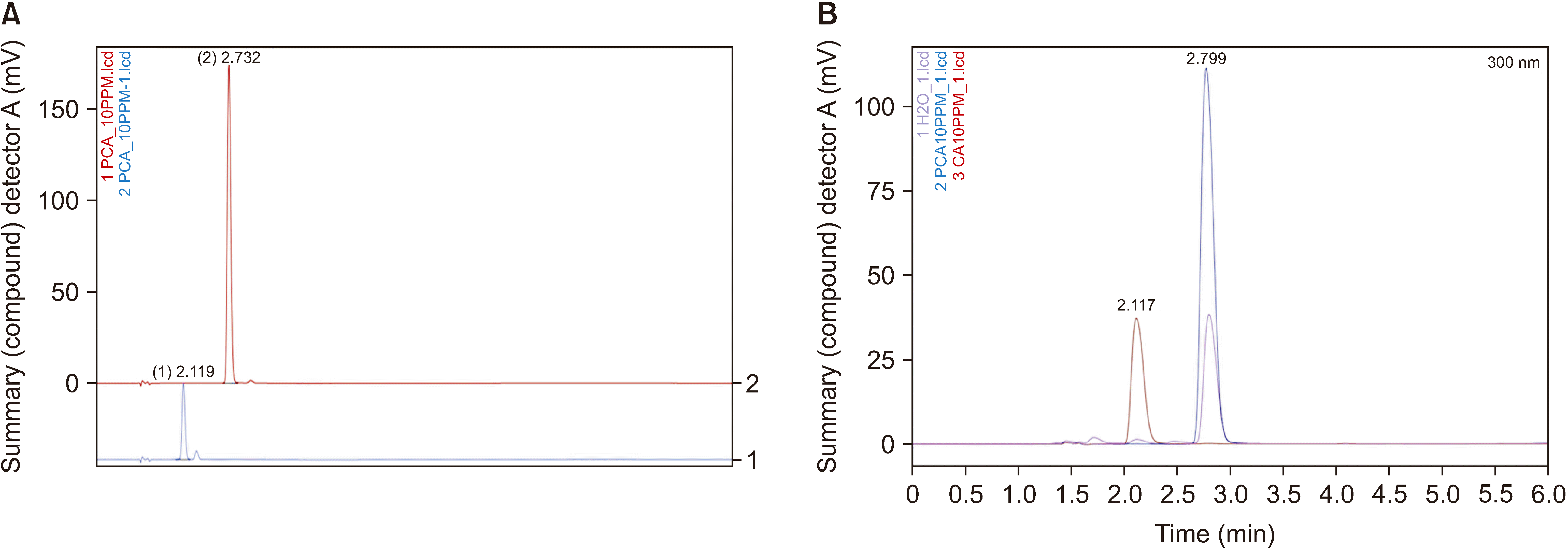
A comparison between the chromatograms of the standard compounds and APE from Trigona thoracica revealed that samples contain p-coumaric acid and caffeic acid with concentrations of 0.385-1.713/100 mg and 0.394-1.723/100 mg, respectively (Fig. 1). The regression equations used to determine p-coumaric acid and caffeic acid concentrations were y = 0.137 × (R2 = 0.999) and y = 0.0315 × (R2 = 0.998), respectively.

2. Acetic acid-induced abdominal writhing test

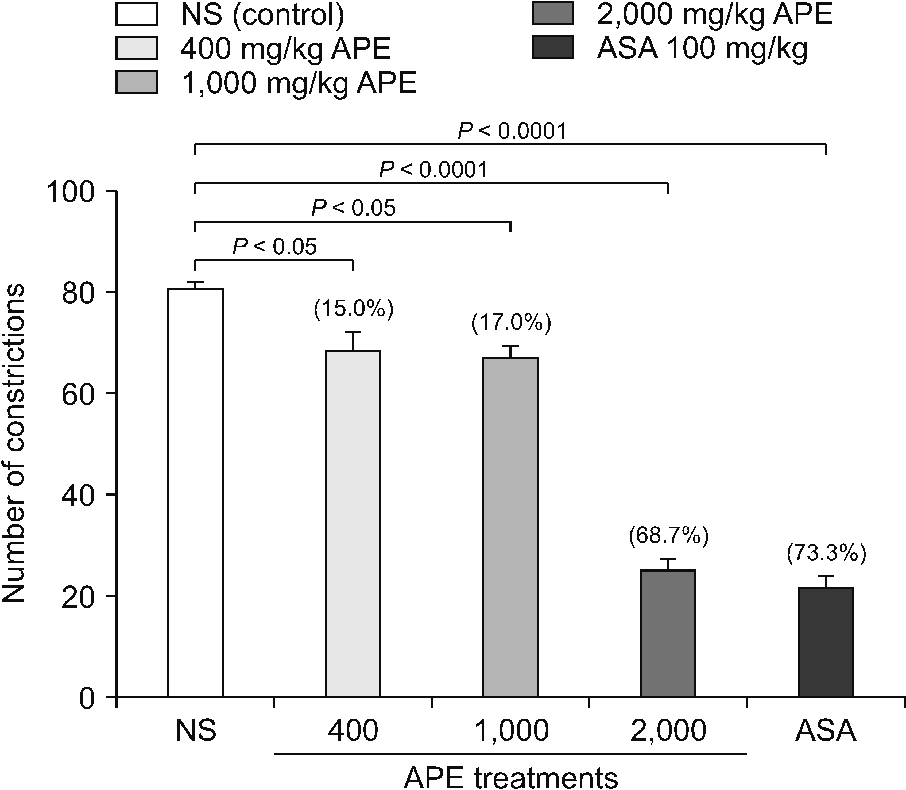
In the acetic acid-induced abdominal constriction test, the oral administration of APE from Trigona thoracica at all doses (400 mg/kg, 1,000 mg/kg, and 2,000 mg/kg) presented significant inhibition of constrictions in acetic acid-induced mice. Their reductions (%) were 15.0%, 17.0%, and 68.7%, respectively, compared to the normal saline injection (Fig. 2). Although the reduction in the number of writhes was detected in all APE dose groups, only 2,000 mg/kg of APE (68.7%) was comparable to the positive control with 100 mg/kg ASA (73.3%).

3. Hot plate test

Regarding the thermally-induced nociception in a hot plate test, APE at a high dose (2,000 mg/kg) caused a significant effect in response latency time at the interval of 60 minutes and 120 minutes (both P < 0.001) when compared to the normal saline (Table 1). On the other hand, 400 mg/kg and 1,000 mg/kg of APE did not prolong the response latency to the thermal stimulus throughout the timescale. Intraperitoneal morphine injection markedly decreased antinociceptive activity (P < 0.0001), which started at the interval of 60 minutes and was prolonged to the end of the experiment at 210 minutes.

4. Formalin-induced paw licking test

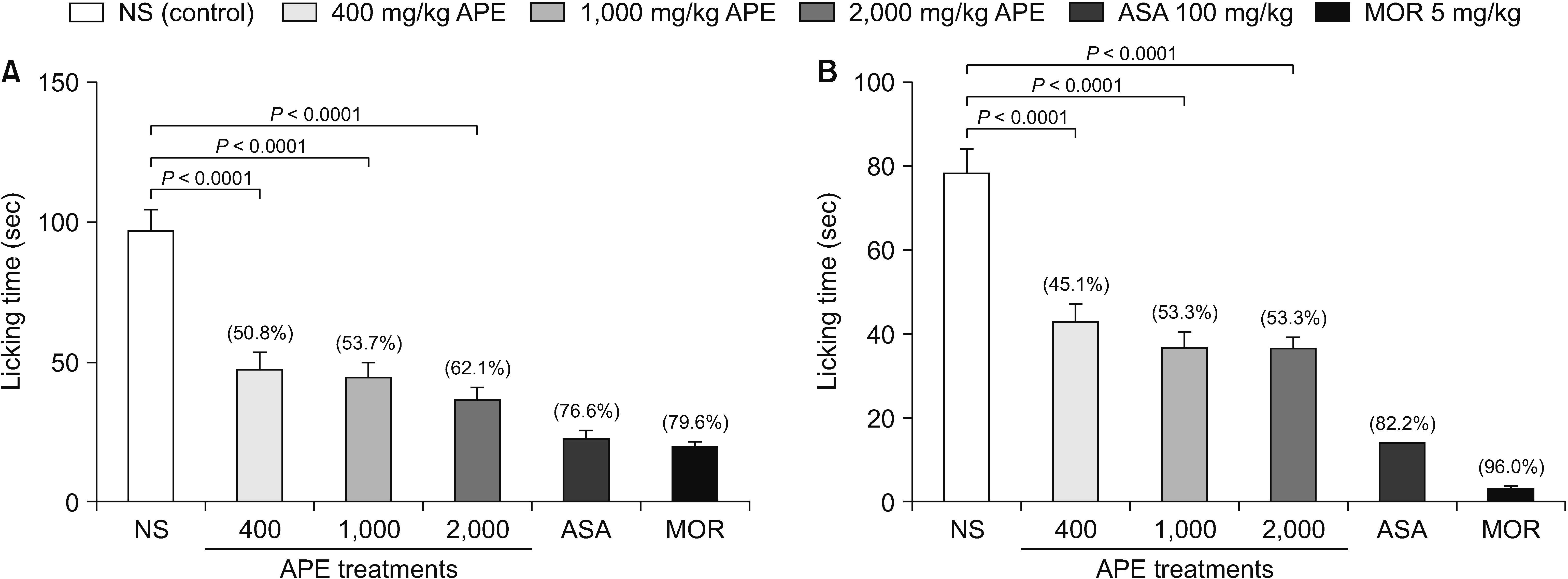
All APE doses significantly reduced the paw licking and biting time (sec) when compared to the normal saline injection during the early and late phases (all P values were < 0.0001). However, the antinociceptive activities of all APE doses were not comparable to ASA and morphine injections at both early and late phases in the formalin-induced paw licking test, which were 76.6% and 79.6% reductions during the early phase and 82.2% and 96.0% reductions during the late phase, respectively (Fig. 3).

DISCUSSION

Most of the compounds identified in propolis belong to flavonoids, quercetin, chrysin (5,7 dihydroxyflavone), and phenolic and caffeic acid derivatives, with various biological activities as free radical scavengers, antimicrobial, or antioxidant effects [4,2630]. Meanwhile, in a previous study, ethanolic extracts of stingless bee propolis from India were reported to contain gallic acid, naringin, p-coumaric acid, and kaempferol [28]. Among the phenolic acids, caffeic acid and p-coumaric acid were the most common compounds identified in propolis [29], which were mainly detected in propolis from Trigona thoracica in the present study. Park et al. [31] noted that caffeic acid (3,4-dihydrocinnamic acid) exerted antinociceptive activity when orally given to mice. The study revealed that the action of caffeic acid was potentially mediated via opioidergic receptors in a dose-dependent manner [31]. Therefore, although further in-vitro and in-vivo studies are necessary, the authors assumed that the antinociceptive activity of APE from Trigona thoracica might involve an opioid analgesic pathway to some degree.
An intraperitoneal injection of irritants in mice illustrates the peripherally mediated nociceptive responses, which have been employed to screen analgesic activity [32,33]. Intraperitoneal injection of acetic acid provokes ‘writhing’ characterized by abdominal contractions and ventral arching of the back and extension of the hind limbs in mice [32]. This behavior involves stimulation of the local peritoneal receptors on the surface of the cells lining the peritoneal cavity, in that acetic acid indirectly induces the release of endogenous substances, such as bradykinin, prostaglandins E2 (PGE2), histamine, and serotonin, into peritoneal fluids, which, in turn, are sensitive to the analgesic effects of NSAIDs [34,35]. Previously, the antinociceptive effect of the aqueous fractions from ethanolic extract of propolis from Melipona scutellaria has been displayed in the same pain model [36]. In the present study, APE from Trigona thoracica successfully reduced the number of writhing motions in the acetic acid-induced abdominal constriction test. In particular, 2,000 mg/kg of APE was comparable (68.7% reduction) to the result of ASA (73.3% reduction). Given that ASA promotes anti-inflammatory activity by inhibiting PG biosynthesis, a high-dose of APE may attenuate inflammatory-mediated peripheral nociceptive response as a peripherally acting analgesic [15]. However, other drugs such as antihistamines, neuroleptics, and adrenergic blockers could also inhibit the abdominal constriction responses in animal models, producing false positive results [33]. Therefore, additional investigation is necessary to verify the antinociceptive activity of APE extracts.
During the hot plate test, direct thermal stimulus elicits two behavioral parts in mice, paw licking and jumping, due to the reflex latency reaction towards the thermal stimulation of non-inflamed paws. In the present study, pre-treatment of a high dose APE from Trigona thoracica (2,000 mg/kg) prolonged the latency of discomfort toward the thermal stimuli. C- and Aδ nerve fibers are involved in the peripheral nociceptive response to thermal cutaneous stimuli. Besides, the hot plate test marks a centrally integrated response, which would be typically affected by opioids in the central pain pathway [32,34,35,37]. Therefore, these results suggest that the APE from Trigona thoracica might affect the opioid receptors and suppress the C-fiber activity in both central and peripheral pain pathways, at least during the 60- and 120-minutes observation period. In a previous study, any compound with a potential analgesic effect during the hot plate test tended to be classified as a strong analgesic [38]. Similar to the results in the authors’ study, Moroccan propolis successfully attenuated the hot plate test, suggesting that the antinociceptive-bearing bioactive compounds are presented in the water extract of Moroccan propolis [39]. Therefore, the APE extracts from Trigona thoracica acted with the same characteristics of the standard analgesic (i.e., morphine) with antinociceptive activity.
The formalin injection test assesses how the mice respond to pain generated by inflammatory mediators such as bradykinin, serotonin, and histamine [40,41]. It may provide more valid information related to clinical pain than the thermal stimuli test by direct tissue inflammation [41]. The formalin injection test is comprised of two phases: 1) the early phase, resulting from the direct chemical nociceptive stimulation [40] and 2) the second phase, resulting from the amplification of inflammatory mediators [40]. These two distinct phases are utilized not only for elucidating the underlying pain mechanism but also for providing information on the effect of drugs on inflammatory- and non-inflammatory-mediated pain [41]. As proof, opioid analgesics as centrally acting drugs inhibited both phases equally, whereas NSAIDs as peripherally acting analgesics, by reducing prostaglandin production, attenuated the second phase alone [42]. Previously, Lima Cavendish et al. [18] verified the antinociceptive effect of the hydroalcoholic extract from Brazilian red propolis measured by the incidence of flinching in the formalin test. In the present study, all doses of APE from Trigona thoracica reduced the duration of biting or licking activities during the early and second phases of the formalin-induced nociception test. Nonetheless, their effects did not reach those of ASA and morphine. Therefore, further studies are necessary regarding the potency of APE compared to conventional analgesics used in clinical practice.
There are several limitations in this study that remain to be explored. Given that both opioid and N-methyl-D-aspartate (NMDA) receptors have been localized in both the dorsal horn of the spinal cord and brain stem in the modulation of acute pain responses. Additional experiments are required to determine involvement of opioid receptors to further delineate the action mechanisms that lie behind these current findings. Although APE, as a crude extract, contains various types of bioactive compounds, flavonoid-based compounds in part have been reported to demonstrate antinociceptive properties. It is in accordance with propolis extracts, which also showed that some minor compounds were suggested to be involved in synergism effects when they are combined. The authors observed the appearance of paw edema between four and five hours after injection of formalin. However, the measurement of the neurochemistry of pain transmission, such as prostaglandin, histamine and bradykinin levels in tissues, were not evaluated. In addition, natural propolis harvested from a specific geographical area does not reflect the actual propolis with specified characteristics features harvested in other places. Further studies are required to overcome the limitations of this study.
In conclusions, the present study provides evidence to establish an antinociceptive profile in APE from the Malaysian stingless bee Trigona thoracica, along with the identification of the chemical constituents of the samples. These results possess significant peripheral and central antinociceptive effects in mice models at specific doses of APE. Further research on the precise mechanism of action for APE extracts will be worthwhile. Compared to conventional medications, this study supports the potential use of propolis extract that targets the prevention or reversal of central and peripheral sensitization that may result from clinical pain conditions. In addition, the long-term outcomes of the use of such natural extracts are encouraged for pragmatic adaptation of APE extracts in clinical practice.

ACKNOWLEDGEMENTS

The authors thank the Faculty of Medicine, Universiti Sultan Zainal Abidin (UniSZA) for providing the facilities.

Notes

DATA AVAILABILITY

After acceptance, the corresponding author of the accepted research article should submit the datasets underlying the results of this paper to the editorial office.

CONFLICT OF INTEREST

Jee Youn Moon is an editorial board member of the Korean Journal of Pain; however, she has not been involved in the peer reviewer selection, evaluation, or decision process of this article. No other potential conflict of interest relevant to this article were reported.

FUNDING

We thanks to Ministry of Higher Education (MOHE) Malaysia for the Fundamental Research Grant Scheme (FRGS/1/2019/WAB11/UNISZA/02/1).

AUTHOR CONTRIBUTIONS

Nurul Alina Muhamad Suhaini: Writing/manuscript preparation; Mohd Faeiz Pauzi: Study conception; Siti Norazlina Juhari: Writing/manuscript preparation; Noor Azlina Abu Bakar: Writing/manuscript preparation; Jee Youn Moon: Writing/manuscript preparation.

REFERENCES

1. Heard TA. 1999; The role of stingless bees in crop pollination. Annu Rev Entomol. 44:183–206. DOI: 10.1146/annurev.ento.44.1.183. PMID: 15012371.
crossref
2. Ruttner F. Ruttner F, editor. 1988. Stingless bees (Meliponinae). In: Biogeography and taxonomy of honeybees. Springer;Berlin, Heidelberg: p. 13–9. DOI: 10.1007/978-3-642-72649-1_2.
crossref
3. Kelly N, Farisya MSN, Kumara TK, Marcela P. 2014; Species diversity and external nest characteristics of stingless bees in meliponiculture. Pertanika J Trop Agric Sci. 37:293–8.
4. Campos JF, dos Santos UP, Macorini LF, de Melo AM, Balestieri JB, Paredes-Gamero EJ, et al. 2014; Antimicrobial, antioxidant and cytotoxic activities of propolis from Melipona orbignyi (Hymenoptera, Apidae). Food Chem Toxicol. 65:374–80. DOI: 10.1016/j.fct.2014.01.008. PMID: 24412556.
crossref
5. Campos JF, Dos Santos UP, da Rocha Pdos S, Damião MJ, Balestieri JB, Cardoso CA, et al. 2015; Antimicrobial, antioxidant, anti-inflammatory, and cytotoxic activities of propolis from the stingless bee Tetragonisca fiebrigi (Jataí). Evid Based Complement Alternat Med. 2015:296186. DOI: 10.1155/2015/296186. PMID: 26185516. PMCID: PMC4491730.
6. Torres AR, Sandjo LP, Friedemann MT, Tomazzoli MM, Maraschin M, Mello CF, et al. 2018; Chemical characterization, antioxidant and antimicrobial activity of propolis obtained from Melipona quadrifasciata quadrifasciata and Tetragonisca angustula stingless bees. Braz J Med Biol Res. 51:e7118. DOI: 10.1590/1414-431x20187118. PMID: 29791598. PMCID: PMC6002130.
crossref
7. Machado JL, Assunção AK, da Silva MC, Dos Reis AS, Costa GC, Arruda Dde S, et al. 2012; Brazilian green propolis: anti-inflammatory property by an immunomodulatory activity. Evid Based Complement Alternat Med. 2012:157652. DOI: 10.1155/2012/157652. PMID: 23320022. PMCID: PMC3541042.
crossref
8. Touzani S, Embaslat W, Imtara H, Kmail A, Kadan S, Zaid H, et al. 2019; In vitro evaluation of the potential use of propolis as a multitarget therapeutic product: physicochemical properties, chemical composition, and immunomodulatory, antibacterial, and anticancer properties. Biomed Res Int. 2019:4836378. DOI: 10.1155/2019/4836378. PMID: 31915694. PMCID: PMC6930758.
9. Ismail TNNT, Sulaiman SA, Ponnuraj KT, Man CN, Hassan NB. 2018; Chemical constituents of Malaysian Apis mellifera propolis. Sains Malaysiana. 47:117–22. DOI: 10.17576/jsm-2018-4701-14.
10. Mohamed WAS, Ismail NZ, Omar EA, Abdul Samad N, Adam SK, Mohamad S. 2020; GC-MS evaluation, antioxidant content, and cytotoxic activity of propolis extract from Peninsular Malaysian stingless bees, Tetrigona Apicalis. Evid Based Complement Alternat Med. 2020:8895262. DOI: 10.1155/2020/8895262. PMID: 33381215. PMCID: PMC7759394.
11. Ibrahim N, Mohd Niza NFS, Mohd Rodi MM, Zakaria AJ, Ismail Z, Mohd KS. 2016; Chemical and biological analyses of Malaysian stingless bee propolis extracts. MJAS. 20:413–22. DOI: 10.17576/mjas-2016-2002-26.
crossref
12. Blakemore PR, White JD. 2002; Morphine, the Proteus of organic molecules. Chem Commun. 2:1159–68. DOI: 10.1039/b111551k. PMID: 12109065.
crossref
13. Allison MC, Howatson AG, Torrance CJ, Lee FD, Russell RI. 1992; Gastrointestinal damage associated with the use of nonsteroidal antiinflammatory drugs. N Engl J Med. 327:749–54. DOI: 10.1056/NEJM199209103271101. PMID: 1501650.
crossref
14. Brodkiewicz Y, Marcinkevicius K, Reynoso M, Salomon V, Maldonado L, Vera N. 2018; Studies of the biological and therapeutic effects of Argentine stingless bee propolis. JDDT. 8:382–92. DOI: 10.22270/jddt.v8i5.1889.
crossref
15. Al-Hariri MT, Abualait TS. 2020; Effects of green Brazilian propolis alcohol extract on nociceptive pain models in rats. Plants (Basel). 9:1102. DOI: 10.3390/plants9091102. PMID: 32867097. PMCID: PMC7570148.
crossref
16. Sun L, Liao L, Wang B. 2018; Potential antinociceptive effects of Chinese propolis and identification on its active compounds. J Immunol Res. 2018:5429543. DOI: 10.1155/2018/5429543. PMID: 30356413. PMCID: PMC6178491.
crossref
17. Tiveron AP, Rosalen PL, Franchin M, Lacerda RC, Bueno-Silva B, Benso B, et al. 2016; Chemical characterization and antioxidant, antimicrobial, and anti-inflammatory activities of South Brazilian organic propolis. PLoS One. 11:e0165588. DOI: 10.1371/journal.pone.0165588. PMID: 27802316. PMCID: PMC5089781.
crossref
18. Lima Cavendish R, de Souza Santos J, Belo Neto R, Oliveira Paixão A, Valéria Oliveira J, Divino de Araujo E, et al. 2015; Antinociceptive and anti-inflammatory effects of Brazilian red propolis extract and formononetin in rodents. J Ethnopharmacol. 173:127–33. DOI: 10.1016/j.jep.2015.07.022. PMID: 26192808.
crossref
19. Aziz MSA, Giribabu N, Rao PV, Salleh N. 2017; Pancreatoprotective effects of Geniotrigona thoracica stingless bee honey in streptozotocin-nicotinamide-induced male diabetic rats. Biomed Pharmacother. 89:135–45. DOI: 10.1016/j.biopha.2017.02.026. PMID: 28222394.
crossref
20. Abdullah NA, Zullkiflee N, Zaini SNZ, Taha H, Hashim F, Usman A. 2020; Phytochemicals, mineral contents, antioxidants, and antimicrobial activities of propolis produced by Brunei stingless bees Geniotrigona thoracica, Heterotrigona itama, and Tetrigona binghami. Saudi J Biol Sci. 27:2902–11. DOI: 10.1016/j.sjbs.2020.09.014. PMID: 33100845. PMCID: PMC7569112.
21. Colucci M, Maione F, Bonito MC, Piscopo A, Di Giannuario A, Pieretti S. 2008; New insights of dimethyl sulphoxide effects (DMSO) on experimental in vivo models of nociception and inflammation. Pharmacol Res. 57:419–25. DOI: 10.1016/j.phrs.2008.04.004. PMID: 18508278.
crossref
22. Mohd Salim NH, Azam Omar E, Wan Omar WA, Mohamed R. 2018; Chemical constituents and antioxidant activity of ethanolic extract of propolis from Malaysian stingless bee Geniotrigona thoracica species. Res J Pharm Biol Chem Sci. 9:646–51.
23. Sani MH, Zakaria ZA, Balan T, Teh LK, Salleh MZ. 2012; Antinociceptive activity of methanol extract of Muntingia calabura leaves and the mechanisms of action involved. Evid Based Complement Alternat Med. 2012:890361. DOI: 10.1155/2012/890361. PMID: 22611437. PMCID: PMC3351243.
24. Hajhashemi V, Khodarahmi G, Asadi P, Rajabi H. 2022; Evaluation of the antinociceptive effects of a selection of triazine derivatives in mice. Korean J Pain. 35:440–6. DOI: 10.3344/kjp.2022.35.4.440. PMID: 36175343. PMCID: PMC9530681.
crossref
25. Muhamad Suhaini NA, Pauzi MF, Juhari SN, Mohd KS, Abu Bakar NA. 2023; In vivo toxicity study on the effects of aqueous propolis extract from Malaysian stingless bee (Geniotrigona thoracica) in mice. Malays Appl Biol. 52:61–9. DOI: 10.55230/mabjournal.v52i2.2646.
crossref
26. Ramos AFN, Miranda JL. 2007; Propolis: a review of its anti-inflammatory and healing actions. J Venom Anim Toxins incl Trop Dis. 13:697–710. DOI: 10.1590/S1678-91992007000400002.
crossref
27. Zhu W, Chen M, Shou Q, Li Y, Hu F. 2011; Biological activities of Chinese propolis and Brazilian propolis on streptozotocin-induced type 1 diabetes mellitus in rats. Evid Based Complement Alternat Med. 2011:468529. DOI: 10.1093/ecam/neq025. PMID: 21785625. PMCID: PMC3135653.
crossref
28. Olczyk P, Ramos P, Komosinska-Vassev K, Stojko J, Pilawa B. 2013; Positive effect of propolis on free radicals in burn wounds. Evid Based Complement Alternat Med. 2013:356737. DOI: 10.1155/2013/356737. PMID: 23762125. PMCID: PMC3676959.
crossref
29. Kasote DM, Pawar MV, Gundu SS, Bhatia R, Nandre VS, Jagtap SD, et al. 2019; Chemical profiling, antioxidant, and antimicrobial activities of Indian stingless bees propolis samples. J Apic Res. 58:617–25. DOI: 10.1080/00218839.2019.1584960.
crossref
30. Bankova VS, Trusheva B, Popova M. 2021; Propolis extraction methods: a review. J Apic Res. 60:734–43. DOI: 10.1080/00218839.2021.1901426.
crossref
31. Park SH, Sim YB, Kim SM, Lee JK, Jung JS, Suh HW. 2011; The effect of caffeic acid on the antinociception and mechanisms in mouse. J Appl Biol Chem. 54:177–82. DOI: 10.3839/jksabc.2011.029.
crossref
32. Vogel HG. 2007. Drug discovery and evaluation: pharmacological assays. Heidelberg;Springer Berlin: DOI: 10.1007/978-3-540-70995-4.
33. Le Bars D, Gozariu M, Cadden SW. 2001; Animal models of nociception. Pharmacol Rev. 53:597–652.
34. Akindele AJ, Ibe IF, Adeyemi OO. 2011; Analgesic and antipyretic activities of Drymaria cordata (Linn.) Willd (Caryophyllaceae) extract. Afr J Tradit Complement Altern Med. 9:25–35. DOI: 10.4314/ajtcam.v9i1.4. PMID: 23983316. PMCID: PMC3746538.
35. Zakaria ZA, Kumar GH, Mat Jais AM, Sulaiman MR, Somchit MN. 2008; Antinociceptive, antiinflammatory and antipyretic properties of Channa striatus fillet aqueous and lipid-based extracts in rats. Methods Find Exp Clin Pharmacol. 30:355–62. DOI: 10.1358/mf.2008.30.5.1236620. PMID: 18806894.
crossref
36. Franchin M, da Cunha MG, Denny C, Napimoga MH, Cunha TM, Koo H, et al. 2012; Geopropolis from Melipona scutellaris decreases the mechanical inflammatory hypernociception by inhibiting the production of IL-1β and TNF-α. J Ethnopharmacol. 143:709–15. DOI: 10.1016/j.jep.2012.07.040. PMID: 22885134.
crossref
37. McDonald J, Lambert DG. 2005; Opioid receptors. Cont Educ Anaesth Crit Care Pain. 5:22–5. DOI: 10.1093/bjaceaccp/mki004.
crossref
38. Vidyalakshmi K, Kamalakannan P, Viswanathan S, Ramaswamy S. 2010; Antinociceptive effect of certain dihydroxy flavones in mice. Pharmacol Biochem Behav. 96:1–6. DOI: 10.1016/j.pbb.2010.03.010. PMID: 20346369.
crossref
39. Mountassir M, Chaib S, Selami Y, Khalki H, Ouachrif A, Moubtakir S, et al. 2014; Antinociceptive activity and acute toxicity of Moroccan black propolis. IJERT. 3:2393–7.
40. Tjølsen A, Berge OG, Hunskaar S, Rosland JH, Hole K. 1992; The formalin test: an evaluation of the method. Pain. 51:5–17. DOI: 10.1016/0304-3959(92)90003-T. PMID: 1454405.
crossref
41. Hunskaar S, Hole K. 1987; The formalin test in mice: dissociation between inflammatory and non-inflammatory pain. Pain. 30:103–14. DOI: 10.1016/0304-3959(87)90088-1. PMID: 3614974.
crossref
42. Shibata M, Ohkubo T, Takahashi H, Inoki R. 1989; Modified formalin test: characteristic biphasic pain response. Pain. 38:347–52. DOI: 10.1016/0304-3959(89)90222-4. PMID: 2478947.
crossref

Fig. 1
Results of high-performance liquid chromatography analysis. (A) Chromatogram of mixture of both (1) caffeic acid (CA) and (2) p-coumaric acid (PCA) reference standard compounds. (B) Comparison of chromatogram between APE from Trigona thoracica, CA and PCA as external standards. It demonstrates two peaks at the same wavelength of 300 nm, which are expressed as CA (retention time = 2.117 min) and p-coumaric acid (retention time = 2.799 min). APE: aqueous propolis extract, PPM: part per million.
kjp-37-2-141-f1.tif
Fig. 2
Effects of APE from Trigona thoracica on acetic acid-induced abdominal constriction test in mice. Each column represents the mean ± standard error of six mice. Statistical analyses are performed using one-way ANOVA followed by Dunnett’s multiple comparisons test. P < 0.05 and P < 0.0001 represent a significant difference when the normal saline group and treated groups (400 mg/kg, 1,000 mg/kg, and 2,000 mg/kg) were compared. Values on top of each column denote the percentage of inhibition. APE: aqueous propolis extract, ASA: acetylsalicylic acid, NS: normal saline.
kjp-37-2-141-f2.tif
Fig. 3
Effects of APE from Trigona thoracica on formalin-induced paw licking test in mice. (A) Early phase. (B) Late phase. Each column represents the mean ± standard error of six mice. Statistical analyses are performed using one-way ANOVA followed by Dunnet’s multiple comparisons test. P < 0.0001 represents a significant difference when the APE (400 mg/kg, 1,000 mg/kg and 2,000 mg/kg, p.o) or positive control group (ASA 100 mg/kg, p.o and morphine 5 mg/kg, i.p) were compared. Values on top of each column denote the percentage of inhibition. APE: aqueous propolis extract, ASA: acetylsalicylic acid, MOR: morphine, p.o: orally, i.p: intraperitoneal, NS: normal saline.
kjp-37-2-141-f3.tif
Table 1
Effect of APE from Trigona thoracica on the hot plate test in male mice
Treatment Dose (mg/kg) Latency of discomfort (sec) at respective time interval (min)
0 min 60 min 90 min 120 min 150 min 180 min 210 min
Normal saline - 4.2 ± 0.4 4.8 ± 0.2 5.1 ± 0.3 4.3 ± 0.2 5.2 ± 0.3 5.0 ± 0.8 5.4 ± 0.1
APE 400 4.0 ± 0.3 5.7 ± 0.6 7.5 ± 0.7 7.0 ± 1.5 5.3 ± 1.1 5.8 ± 0.8 6.5 ± 0.4
1,000 3.8 ± 0.6 5.0 ± 0.4 5.9 ± 0.5 6.5 ± 0.3 7.3 ± 0.9 7.3 ± 0.8 5.6 ± 0.1
2,000 4.5 ± 0.4 7.6 ± 0.6** 7.7 ± 0.8 9.2 ± 1.0** 8.9 ± 0.9* 8.5 ± 0.6* 6.8 ± 0.6
Morphine 5 3.9 ± 0.2 9.3 ± 0.8*** 12.6 ± 1.2**** 13.1 ± 1.2**** 11.7 ± 1.3**** 10.9 ± 1.0*** 10.7 ± 0.1***

Data indicated as mean ± standard error of the reaction time (sec) of six mice.

Mice were treated with normal saline (10 mL/kg, p.o), APE (400 mg/kg, 1,000 mg/kg, and 2,000 mg/kg, p.o), or morphine (5 mg/kg, i.p). Statistical analyses were performed using one-way ANOVA followed by Dunnett’s multiple comparisons test.

APE: aqueous propolis extract, p.o: orally, i.p: intraperitoneal.

*P < 0.05, and **P < 0.001, ***P < 0.0001, and ****P = 0.0001 represent a significant difference compared to the normal saline group.

TOOLS
Similar articles