

 PDF
PDF Citation
 Print
Print


INTRODUCTION
Glycated albumin (glycoalbumin, GA) is a marker of glycemic status of the previous two to four weeks, whereas HbA1c reflects that of the previous one to two months [1, 2]. Compared to HbA1c, GA reflects rapid changes in glycemic control and is not affected by hematologic diseases [2]. While GA has been widely adopted in Asian countries, its clinical significance in Caucasians was only recently validated [3]. Our institute has been measuring GA with the Norudia GA assay (Sekisui Medical Co., Ltd., Tokyo, Japan) since 2018. When using the original version of the Norudia GA, the researchers noted certain cases with extremely low GA results inconsistent with the glycemic status of the corresponding patients. This phenomenon led to the development of a modified version of the Norudia GA assay. This study evaluated the analytical performance of the modified version of the Norudia GA assay (Sekisui Medical Co., Ltd.) that aimed to resolve false negative results.
Go to : 
MATERIALS AND METHODS
GA was measured with the modified version of the Norudia GA assay using Cobas c702 (Roche Diagnostics International, Rotkreuz, Switzerland). The Norudia GA assay is capable of measuring GA levels from both serum and plasma, requiring approximately 10 minutes per test. Precision was evaluated following CLSI EP05-A3 [4]. Accordingly, two Norudia GA control materials (Lot 903RCT, Sekisui Medical Co., Ltd.) with estimated GA levels of 13.2% and 31.0% and one pooled serum sample with an estimated level of 20.0% were used. The samples were measured in duplicate, twice daily, for 30 days to assess repeatability and within-laboratory precision. The goal for imprecision was 2.6% or less, which is the allowed imprecision in the Westgard database [5]. Precision was considered acceptable if the coefficient of variation (CV) was less than the total allowable error (7.2%) listed in the Westgard database [5]. Linearity was evaluated following CLSI EP06-A [6]. To produce a high-concentration GA solution, pooled normal serum with a glucose level of 5 g/dL was created by adding D-(+)-glucose powder (Sigma-Aldrich, Saint Louis, MO, USA). The solution was incubated for three days at 37°C and then diluted to five concentration levels admixed with 0.85% saline. For method comparison, 539 serum samples, of which 39 were false negative samples, collected in a serum separating tube (Vacutainer SST II Tube 8.5 mL, #368972; Becton Dickinson, Sunnyvale, CA, USA) were used. The false negative samples were initially identified by their significant deviation from the corresponding patient’s previous GA result. To confirm false negativity, these samples were retested using the Lucica GA-L assay (Asahi Kasei Pharma Corporation, Tokyo, Japan). Negative interference was defined as a % difference between the GA results of the Norudia GA and Lucica GA-L assays of <−14.4%(Norudia GA−Lucica GA-LLucica GA-L<−0.144). The % differences were calculated relative to the results of the Lucica GA-L assay, rather than the mean values. Considering the total allowable error for GA (7.2%) suggested by Ricos et al. [7], the cutoff for negative interference was set as twice the total allowable error (14.4%), assuming that the error in the compared assays in the opposite direction from the ground truth would be the maximum allowable error. During the comparison study, three assays were utilized: 1) Lucica GA-L, 2) Original Norudia GA, and 3) Modified Norudia GA. The Lucica GA-L assay was used as the reference in the comparison study of both the original and modified versions of the Norudia GA assay since the Lucica GA-L assay showed results consistent with the glycemic status of each patient. Furthermore, the Diabetes Mellitus Indices Committee of the Japanese Society of Clinical Chemistry reported that the Lucica GA-L assay correlates well with the isotope dilution–tandem mass spectrometry reference method in both the reference material (JCCRM611) and patient sera [8]. The Passing-Bablok regression and Bland-Altman plots were used to compare the methods according to CLSI EP09-A3 [9]. To avoid a biased positive correlation, the % differences in the Bland-Altman plots were plotted against the results of the Lucica GA-L assay, not the mean values [10]. Sample stability at 4°C and -70°C was evaluated using seven specimens, including one false negative specimen. The initial values and those after 7, 14, and 28 days of storage were evaluated. To verify the current GA reference interval of our institute, samples from 20 healthy controls were used following CLSI EP28-A3C [11]. The healthy control group was selected from individuals who visited our institute for a routine medical check-up and had normal fasting glucose levels. All statistical analyses were conducted using Statistics 1.9.0, Polynomials 3.2.2, and GLM 1.8.3 on Julia 1.9. All plots were created using Gadfly 1.3.4 on Julia 1.9. The Institutional Review Board of Samsung Medical Center, Seoul, Korea, approved the study (IRB No. 2020-08-081-006) and waived the need for informed consent.
Go to :
RESULTS
Precision analysis showed acceptable results. The ranges of repeatability and within-laboratory CV were 1.5–2.2% and 3.1–5.3%, respectively (Table 1). The repeatability CVs were within the desirable imprecision (2.6%), and the within-laboratory CVs were less than the total allowable error (7.2%). Linearity analysis demonstrated the best fit with first-order polynomial regression with a slope of 1.040 (95% CI, 1.004–1.075) and a coefficient of determination (R2) of 0.998 (Fig. 1). Method comparison of the modified version of the Norudia GA assay (Fig. 2A and 2B) and the original version of the Norudia GA assay (Fig. 2C and 2D) exhibited a correlation coefficient (r) of 0.973 and 0.892, respectively, with reference to the Lucica GA-L assay. The Bland-Altman plots revealed that the samples with falsely low results obtained using the original Norudia GA produced a higher GA result when measured with the modified version (Fig. 2B and 2D). Nonetheless, certain samples had falsely depressed results in the modified version of the Norudia GA assay compared with the results from the Lucica GA-L assay (Fig. 2B). According to the current definition of negative interference, 14 samples demonstrated negative interference in the modified version of the Norudia GA assay. Table 2 illustrates the false negative samples and their GA results obtained with all three assays. Sample stability revealed GA levels tended to increase with storage duration at both 4°C and -70°C (Fig. 3). The 20 healthy control samples showed a GA range of 12.5–15.7%, all within the reference interval (11.2–17.5%) [12], verifying transference of this reference interval (Fig. 4). No outliers existed among the 20 healthy control samples as determined by the Tukey method.
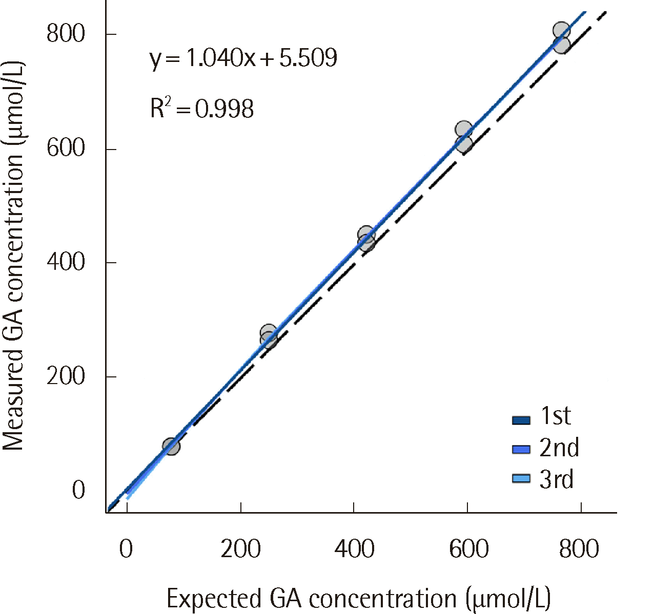 | Fig. 1Linear and polynomial regressions of GA measured with the modified version of the Norudia GA assay.
|
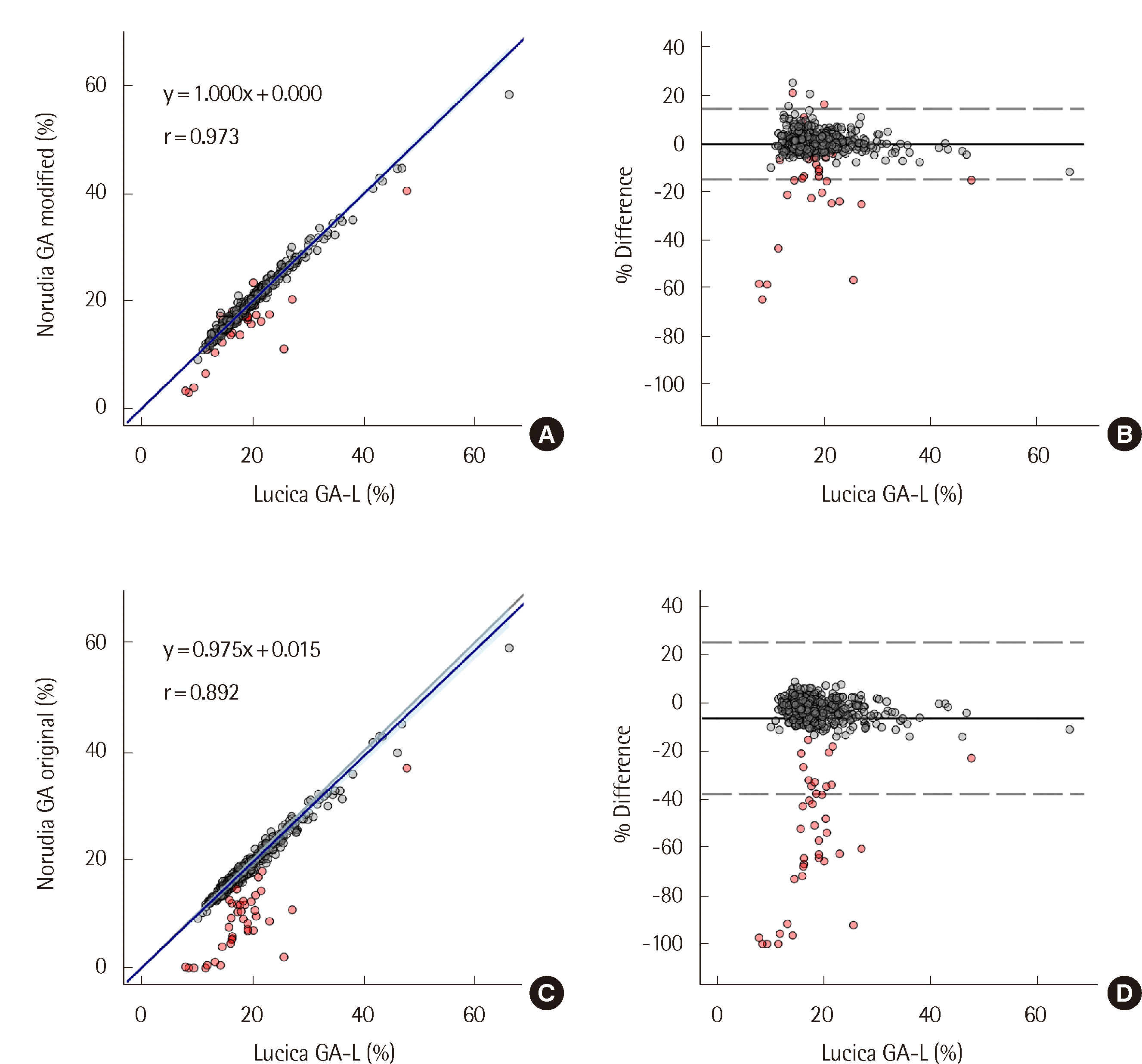 | Fig. 2(A) Scattergram with Passing-Bablok regression and (B) Bland-Altman plot showing the results of comparison between Norudia GA (modified) and Lucica GA-L, and (C) Scattergram with Passing-Bablok regression and (D) Bland-Altman plot showing the results of comparison between Norudia GA (original) and Lucica GA-L. The solid navy blue line indicates the fitted Passing-Bablok regression, and the blue-colored area represents the 95% CI. In the Bland-Altman plots, the solid black line indicates the mean % difference, and the dashed lines represent the 95% CI. The red dots indicate the samples with negative interference demonstrating a % difference of <-14.4% between the original version of the Norudia GA and Lucica GA-L assays.
|
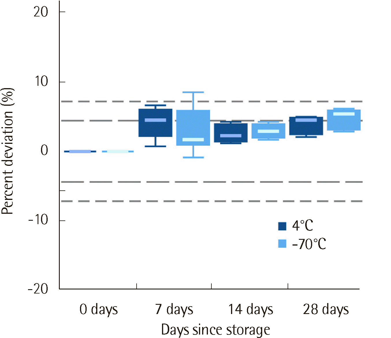 | Fig. 3Temperature stability of glycated albumin (GA) measured with the modified version of the Norudia GA assay after storage at either 4°C or -70°C for 7, 14, and 28 days. The long-dashed grey lines indicate the stability limit (±4.4%), whereas the short-dashed grey lines indicate the total allowable error (±7.2%) as designated by Westgard.
|
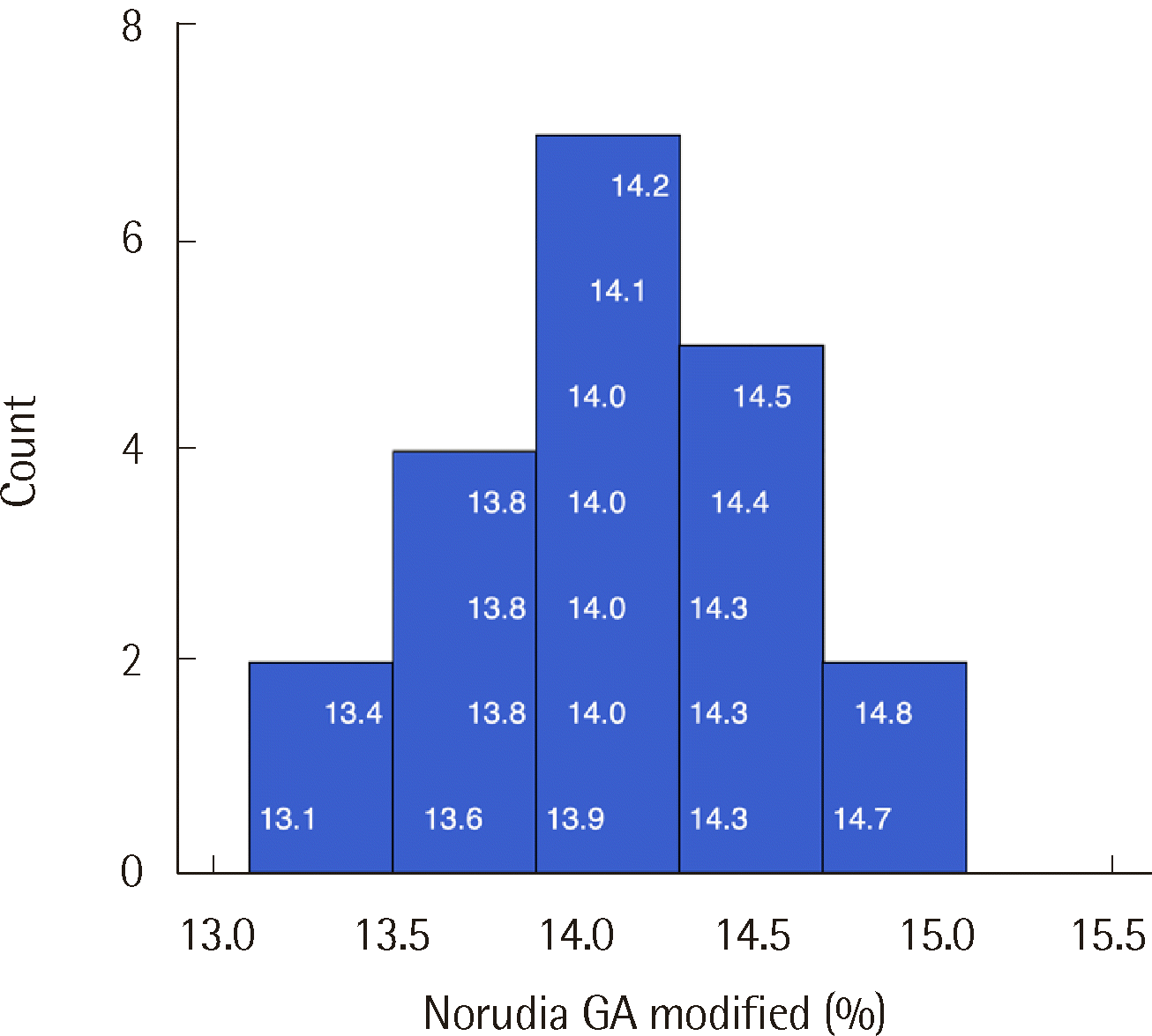 | Fig. 4Histogram of GA results from 20 healthy control samples used for reference interval transference. The values overlaid on the histogram represent the GA results.
|
Table 1
Precision of the modified version of the Norudia GA assay
| Material | Mean | SD | Repeatability | Within-laboratory CV |
|---|---|---|---|---|
| QC 1 | 13.88 | 0.151 | 1.8% | 5.3% |
| QC 2 | 31.81 | 0.199 | 2.2% | 3.7% |
| Pooled serum | 19.79 | 0.116 | 1.5% | 3.1% |
![]()
Table 2
Results of specimens with negative interference in the original version of the Norudia GA assay
![]()
Go to :
DISCUSSION
GA has been widely adopted in clinical practice recently to estimate short-term glycemic control. Previous publications evaluating the original version of the Norudia GA assay demonstrated that the assay is comparable with the Lucica GA-L assay in measuring GA [12, 13]. While our institute had a total of 29,813 GA orders in 2022, only 39 samples obtained from 34 different patients were false negatives as per our definition of negative interference from August 2021 to February 2023. This phenomenon is estimated to occur in approximately 0.08% of the GA orders, thus capturing this during performance evaluation is practically infeasible given the scarcity of this phenomenon.
During an investigation in 2019 when the researchers first encountered an extremely low GA result, monoclonal paraprotein precipitation was hypothesized to interfere with light absorbance. Sparse literature suggests paraprotein precipitation as the cause of interference resulting in negative results of various chemistry assays [14-17]. Lithium-heparin was the proposed cause of the precipitation [15, 17]. In the present study, two patients with monoclonal paraproteinemia showed prominent turbidity in samples stored in a lithium-heparin tube. Furthermore, M-protein and lithium- heparin-induced negative interference occurred in the original version of the Norudia GA assay. However, most cases with falsely low GA results did not have monoclonal paraproteinemia, which implies that not all interferences could be explained by this phenomenon.
Another hypothesis was that anti-oxidative agents such as ascorbic acid could interfere with the Trinder reaction [18, 19]. The Norudia GA assay measures GA based on the absorbance of purple- red pigment produced through oxidization [20], and anti-oxidative agents could interfere with the reaction. Among the thirtyfour patients who provided negative interference samples, seven were taking thioctic acid, which shows anti-oxidative activity. Notably, the number of patients taking anti-oxidative agents could be underestimated since the intake of over-the-counter medications such as vitamin supplements cannot be determined with prescription records.
The manufacturer implemented a new formula for their Norudia GA assay to overcome negative interference. The revised formula increased the amount of substance that eliminates the reduction agent, decreasing negative interference. While details regarding the formula have not been disclosed by the manufacturer, the improvement in results supports the hypothesis that anti-oxidative agents interfere with the reaction. For all negatively affected samples, the modified version of the Norudia GA assay showed results higher than the original version of the Norudia GA assay. However, some samples still demonstrated negative interference in the modified version of the Norudia GA assay, albeit to a lesser degree than the original version. Given that negative interference remains despite the revised formula and that not all patients prescribed with anti-oxidative agents displayed falsely low GA levels, the exact mechanism of the negative interference is yet to be elucidated. Further research should investigate the underlying mechanism causing falsely low results and minimize the remaining negative interference.
In summary, the modified version of the Norudia GA assay demonstrated fair analytical performance. Given that it reduces the negative interference, putatively caused by anti-oxidative agents, observed in the original version of the Norudia GA assay, the modified assay would be beneficial in clinical laboratories experiencing negative interference using the original version of the Norudia GA assay.
Go to :
 XML Download