Journal List > Korean J Orthod > v.54(2) > 1516086807

Yurdakul and Karsli: Evaluating anchorage loss in upper incisors during distalization of maxillary posterior teeth using clear aligners in adult patients: A prospective randomized study

Abstract

Objective

To evaluate the effect of clear aligner treatment and differential sequence distalization of maxillary posterior teeth on anchorage loss in the upper incisors (U1s).

Methods

This study used lateral cephalometries and digital models of 12 patients treated with 33% sequential distalization (group 1, mean age 22.9 ± 0.7 years, five males, seven females) and 12 treated with 50% sequential distalization (group 2, mean age 25.83 ± 0.5 years, three males, nine females) acquired before and after distalization of upper second premolars (U5) and upper first molars (U6) and upper second molars (U7). The amount of distalization was determined as 2.5 mm in both the groups. Independent Samples t test was used to compare normally distributed parameters. Mann–Whitney U and Wilcoxon tests were used to compare parameters that were not normally distributed.

Results

In both groups, the posterior teeth significantly moved by tipping distally and the U1s were displaced anteriorly. Increase in maxillary posterior transverse width (P < 0.001) and distopalatal rotation were observed in U5, U6, and U7 after distalization. It was also observed that U1 was significantly more proclined (1.82°; P < 0.001) and protruded (0.62 mm; P < 0.001), and the overjet (0.45 mm; P < 0.001) increased more in group 1 than in group 2.

Conclusions

After sequential distalization of maxillary posterior teeth, more anchorage loss was observed in the anterior region in group 1 than in group 2.

INTRODUCTION

Maxillary molar distalization is one of the most common method used to correct Class II molar relationship in the treatment of patients with increased overjet without extraction.1,2 Intraoral distalization appliances that minimize patient compliance have become popular in the recent times.1-3 However, these appliances may cause unwanted effects, such as clockwise rotation of the mandible, extrusion of primary premolars, and anchorage loss in the anterior teeth.3-7
In the recent years, more esthetic and comfortable options have become popular instead of traditional treatments. Clear aligner (CA) systems have been introduced for the treatment of mild dental crowding and diastema.8-10 As a result of application of forces that comply with attachments of different sizes and shapes placed on the teeth surface, complex movements have become more foreseeable.11
Previous studies reported that CA could provide the force system required to achieve bodily tooth movement.1,12-14 Samoto and Vlaskalic15 stated that sequential distalization protocol minimizes anchorage loss in anterior teeth and uncontrolled tipping in posterior area by maintaining maximum contact between teeth and aligner, compared to en masse movements. The frequently applied protocols in sequenced distalization treatment using CA are as follows:
  • 33% sequenced distalization: When upper second molars (U7) are moved 33% of the total distance to be distalized, distalization of upper first molars (U6) starts.

  • 50% sequenced distalization: When U7 are moved 50% of the total distance to be distalized, distalization of U6 starts.

To the best of our knowledge, comparison of the clinical effects of distalization performed using the aforementioned two different sequencing protocols has not been conducted. Therefore, this study aimed to compare the amount of anchorage loss occurring in upper incisors (U1s) after the implementation of the two sequenced distalization protocols using CA in patients with Class II malocclusion.
The null hypotheses of our study are:
1) There are no differences between the two groups in terms of anchorage loss in U1s.
2) There are no differences between the two groups in terms of distalization amount and tipping of upper second premolars (U5), U6, and U7.

MATERIALS AND METHODS

Ethical considerations

This prospective study was approved by the Clinical Research Ethics Committee of Karadeniz Technical University Faculty of Medicine (protocol number: 2021/2) and the Turkish Pharmaceuticals and Medical Devices Agency (approval number: 2022-074, E-68869993-511.06-820892) and conducted in accordance with the tenets of Declaration of Helsinki. Informed consent was obtained from all patients.

Sample size calculation

GPower 3.1.0 software package (Universitat Düsseldorf, Düsseldorf, Germany) was used to determine the number of individuals required for this study. Sample size evaluation was based on the standard deviation of a similar study performed by Saif et al.16 The adequate minimum number of patients required was 22, which was calculated by setting the alpha error to 0.05, beta error to 0.20, and effect size to 0.65. Ultimately, 24 patients were included in this study to increase the strength of the study. All patients underwent therapy using the Orthero Clear Aligner (İstanbul, Turkey) system.

Study sample

A total of 24 patients (16 females and 8 males) who underwent orthodontic treatment at Karadeniz Technical University Faculty of Dentistry Hospital between July 2022 and August 2023 formed our study population. The patients were randomly divided into two groups and randomization was performed using www.randomization.com. In total, 12 patients (five males and seven females) were assigned to the 33% sequenced distalization group (group 1), and 12 (three males and nine females) to the 50% sequenced distalization group (group 2). The mean age of study participants belonging to groups 1 and 2 were 22.9 ± 0.7 years and 25.83 ± 0.5 years, respectively.
Inclusion criteria were:
  • Patients with half-cusp Class II molar relationship

  • Those without maxillary transverse discrepancy

  • Those with all permanent teeth intact, except the third molars

  • Patients cooperating enough in use of CA and complying with the treatment

Exclusion criteria were:
  • Existence of skeletal malocclusion

  • Cases requiring unilateral distalization

  • Existence of temporomandibular joint problems

  • Existence of periodontal disease

  • Patients with a systemic disorder

Treatment protocol

The treatment plans for this prospective study were designed by a single orthodontist, who also performed the treatments. The same technician assisted with the planning stage.
Distalization in individuals belonging to group 1 was performed by means of 33% sequential alignment using CAs. Vertical rectangular attachments were added on U5, U6, and U7. The average number of aligners used in group 1 was 16.27 and the mean treatment duration was 8.12 ± 0.75 months. For individuals belonging to group 2, distalization of U5, U6, and U7 was performed in a 50% sequence with vertical rectangular attachments and CAs. The average number of aligners used in group 2 was 22.75 and the mean treatment duration was 10.6 ± 0.78 months.
In particular, sequential distalization of U5, U6, and U7 was planned for all patients, and the total amount of distalization planned for both groups was determined as 2.5 mm. To control the distalization movement, rectangular vertical attachments were placed on U5, U6, and U7, and the movement of only these teeth was planned throughout the distalization process in the upper arch. To strengthen the anchorage, rectangular horizontal attachments were also placed on upper first premolars. No attachments were added to any other tooth, and no movement of any other tooth was planned.
In this study, we evaluated the anchorage loss that may occur in U1 after the distalization of U5, U6, and U7. Therefore, Class II intermaxillary elastics or any additional mechanics were not used during distalization. Similarly, no attachments were placed on the anterior teeth, and interproximal reduction was not included in the treatment plan. All patients were treated with this standard procedure without any other auxiliary mechanics and were required to wear aligners for at least 22 hours per day.
Cephalometric radiographs and digital model recordings of all patients before treatment (T0) and after completion of distalization (T1) were obtained.

Cephalometric analysis

Reference planes used in this study included:
  • Horizontal plane (HR): constructed at a 7° angle to the sella-nasion plane

  • Vertical plane (VR): constructed perpendicular to the horizontal reference plane at the sella point

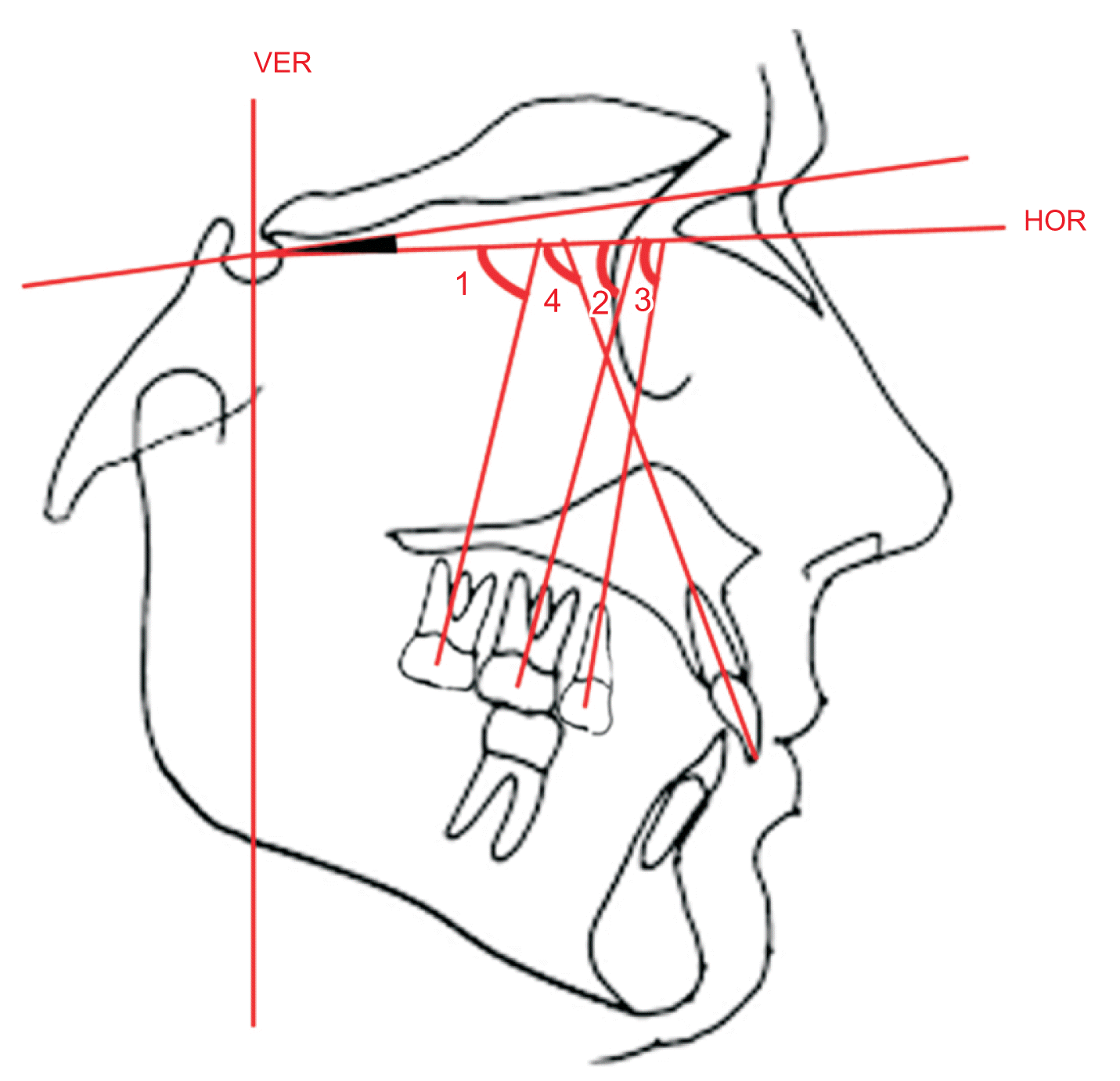
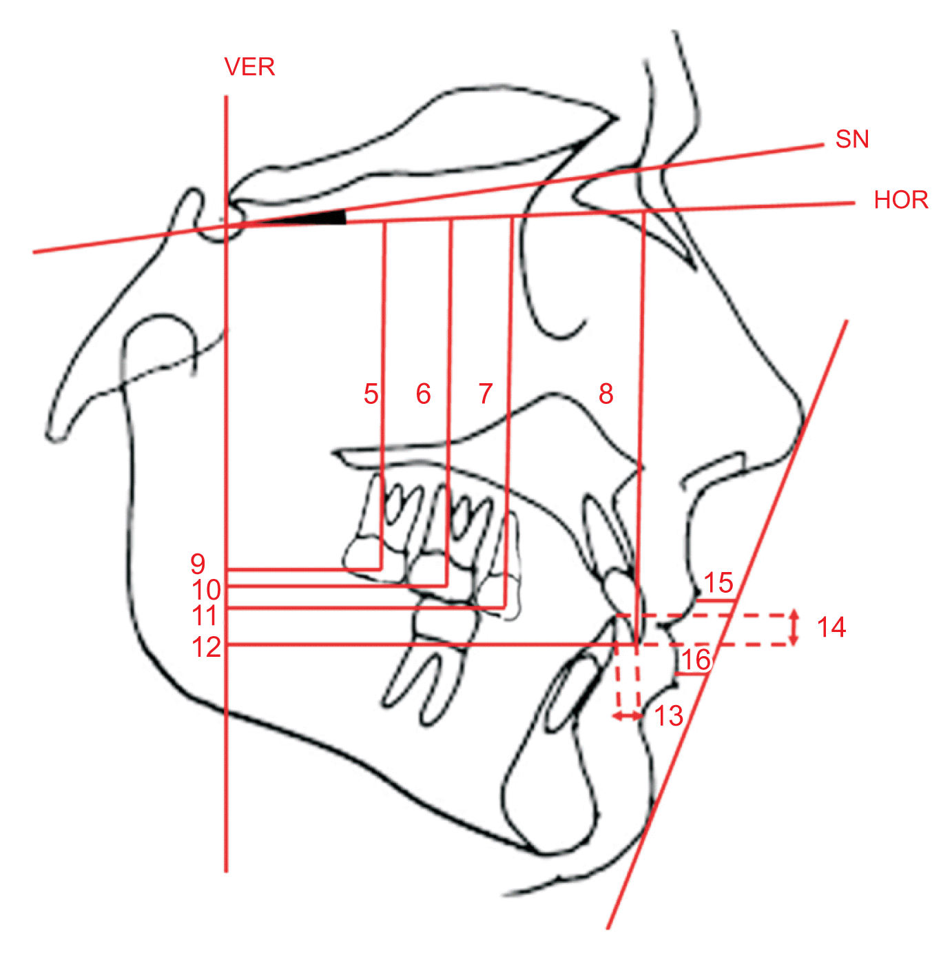
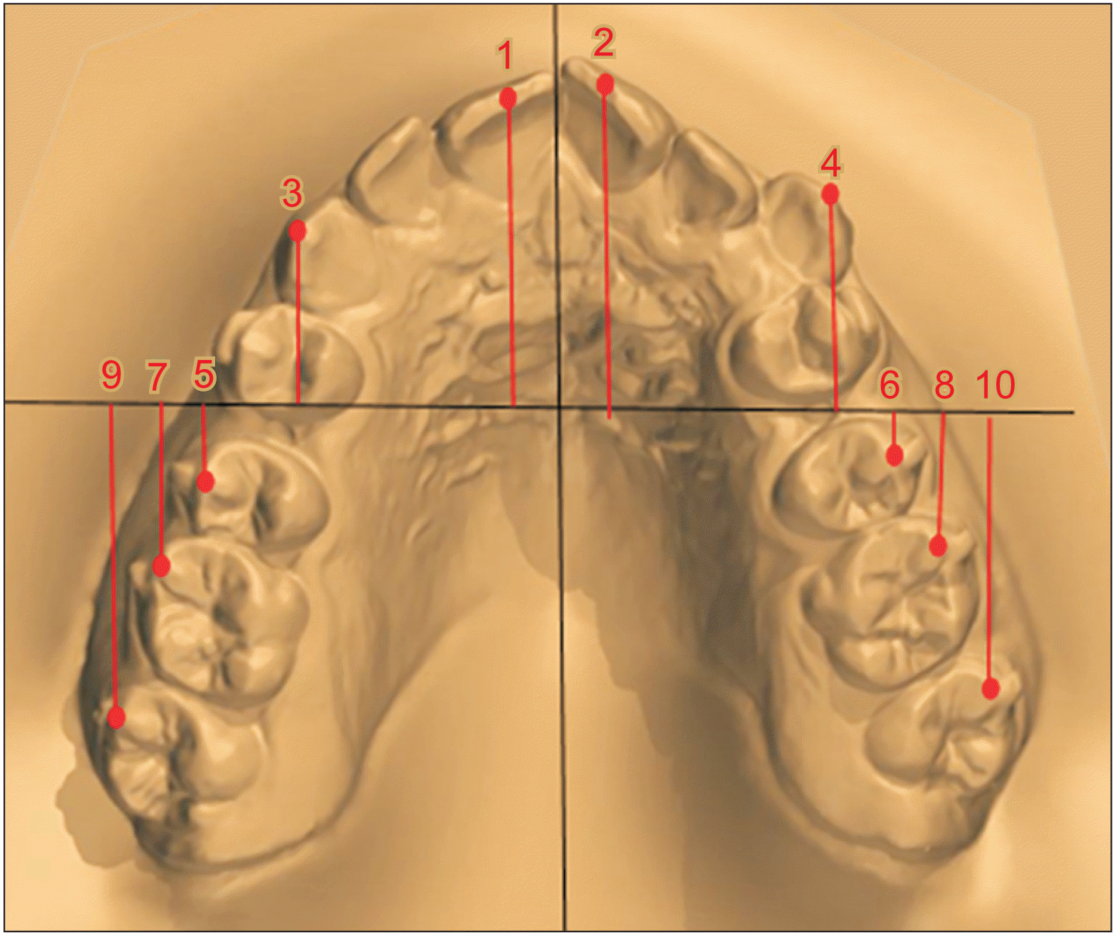
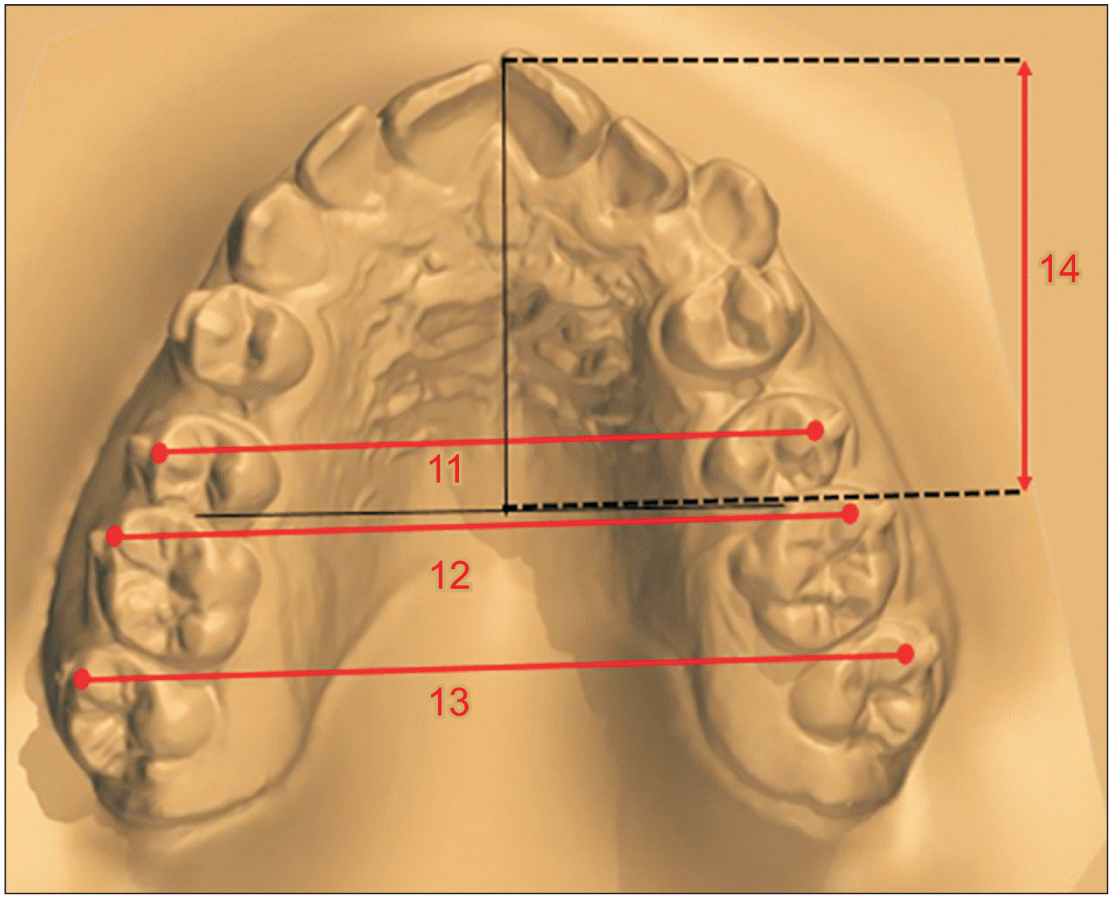
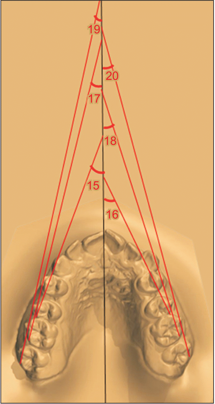
Twelve linear dentoalveolar and four angular measurements were performed on the cephalometric radiographs (Figures 1 and 2). Fourteen linear and six angular measurements were performed using digital models (Figures 35).

Statistical analysis

All the statistical analyses were performed using SPSS software (SPSS v23; IBM Corp., Armonk, NY, USA). Conformity to normal distribution was examined using Shapiro–Wilk test. Independent samples t test was used to compare the parameters normally distributed by groups. Mann–Whitney U test (for intergroup comparisons) and Wilcoxon test (for intragroup comparisons) were used to compare the parameters that were not distributed normally. Intraclass correlation coefficient was used to examine compliance between parameter and repeat measurements. Level of significance was set at P < 0.05.

RESULTS

To test for intraobserver method error, ten lateral cephalometric radiographs and digital models were subjected to the same analyses and measurements by the same researcher after approximately two weeks. All evaluations made by the observer at two-time intervals ranged between 95% confidence interval 0.83–0.99, and the measurements were found to be quite consistent.

Cephalometric measurements

Table 1 shows the comparison between the two groups in terms of cephalometric parameters during pre-treatment (T0) periods, time-dependent change differences (T1-T0), and changes during post- (T1) and pre-observation (T0) periods. No significant differences were observed between the two groups at T0 (P > 0.05). Analysis of changes in cephalometric dentoalveolar measurements from T0 to T1 in group 1 revealed an increase in U1/HR (7.15°; P < 0.001) and a decrease in U5/HR (5.92°; P < 0.01), U6/HR (8.19°; P < 0.001), and U7/HR (11.35°; P < 0.01) angles. In dentoalveolar linear measurements, there were significant decreases in U5-VR (1.91 mm; P < 0.001), U6-VR (2.30 mm; P < 0.001), U7-VR (2.41 mm; P < 0.001), and U1-HR (0.19 mm; P < 0.05) along with a significant increase in U1 VR (1.56 mm; P < 0.001). There was also an increase in overjet (1.30 mm; P < 0.001) and a decrease in overbite (0.15 mm; P < 0.05) after distalization. Additionally, it was determined that the upper lip was protruded (0.45 mm; P < 0.01) (Table 1).
Analysis of the changes in cephalometric dentoal veolar angular measurements from T0 to T1 in group 2 revealed an increase in U1/HR (5.33°; P < 0.001) and a decrease in U5/HR (5.96°; P < 0.001), U6/HR (8.45°; P < 0.001), and U7/HR (10.96°; P < 0.001) angles. Linear measurements showed significant decreases in U5-VR (1.86 mm; P < 0.001), U6-VR (2.32 mm; P < 0.001), U7-VR (2.42 mm; P < 0.001), and U7-HR (0.15 mm; P < 0.05), along with a significant increase in U1-VR (0.94 mm; P < 0.001). There was also an increase in the overjet value (0.85 mm; P < 0.001). Meanwhile, it was determined that the upper lip was protruded (0.29 mm; P < 0.01) (Table 1).
When the differences in time-dependent changes in cephalometric measurements between the two groups were examined, it was found that there were greater in creases in U1/HR (1.82°; P < 0.001), U1-VR (0.62 mm; P < 0.001), and overjet (0.45 mm; P < 0.001) values in group 1 (Table 1).

Digital model measurements

Table 2 shows the comparison of digital model parameters between the two groups in terms of pre-treatment (T0) periods, time-dependent change differences (T1-T0), and changes during post- (T1) and pre-observation (T0) periods. No significant differences were observed between the two groups at T0 (P > 0.05). In the context of sagittal model parameters for groups 1 and 2, significant increases were observed in all the sagittal and transverse (P < 0.001) measurements from T0 to T1. In contrast, for angular model measurements, significant decreases were found from T0 to T1 in both the groups (Table 2).
When the differences in time-dependent changes in cephalometric measurements between the groups were examined, although greater increases were found for group 1 in the distance from incisor tip of 11 to G line (0.16 mm) and the distance from incisor tip of 21 to G line (0.01 mm), the difference was not statistically significant (P > 0.05). In addition, there were no significant differences between the two groups over time in terms of all the model measurements (P > 0.05) (Table 2).

DISCUSSION

This was a prospective randomized clinical trial, and this study design has been deemed to provide the highest level of scientific evidence.17 Other strengths of this study include: (1) use of power analysis for sample size calculation; (2) application of inclusion and exclusion criteria for selection of the study participants; (3) treatment of all patients by the same clinician; (4) working with the same technician throughout the study; (5) having the same operator perform all the lateral cephalometric radiography using the same equipment and under the same conditions; (6) having the same researcher measure all the lateral cephalometric radiographs and digital models; and (7) estimation of intraexaminer error of the method.
In the present study, the effects of maxillary molar distalization treatment using CAs on skeletal, dentoalveolar, and soft tissue were evaluated. The results showed that the distalization movement of 2–3 mm using CAs was effective; however, the amount of anterior anchorage loss differed according to the sequence protocol. Thus, the study hypotheses were rejected.

Molar distalization and distal tipping

Ravera et al.4 reported that the maxillary first and second molars moved 2.25 mm and 2.52 mm distally, respectively, without any significant distal tipping. They showed that the presence of rectangular vertical attachments on the buccal surface of the teeth were responsible for small amount of tipping. Similarly, Caruso et al.14 demonstrated that the maxillary molars moved distally with bodily movement of the CAs. These findings are consistent with those of Simon et al.,18 who showed distal displacement of the molars by 2–3 mm. However, studies on traditional distalization applications indicate that molar teeth move significantly by tipping distally.19-28 In our study, distal tipping was also observed during distalization of U5, U6, and U7 in both the groups, and these findings are consistent with those of previous studies that investigated distalization using CAs.4,12-18 At the same time, we did not observe any significant difference between the two groups in terms of amount of distalization. Additionally, in this study, 2.5 mm distalization was planned for each tooth in all patients, and it was determined that comparatively less distal movement occurred in the posterior teeth than originally planned in both the groups. In both groups, the amount of distal movement occurred the most in U7 and the least in U5. This can be explained by two factors. First, the mechanical forces produced by CAs are typically distributed between the teeth and along the periodontal ligament. The periodontal ligament area of maxillary first molars is generally larger than that of maxillary second molars, and the need for anchorage during movement is greater. Second, aligners are made of plastic, and as the number of teeth to be moved distally increases, the deformation of aligners increases, which may cause subsequent movements to be less than predicted.4
Moreover, in our study, cephalometric measurements in the sagittal plane and digital model measurements were consistent. Moreover, the amount of distalization measured in the digital model and cephalometric radiography was compatible in both the study groups. These findings are consistent with those of previous studies.22,24-27

Anterior loss of anchorage

In treatments involving CAs, anchorage loss may occur in anterior region due to reciprocal force that arise in response to distalization force.4 Caruso et al.14 investigated the effect of distalization of molars using CAs and found no anchorage loss during treatment and that root torque control of U1s were perfectly ensured. Saif et al.16 assessed digital models and reported different amounts of anchorage loss in almost every patient. Patterson et al.29 stated that Class II elastics should be used to avoid overjet increase during upper molar distalization. Other researchers have also confirmed that Class II elastics prevent uncontrolled proclination of anterior teeth.4,15
The findings of this study showed that 33% sequence distalization caused more anchorage loss in anterior teeth than 50% sequence distalization. This means that more proclination and protrusion of U1s were observed in the 33% sequence group. This finding can be explained as follows. While a maximum of two teeth moved simultaneously when 50% sequence was applied to the posterior teeth, three or more teeth participated in simultaneous movement when 33% sequence was applied. This complicates anchorage control and causes more opposing inverse force in the anterior teeth, which serves as the anchorage unit, while the posterior teeth move. In this study, the faster 33% sequence distalization protocol was found to be preferable because Class II elastics were not used to evaluate the absolute response of anterior tooth movements. Moreover, it is likely that these posterior anchorage losses were also seen with increased protrusion and proclination in the anterior region, since the molar teeth (which were initially distalized in the posterior segment) lost anchorage during the movement of U5 and also slightly mesialized and preserved the length of the CAs.
In summary, in cases requiring upper molar distalization, Class II intermaxillary elastics can be used in early stages to prevent loss of anchorage. If distalization with CAs is to be performed in patients with Class II malocclusion and greater overjet, the 50% sequence protocol would be preferable.

Vertical changes

It has been reported that mandible rotates posteriorly with downward movement of the mesial cusp and that the vertical dimensions and angles increase when the molars are tipped distally by traditional intraoral distalization appliances.26-28 Studies conducted using CAs have reported no significant change in vertical direction values after distalization.4,14,30 Thickness of the CAs in the occlusal plane and the ‘bite block effect’ that occurs with it are responsible for the absence of change in the vertical dimension. Traditional applications have been reported to decrease the overbite with intrusion of U1s after distalization. It has been found that the resulting intrusion develops due to the proclination of U1s.4,14,16,18
In the current study, in accordance with the literature, no significant change was observed in the angle between SN and Steiner's mandibular plane in either group, and no significant vertical movement occurred after distalization in U5, U6, or U7. In addition, it was confirmed that the right-direction angle of the aligners did not change due to the bite-block effect. Therefore, we found that CAs could provide an effective alternative for average distalization of posterior teeth by 2–3 mm, especially in patients with hyperdivergent growth pattern or open bite. In addition, in the present study, a small amount of U1 intrusion was observed in group 1, but this value was not statistically significant in group 2. This intrusion in group 1 was thought to be related to the amount of tooth proclination.

Transverse changes

In a normal arch form, the width of the posterior region in the transverse direction gradually increases distally. However, if the normal arch form is not followed, a posterior crossbite may occur due to distal movement of posterior teeth. It can be argued that an increase in the transverse widths between the teeth during distalization of upper posterior teeth using CAs is advantageous in terms of harmony of the arches.31
In this study, we observed that the width between U5, U6, and U7 increased in the transverse direction in both the groups. Therefore, distalization using CAs may be preferred, especially in Class II cases with a tendency towards posterior crossbite.

Rotation changes of the molars

In this study, we found that U5, U6, and U7 underwent distopalatal rotation in both the groups. The post-distalization rotation amounts were similar in both the groups, with least and maximum amount of rotation occurring in U5 and U7, respectively.
Previous studies using traditional intraoral distalization headgears have reported that rotations in molars occur in accordance with the size and direction of application of the distalization force.27,28,32,33 Gulati et al.32 reported 2.4° distopalatal rotation in molar teeth after performing distalization using the Jones Jig appliance. On the other hand, Wilmes and Drescher33 elicited 3.4° distobuccal rotation with the Beneslider Appliance. Henry34 in the year 1956 stated that the permanent U6 in malocclusions are rotated in 83% of the cases and that the axis of rotation passes through the palatal root and mesiopalatal cusp. In CA systems, it is important to apply distopalatal rotation to the posterior teeth during digital planning.35 Therefore, distopalatal rotation occurred in teeth that moved distally, and this was in accordance with the virtual planning in this study.

Soft tissue changes

Previous studies using intraoral molar distalization appliances have reported a significant level of protrusion in the upper lip after distalization, which may be due to increased U1 proclination resulting from the loss of anchorage in the anterior region.28,36 In our study, significant amount of protrusion was observed in the upper lip in both the groups, which was associated with the movement of U1s toward the lips.

Limitations

As in all dynamic appliance studies, an obvious limitation of this study was that it required excellent patient cooperation. In terms of controlling aligner–patient compliance, it would be beneficial to place indicators on aligners in the future. In addition, long-term results should be evaluated in subsequent studies. Although our results are encouraging, this issue needs to be further investigated in future randomized clinical trials.

CONCLUSIONS

  • During sequential distalization of posterior teeth using aligners, distal tipping and movement were also observed.

  • The 33% sequenced distalization protocol caused significantly more anchorage loss in the anterior region than the 50% sequenced protocol.

  • Clinicians should be aware of the counteracting effects of maxillary molar distalization in the anterior region.

ACKNOWLEDGEMENTS

The authors thank Prof. Dr. Hakan Gögen for his knowledge and experience.

Notes

AUTHOR CONTRIBUTIONS

Conceptualization: NK. Data curation: ZY. Investigation: NK. Methodology: NK. Resources: NK. Supervision: NK. Validation: NK. Visualization: ZY. Writing–original draft: all authors. Writing–review & editing: all authors.

CONFLICTS OF INTEREST

No potential conflict of interest relevant to this article was reported.

REFERENCES

1. Lione R, Franchi L, Laganà G, Cozza P. 2015; Effects of cervical headgear and pendulum appliance on vertical dimension in growing subjects: a retrospective controlled clinical trial. Eur J Orthod. 37:338–44. https://doi.org/10.1093/ejo/cju061. DOI: 10.1093/ejo/cju061. PMID: 25316493.
crossref
2. Park CO, Sa'aed NL, Bayome M, Park JH, Kook YA, Park YS, et al. 2017; Comparison of treatment effects between the modified C-palatal plate and cervical pull headgear for total arch distalization in adults. Korean J Orthod. 47:375–83. https://doi.org/10.4041/kjod.2017.47.6.375. DOI: 10.4041/kjod.2017.47.6.375. PMID: 29090125. PMCID: PMC5653686.
crossref
3. Clemmer EJ, Hayes EW. 1979; Patient cooperation in wearing orthodontic headgear. Am J Orthod. 75:517–24. https://doi.org/10.1016/0002-9416(79)90070-8. DOI: 10.1016/0002-9416(79)90070-8. PMID: 286553.
crossref
4. Ravera S, Castroflorio T, Garino F, Daher S, Cugliari G, Deregibus A. 2016; Maxillary molar distalization with aligners in adult patients: a multicenter retrospective study. Prog Orthod. 17:12. https://doi.org/10.1186/s40510-016-0126-0. DOI: 10.1186/s40510-016-0126-0. PMID: 27041551. PMCID: PMC4834290. PMID: 970deba192ee417f921a2b9ccfcdac13.
crossref
5. Bolla E, Muratore F, Carano A, Bowman SJ. 2002; Evaluation of maxillary molar distalization with the distal jet: a comparison with other contemporary methods. Angle Orthod. 72:481–94. https://pubmed.ncbi.nlm.nih.gov/12401059/.
6. Fontana M, Cozzani M, Caprioglio A. 2012; Non-compliance maxillary molar distalizing appliances: an overview of the last decade. Prog Orthod. 13:173–84. https://doi.org/10.1016/j.pio.2011.10.002. DOI: 10.1016/j.pio.2011.10.002. PMID: 23021121.
crossref
7. Yu IJ, Kook YA, Sung SJ, Lee KJ, Chun YS, Mo SS. 2014; Comparison of tooth displacement between buccal mini-implants and palatal plate anchorage for molar distalization: a finite element study. Eur J Orthod. 36:394–402. https://doi.org/10.1093/ejo/cjr130. DOI: 10.1093/ejo/cjr130. PMID: 22051536.
crossref
8. Fuziy A, Rodrigues de Almeida R, Janson G, Angelieri F, Pinzan A. 2006; Sagittal, vertical, and transverse changes consequent to maxillary molar distalization with the pendulum appliance. Am J Orthod Dentofacial Orthop. 130:502–10. https://doi.org/10.1016/j.ajodo.2004.12.031. DOI: 10.1016/j.ajodo.2004.12.031. PMID: 17045150.
crossref
9. Fontana M, Cozzani M, Caprioglio A. 2012; Soft tissue, skeletal and dentoalveolar changes following conventional anchorage molar distalization therapy in class II non-growing subjects: a multicentric retrospective study. Prog Orthod. 13:30–41. https://doi.org/10.1016/j.pio.2011.07.002. DOI: 10.1016/j.pio.2011.07.002. PMID: 22583585.
crossref
10. Mariani L, Maino G, Caprioglio A. 2014; Skeletal versus conventional intraoral anchorage for the treatment of class II malocclusion: dentoalveolar and skeletal effects. Prog Orthod. 15:43. https://doi.org/10.1186/s40510-014-0043-z. DOI: 10.1186/s40510-014-0043-z. PMID: 25138818. PMCID: PMC4138549.
crossref
11. Melsen B. 2011; Northcroft lecture: how has the spectrum of orthodontics changed over the past decades? J Orthod. 38:134–43. quiz 145https://doi.org/10.1179/14653121141362. DOI: 10.1179/14653121141362. PMID: 21677105.
crossref
12. Schupp W, Haubrich J, Neumann I. 2010; Treatment of anterior open bite with the Invisalign system. J Clin Orthod. 44:501–7. https://pubmed.ncbi.nlm.nih.gov/21105589/.
13. Joffe L. 2003; Invisalign: early experiences. J Orthod. 30:348–52. https://doi.org/10.1093/ortho/30.4.348. DOI: 10.1093/ortho/30.4.348. PMID: 14634176.
crossref
14. Caruso S, Nota A, Ehsani S, Maddalone E, Ojima K, Tecco S. 2019; Impact of molar teeth distalization with clear aligners on occlusal vertical dimension: a retrospective study. BMC Oral Health. 19:182. https://doi.org/10.1186/s12903-019-0880-8. DOI: 10.1186/s12903-019-0880-8. PMID: 31409348. PMCID: PMC6692944. PMID: a52f54f493354496bfddc508889c341b.
crossref
15. Samoto H, Vlaskalic V. 2014; A customized staging procedure to improve the predictability of space closure with sequential aligners. J Clin Orthod. 48:359–67. https://pubmed.ncbi.nlm.nih.gov/25083756/.
16. Saif BS, Pan F, Mou Q, Han M, Bu W, Zhao J, et al. 2022; Efficiency evaluation of maxillary molar distalization using Invisalign based on palatal rugae registration. Am J Orthod Dentofacial Orthop. 161:e372–9. https://doi.org/10.1016/j.ajodo.2021.11.012. DOI: 10.1016/j.ajodo.2021.11.012. PMID: 34974928.
crossref
17. Papadopoulos MA. 2003; Meta-analysis in evidence-based orthodontics. Orthod Craniofac Res. 6:112–26. https://pubmed.ncbi.nlm.nih.gov/12809274/. DOI: 10.1034/j.1600-0854.2003.3r275.x. PMID: 12809274.
crossref
18. Simon M, Keilig L, Schwarze J, Jung BA, Bourauel C. 2014; Treatment outcome and efficacy of an aligner technique--regarding incisor torque, premolar derotation and molar distalization. BMC Oral Health. 14:68. https://doi.org/10.1186/1472-6831-14-68. DOI: 10.1186/1472-6831-14-68. PMID: 24923279. PMCID: PMC4068978.
crossref
19. Angelieri F, de Almeida RR, Janson G, Castanha Henriques JF, Pinzan A. 2008; Comparison of the effects produced by headgear and pendulum appliances followed by fixed orthodontic treatment. Eur J Orthod. 30:572–9. https://doi.org/10.1093/ejo/cjn060. DOI: 10.1093/ejo/cjn060. PMID: 19054813.
crossref
20. Kirjavainen M, Kirjavainen T, Hurmerinta K, Haavikko K. 2000; Orthopedic cervical headgear with an expanded inner bow in class II correction. Angle Orthod. 70:317–25. https://pubmed.ncbi.nlm.nih.gov/10961782/.
21. Mossaz CF, Byloff FK, Kiliaridis S. 2007; Cervical headgear vs pendulum appliance for the treatment of moderate skeletal class II malocclusion. Am J Orthod Dentofacial Orthop. 132:616–23. https://doi.org/10.1016/j.ajodo.2005.11.043. DOI: 10.1016/j.ajodo.2005.11.043. PMID: 18005835.
crossref
22. Caprioglio A, Fontana M, Longoni E, Cozzani M. 2013; Long-term evaluation of the molar movements following Pendulum and fixed appliances. Angle Orthod. 83:447–54. https://doi.org/10.2319/050812-378.1. DOI: 10.2319/050812-378.1. PMID: 23075060. PMCID: PMC8763082.
crossref
23. Al-Thomali Y, Basha S, Mohamed RN. 2017; Pendulum and modified pendulum appliances for maxillary molar distalization in class II malocclusion - a systematic review. Acta Odontol Scand. 75:394–401. https://doi.org/10.1080/00016357.2017.1324636. DOI: 10.1080/00016357.2017.1324636. PMID: 28502196.
crossref
24. Schütze SF, Gedrange T, Zellmann MR, Harzer W. 2007; Effects of unilateral molar distalization with a modified pendulum appliance. Am J Orthod Dentofacial Orthop. 131:600–8. https://doi.org/10.1016/j.ajodo.2005.09.031. DOI: 10.1016/j.ajodo.2005.09.031. PMID: 17482078.
crossref
25. Acar AG, Gürsoy S, Dinçer M. 2010; Molar distalization with a pendulum appliance K-loop combination. Eur J Orthod. 32:459–65. https://doi.org/10.1093/ejo/cjp136. DOI: 10.1093/ejo/cjp136. PMID: 20231213.
crossref
26. Chiu PP, McNamara JA Jr, Franchi L. 2005; A comparison of two intraoral molar distalization appliances: distal jet versus pendulum. Am J Orthod Dentofacial Orthop. 128:353–65. https://doi.org/10.1016/j.ajodo.2004.04.031. DOI: 10.1016/j.ajodo.2004.04.031. PMID: 16168332.
crossref
27. Ngantung V, Nanda RS, Bowman SJ. 2001; Posttreatment evaluation of the distal jet appliance. Am J Orthod Dentofacial Orthop. 120:178–85. https://doi.org/10.1067/mod.2001.114645. DOI: 10.1067/mod.2001.114645. PMID: 11500660.
crossref
28. Brickman CD, Sinha PK, Nanda RS. 2000; Evaluation of the Jones jig appliance for distal molar movement. Am J Orthod Dentofacial Orthop. 118:526–34. https://doi.org/10.1067/mod.2000.110332. DOI: 10.1067/mod.2000.110332. PMID: 11094366.
crossref
29. Patterson BD, Foley PF, Ueno H, Mason SA, Schneider PP, Kim KB. 2021; Class II malocclusion correction with Invisalign: is it possible? Am J Orthod Dentofacial Orthop. 159:e41–8. https://doi.org/10.1016/j.ajodo.2020.08.016. DOI: 10.1016/j.ajodo.2020.08.016. PMID: 33223374.
crossref
30. Cui JY, Ting L, Cao YX, Sun DX, Bing L, Wu XP. 2022; Morphology changes of maxillary molar distalization by clear aligner therapy. Int J Morphol. 40:920–6. https://doi.org/10.4067/S0717-95022022000400920. DOI: 10.4067/S0717-95022022000400920.
crossref
31. Hu YR, Song BL, Li B, Shi RY, Liu SY, Gu ZX. 2023; Three-dimensional analysis of maxillary dentition during molar distalization with clear aligners under different movement designs: an in vitro experiment. Zhonghua Kou Qiang Yi Xue Za Zhi. 58:265–70. Chinese. https://doi.org/10.3760/cma.j.cn112144-20220731-00431.
32. Gulati S, Kharbanda OP, Parkash H. 1998; Dental and skeletal changes after intraoral molar distalization with sectional jig assembly. Am J Orthod Dentofacial Orthop. 114:319–27. https://doi.org/10.1016/s0889-5406(98)70215-x. DOI: 10.1016/S0889-5406(98)70215-X. PMID: 9743138.
crossref
33. Wilmes B, Drescher D. 2010; Application and effectiveness of the Beneslider: a device to move molars distally. World J Orthod. 11:331–40. https://pubmed.ncbi.nlm.nih.gov/21490998/.
34. Henry RG. 1956; Relationship of the maxillary first permanent molar in normal occlusion and malocclusion: an intraoral study. Am J Orthod. 42:288–306. https://doi.org/10.1016/0002-9416(56)90128-2. DOI: 10.1016/0002-9416(56)90128-2.
crossref
35. Lione R, Paoloni V, De Razza FC, Pavoni C, Cozza P. 2022; The efficacy and predictability of maxillary first molar derotation with Invisalign: a prospective clinical study in growing subjects. Appl Sci. 12:2670. https://doi.org/10.3390/app12052670. DOI: 10.3390/app12052670. PMID: ff1dd15a801a40029372e2330d21c639.
crossref
36. Ghosh J, Nanda RS. 1996; Evaluation of an intraoral maxillary molar distalization technique. Am J Orthod Dentofacial Orthop. 110:639–46. https://doi.org/10.1016/s0889-5406(96)80041-2. DOI: 10.1016/S0889-5406(96)80041-2. PMID: 8972811.
crossref

Figure 1
Dental angular measurements on cephalometric radiography. (1) U7/HR, (2) U6/HR, (3) U5/HR, (4) U1/HR. Determination of the long-axis of the teeth: crown tip of U1 and root apex point were marked, crown centroid point of U5 and root apex point were marked, crown centroid point of U6 and U7 and trifurcation point were marked. Centroid point of crown: the midpoint of the mesial and distal convexity of the crown of the molar tooth was accepted as the centroid point of crown.
U7, upper second molars; U6, upper first molars; U5, upper second premolars; U1, upper incisor; HR, horizontal plane; VER, vertical plane; HOR, horizontal plane.
kjod-54-2-117-f1.tif
Figure 2
Dental linear measurements on cephalometric radiography. (5) The distance from crown centroid point of U7 to the horizontal plane (U7-HR), (6) the distance from crown centroid point of U6-HR, (7) the distance from crown centroid point of U5-HR, (8) the distance from crown tip point of U1-HR, (9) the distance from crown centroid point of U7 to the vertical plane (U7-VR), (10) the distance from crown centroid point of U6-VR, (11) the distance from crown centroid point of U5-VR, (12) the distance from crown tip point of U1-VR, (13) overjet, (14) overbite, (15) Ls-E, (16) Li-E.
U7, upper second molars; U6, upper first molars; U5, upper second premolars; U1, upper incisor; Ls, labrale superius; Li, labrale inferius; E, the line between the soft tissue pogonion and tip of the nose; VER, vertical plane; SN, sella-nasion plane; HOR, horizontal plane.
kjod-54-2-117-f2.tif
Figure 3
Sagittal linear measurements on the digital model. Line G: line tangent to inferior border of palatal rugae and perpendicular to midpalatal raphe. (1) The distance from incisor tip of 11 to G line, (2) the distance from incisor tip of 21 to G line, (3) the distance from incisor tip of 13 to G line, (4) the distance from crown tip of 23 to G line, (5) the distance from buccal cusp tip of 15 to G line, (6) the distance from buccal cusp tip of 25 to G line, (7) the distance from mesiobuccal cusp tip of 16 to G line, (8) the distance from mesiobuccal cusp tip of 26 to G line, (9) the distance from mesiobuccal cusp tip of 17 to G line, (10) the distance from mesiobuccal cusp tip of 27 to G line.
kjod-54-2-117-f3.tif
Figure 4
Transverse measurements and arc length (mm) on the digital model. (11) Transverse width between 15–25, (12) transverse width between 16–26, (13) transverse width between 17–27, (14) arch length: the distance between the contact point of the upper incisors and the mesial contact points of the upper first molars.
kjod-54-2-117-f4.tif
Figure 5
Rotation measurements on the digital model. Upper right second premolar: the angle between the mid-palatal raphe and the line passing through the mesial and distal convex ridges of right maxillary second premolar. Upper left second premolar: the angle between the mid-palatal raphe and the line passing through the mesial and distal convex ridges of left maxillary second premolar. Upper right first molar: the angle between the mid-palatal raphe and the line passing through the mesiobuccal and distobuccal cusps of the right maxillary first molar. Upper left first molar: the angle between the mid-palatal raphe and the line passing through the mesiobuccal and distobuccal cusps of the left maxillary first molar. Rotation measurement of upper right second molar: the angle between the mid-palatal raphe and the line passing through the mesiobuccal and distobuccal cusps of the right maxillary second molar. Upper left second molar: the angle between the mid-palatal raphe and the line passing through the mesiobuccal and distobuccal cusps of the left maxillary second molar.
kjod-54-2-117-f5.tif
Table 1
Comparison between the two groups in terms of cephalometric parameters during pre-treatment periods, time-dependent change differences, and changes during post- and pre-observation periods
Group 1 Group 2 P value
(Group 1- Group 2)
Group 1 Group 2 Group 1 Group 2 Group 1 Group 2 P value
(Group 1- Group 2)
T0 T0 T0 T1 T1 T1-T0 T1-T0 Differences T1-T0 Differences T1-T0 Differences T1-T0
Skeletal measurements (°)
SNA 81.06 ± 3.60 78.50 ± 2.43 0.056a 81.29 ± 3.53 78.70 ± 2.47 0.104c 0.102c 0.23 ± 0.15 0.20 ± 0.10 0.573a
SNB 75.38 ± 2.96 74.72 ± 3.23 0.651a 75.51 ± 2.96 74.89 ± 3.24 0.232c 0.221c 0.13 ± 0.04 0.17 ± 0.04 0.113a
ANB 5.67 ± 2.36 3.79 ± 2.50 0.084a 5.78 ± 2.27 3.81 ± 2.51 0.423c 0.475c 0.10 ± 0.16 0.03 ± 0.11 0.164a
GoGn/SN 36.42 ± 6.65 36.58 ± 6.79 0.952a 36.25 ± 6.68 36.42 ± 6.80 0.438c 0.166c –0.17 ± 0.72 –0.17 ± 0.39 0.889b
Dental angular measurements (°)
U1/HR 100.10 ± 11.13 103.55 ± 9.68 0.426a 107.25 ± 11.31 108.89 ± 9.66 < 0.001c < 0.001c 7.15 ± 0.79 5.33 ± 0.57 < 0.001a
U5/HR 83.64 ± 3.41 84.64 ± 7.97 0.695a 77.71 ± 3.25 78.67 ± 7.79 0.002d < 0.001c –5.92 ± 0.72 –5.96 ± 0.53 0.873a
U6/HR 78.50 ± 4.36 82.46 ± 9.52 0.208a 70.30 ± 3.96 74.01 ± 9.04 < 0.001c < 0.001c –8.19 ± 0.75 –8.45 ± 1.06 0.502a
U7/HR 74.76 ± 6.72 80.31 ± 14.58 0.564b 63.41 ± 6.48 69.35 ± 14.83 0.002d < 0.001c –11.35 ± 0.84 –10.96 ± 0.82 0.269a
Dental linear measurements (mm)
U1-VR 62.16 ± 3.72 62.88 ± 5.59 0.716a 63.73 ± 3.65 63.78 ± 3.65 < 0.001c < 0.001c 1.56 ± 0.25 0.94 ± 0.35 0.001b
U5-VR 40.34 ± 3.47 41.14 ± 5.01 0.653a 38.43 ± 3.41 39.28 ± 4.99 < 0.001c < 0.001c –1.91 ± 0.22 –1.86 ± 0.16 0.419b
U6-VR 32.91 ± 3.50 33.08 ± 4.92 0.923a 30.60 ± 3.50 30.75 ± 4.93 < 0.001c < 0.001c –2.30 ± 0.09 –2.32 ± 0.07 0.506a
U7-VR 24.56 ± 3.40 24.46 ± 3.89 0.946a 22.15 ± 2.86 22.04 ± 4.04 < 0.001c < 0.001c –2.41 ± 1.74 –2.42 ± 0.41 0.817b
U1-HR 70.64 ± 5.07 71.79 ± 3.08 0.508a 70.45 ± 5.15 72.43 ± 3.77 0.013c 0.247c –0.19 ± 0.22 0.64 ± 1.81 0.109b
U5-HR 62.68 ± 5.98 62.94 ± 3.55 0.899a 62.73 ± 5.98 62.91 ± 3.52 0.494c 0.703c 0.05 ± 0.24 –0.03 ± 0.25 0.446a
U6-HR 59.88 ± 5.93 60.44 ± 3.56 0.781a 59.88 ± 5.81 60.39 ± 3.55 0.950c 0.407c 0.01 ± 0.28 –0.06 ± 0.22 0.565a
U7-HR 56.69 ± 5.36 57.81 ± 3.95 0.569a 56.69 ± 5.36 57.66 ± 4.00 0.998c 0.025c 0.00 ± 0.31 –0.15 ± 0.20 0.204b
Overjet 4.57 ± 1.77 4.09 ± 1.10 0.603b 5.87 ± 1.79 4.94 ± 1.05 < 0.001c < 0.001c 1.30 ± 0.10 0.85 ± 0.13 < 0.001a
Overbite 2.81 ± 2.48 2.30 ± 2.77 0.314a 2.66 ± 2.49 2.20 ± 2.56 0.015c 0.107c –0.15 ± 0.15 –0.10 ± 0.31 0.976b
Soft tissue measurements (mm)
Ls-E 4.51 ± 2.97 4.31 ± 1.75 0.844a 4.06 ± 3.00 4.02 ± 1.72 0.002 0.001 –0.45 ± 0.39 –0.29 ± 0.22 0.226a
Li-E 3.53 ± 3.02 1.69 ± 0.92 0.143a 3.52 ± 3.02 1.51 ± 1.06 0.498 0.137 –0.01 ± 0.05 –0.18 ± 0.38 0.643b

Values are presented as mean ± standard deviation.

T0, before treatment; T1, after completion of distalization; SNA, angle between sella-nasion plane and subspinale point A; SNB, the angle between sella-nasion plane and subspinale point B; ANB, the angle between the maxilla and the mandible; GoGn/SN, the angle between SN and Steiner's mandibular plane; U1, upper incisor; U5, upper second premolar; U6, upper first molar; U7, upper second molar; HR, horizontal plane; VR, vertical plane; Ls, labrale superius; Li, labrale inferius; E, the line between the soft tissue pogonion and tip of the nose.

aIndependent samples t test; bMann–Whitney U test; cPaired two sample t test; dWilcoxon test.

Table 2
Comparison of the digital model parameters between the two groups in terms of pre-treatment periods, time-dependent change differences, and changes during post- and pre-observation periods
Group 1 Group 2 P value
(Group 1- Group 2)
Group 1 Group 2 P value
(Group 1)
P value
(Group 2)
Group 1 Group 2 P value
(Group 1- Group 2)
T0 T0 T0 T1 T1 T1-T0 T1-T0 Differences T1-T0 Differences T1-T0 Differences T1-T0
Sagittal measurements (°)
UR1-G 18.25 ± 2.57 18.82 ± 1.95 0.419b 19.37 ± 2.44 19.79 ± 1.95 0.002d < 0.001c 1.13 ± 0.27 0.97 ± 0.15 0.082b
UL1-G 18.47 ± 2.58 19.15 ± 2.15 0.492a 19.51 ± 2.57 20.18 ± 2.14 < 0.001c < 0.001c 1.04 ± 0.28 1.03 ± 0.25 0.954b
UR3-G 12.14 ± 2.43 11.36 ± 1.54 0.355b 12.80 ± 2.40 12.03 ± 1.56 0.002d < 0.001c 0.66 ± 0.11 0.67 ± 0.07 0.915a
UL3-G 12.45 ± 3.22 11.81 ± 2.01 0.686b 13.14 ± 3.19 12.51 ± 1.99 < 0.001c 0.002d 0.69 ± 0.08 0.70 ± 0.09 0.707b
UR5-G 2.34 ± 1.69 2.42 ± 0.97 0.877a 4.14 ± 1.72 4.27 ± 1.01 < 0.001c < 0.001c 1.81 ± 0.07 1.84 ± 0.10 0.295a
UL5-G 2.08 ± 1.85 1.55 ± 1.19 0.563b 3.91 ± 1.84 3.43 ± 1.26 0.002d < 0.001c 1.82 ± 0.09 1.88 ± 0.16 0.564b
UR6-G 7.87 ± 2.17 7.91 ± 1.67 0.876a 10.13 ± 2.18 10.19 ± 1.67 < 0.001c < 0.001c 2.26 ± 0.08 2.28 ± 0.06 0.492a
UL6-G 6.66 ± 3.11 7.08 ± 1.75 0.687a 8.92 ± 3.07 9.34 ± 1.72 < 0.001c < 0.001c 2.26 ± 0.10 2.26 ± 0.06 0.867a
UR7-G 18.39 ± 2.48 18.36 ± 2.10 0.644b 20.79 ± 2.33 20.78 ± 2.08 < 0.001c 0.002d 2.40 ± 0.34 2.42 ± 0.07 0.119b
UL7-G 16.61 ± 3.36 17.49 ± 1.98 0.446a 19.01 ± 3.37 19.90 ± 1.96 < 0.001c < 0.001c 2.40 ± 0.09 2.41 ± 0.05 0.691a
Arch length 24.32 ± 3.15 24.61 ± 2.75 0.811a 27.84 ± 3.14 28.19 ± 2.73 < 0.001c < 0.001c 2.52 ± 0.13 2.58 ± 0.15 0.434b
Transverse measurements (mm)
UR5-UL5 44.27 ± 2.61 44.22 ± 3.63 0.973a 46.07 ± 2.71 46.25 ± 3.58 < 0.001c < 0.001c 1.81 ± 0.62 2.03 ± 0.71 0.470b
UR6-UL6 50.30 ± 3.10 49.89 ± 4.66 0.803a 52.13 ± 3.25 51.48 ± 4.40 < 0.001c < 0.001c 1.84 ± 0.60 1.59 ± 0.45 0.267a
UR7-UL7 57.17 ± 3.93 57.80 ± 5.30 0.741a 58.08 ± 3.96 58.77 ± 5.39 < 0.001c < 0.001c 0.91 ± 0.35 0.96 ± 0.20 0.644a
Angular measurements (mm)
UR5/midpalatal raphe 21.26 ± 5.99 23.43 ± 7.28 0.432a 17.03 ± 5.79 17.69 ± 6.92 < 0.001c < 0.001c –4.22 ± 1.92 –5.75 ± 2.03 0.072a
UL5/midpalatal raphe 23.31 ± 8.33 20.00 ± 5.63 0.266a 17.20 ± 7.62 14.20 ± 5.87 < 0.001c < 0.001c –6.11 ± 1.55 –5.80 ± 1.34 0.604a
UR6/midpalatal raphe 14.64 ± 4.90 19.80 ± 8.96 0.248b 6.71 ± 4.77 12.20 ± 9.23 < 0.001c 0.002d –7.93 ± 1.33 –7.60 ± 1.08 0.511a
UL6/midpalatal raphe 16.63 ± 5.44 16.92 ± 5.84 0.901a 8.19 ± 5.60 8.54 ± 5.07 < 0.001c < 0.001c –8.44 ± 0.86 –8.39 ± 1.14 0.894a
UR7/midpalatal raphe 15.37 ± 2.21 15.40 ± 2.78 0.974a 5.39 ± 2.34 5.98 ± 2.62 < 0.001c < 0.001c –9.98 ± 1.08 –9.42 ± 1.06 0.214a
UL7/midpalatal raphe 16.16 ± 4.07 15.15 ± 2.87 0.488a 6.44 ± 4.36 5.32 ± 2.71 < 0.001c < 0.001c –9.72 ± 0.94 –9.83 ± 1.00 0.791a

Values are presented as mean ± standard deviation.

T0, before treatment; T1, after completion of distalization; UR1, upper right central incisor; UL1, upper left central incisor; UR3, upper right canine; UL3, upper left canine; UR5, upper right second premolar; UL5, upper left second premolar; UR6, upper right first molar; UL6, upper left first molar; UR7, upper right second molar; UL7, upper left second molar; G, the line perpendicular to the midpalatal raphe and tangent to the distal third rugae.

aIndependent samples t test; bMann–Whitney U test; cPaired two sample t test; dWilcoxon test.

TOOLS
Similar articles