

 PDF
PDF Citation
 Print
Print


INTRODUCTION
Pseudo-pseudo Meigs’ syndrome (PPMS) or Tjalma syndrome is a rare condition in patients with systemic lupus erythematosus (SLE) and characterized by the presence of increased cancer antigen-125 (CA-125), pleural effusion, and ascites without an underlying ovarian tumor [1]. Usually, renal involvement accompanies this triad, and management of SLE with organ involvement requires effective immunosuppressive treatment to control both effusion and overall manifestations of SLE [2]. Here we reported a case of undiagnosed SLE with PPMS presenting with massive ascites.
The patient gave written informed consent both the use of treatment and documentation of case.
CASE REPORT
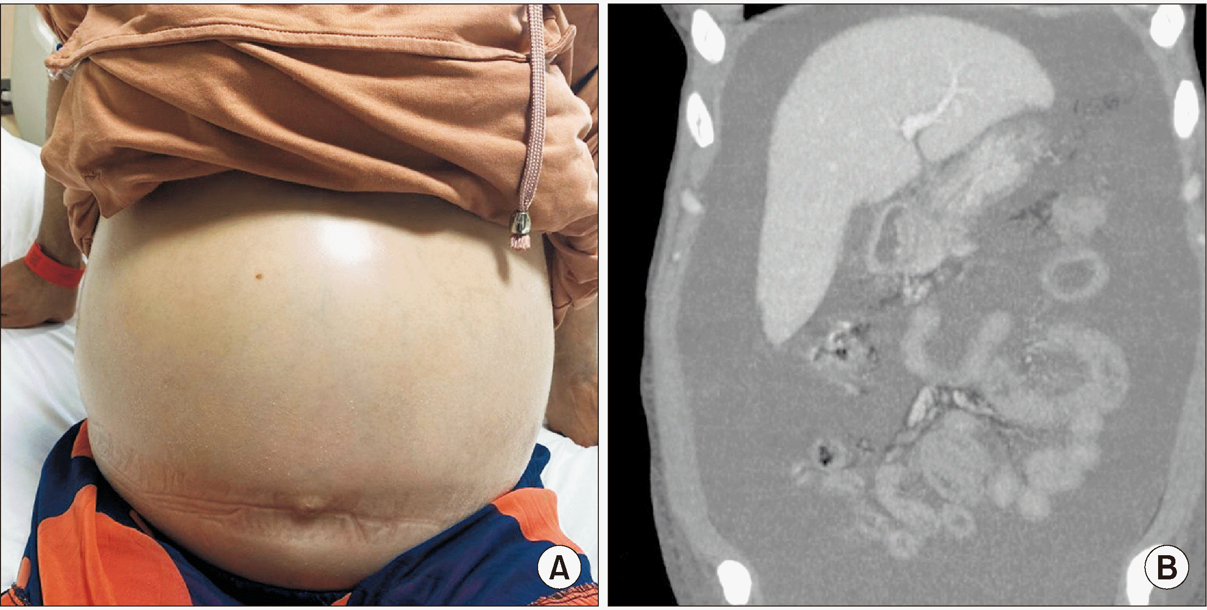
A 33-year-old female patient admitted with abdominal distension that had persisted for three months and had been worsening over the past month. She was hospitalized at the internal medicine clinic to be investigated for the etiology of ascites. She had a history of hypothyroidism and primary infertility. Three years ago, she was evaluated for bilateral pleural effusion, transudate in nature, and both tuberculosis and malignancy were excluded. While the patient had a 15-pack-year smoking history, she denied the use of any other drug or alcohol. On physical examination, she had cachexia, pleural effusion, massive ascites, and pretibial pitting edema (Figure 1A). Laboratory showed lymphopenia, normocytic anemia, and marked hypoalbuminemia (Table 1). In the diagnostic paracentesis, serum ascites albumin gradient was <1.1, with scarce cells, sterile culture, and benign cytology. Echocardiography showed mild pericardial effusion with normal ejection fraction and valve function. Considering the increased CA-125 value of 70 U/mL (0~35 U/mL), detailed gynecological examination, abdominal ultrasonography, tomography, magnetic resonance imaging, upper gastrointestinal endoscopy and colonoscopy, and PET/CT were performed. They showed no gynecological or other malignancy but massive ascites with normal bilateral ovaries, increased number, and engorgement of mesenteric vessels exhibiting comb sign were found (Figure 1B). With respect to the creatinine progression from 1.0 to 1.5 mg/dL and proteinuria of 11 grams/day, additional serological tests were requested and the patient was referred to us. She described Raynaud's phenomenon and arthralgia without any other findings for connective tissue disease. While C-reactive protein was in the normal range, erythrocyte sedimentation rate (ESR) was 80 mm/h, and blood creatinine was 1.6 mg/dL (normal range 0.5~0.9 mg/dL). Also, microscopic hematuria and pyuria in addition to nephrotic range proteinuria were found on urine analysis. Antinuclear antibody test with a titer of 1/3,200 and speckled pattern, anti-SM, anti-SM/RNP, anti-dsDNA, direct coombs positivity, and hypocomplementemia were detected. Antiphospholipid antibodies were negative. She got the diagnosis of SLE with renal involvement and PPMS. Intravenous methylprednisolone of 1,000 mg/day for 3 days followed by 60 mg/day was started. Upon detection of class-V membranous lupus nephritis in renal biopsy, hydroxychloroquine, and mycophenolate mofetil were added to the treatment and titrated to maximum effective daily dosage when methylprednisolone was tapered. While lymphopenia improved in the first week of treatment, massive ascites began to regress, and creatinine and ESR were decreased to 0.6 mg/dL and 23 mm/h, respectively (Table 1).
DISCUSSION
SLE is a chronic multisystemic autoimmune disease. Although serositis as pleural and pericardial effusion is an expected and one of the diagnostic criteria of SLE, massive ascites are rare in the absence of any other accompanying condition like nephrotic syndrome, lupus peritonitis, constrictive pericarditis, autoimmune hepatitis or primary biliary cholangitis, protein-losing enteropathy, or thrombosis of intrabdominal vessels namely Budd-Chiari syndrome or portal vein thrombosis [3,4]. Although CA-125 is attributed to the presence of gynecological malignancies, raised levels of it reveal an overall mesothelial cell activation in the abdominal cavity resulting in peritoneal effusion occurring for several reasons [5,6].
In literature, a few cases, all female, of SLE with initial presentation of massive ascites were reported. A special entity of Tjalma or PPMS was named as the triad of massive ascites, pleural effusion, and the raised level of CA-125 in SLE patients in attribution the Meig’s syndrome, the co-existence of the same triad occurred in the presence of a benign ovarian tumor [7]. PPMS usually occurs in SLE patients with major organ, especially renal, involvement, appearing before, concomitantly, or after the development of effusions, but this is not an obligation [8,9]. Similarly, systemic complement activation leading to the consumption of C3 and C4 is expected and low levels are found as a biomarker of disease severity [4,9].
Although components of PPMS are an uncommon remarkable manifestation of SLE needing further clinical attention in both diagnosis and follow-up of patients, it does not need to require special treatment rather than SLE itself. The prognosis of PPMS is good and the outcomes of patients correlate with the overall disease activity of SLE. Both regression of pleural and peritoneal effusions and normalization of CA-125 levels are expected with the administration of immunosuppressive agents, mainly corticosteroids, cyclophosphamide, hydroxychloroquine, mycophenolate mofetil or rituximab [9,10].
SUMMARY
In conclusion, if SLE is not considered in patients with ascites, pleural effusion, and high CA-125 levels, this may lead to unnecessary invasive investigations for benign or malignant etiologies and delay in the administration of immunosuppressive therapies for the possible organ involvement, especially in patients with undiagnosed SLE.
 XML Download