Journal List > J Korean Med Sci > v.39(11) > 1516086592

Lee, Do, Kim, Choe, Sung, Choi, and Kim: Comparative Analysis of CT Findings and Clinical Outcomes in Adult Patients With Disseminated and Localized Pulmonary Nocardiosis

Abstract

Background

Pulmonary nocardiosis is a rare opportunistic infection with occasional systemic dissemination. This study aimed to investigate the computed tomography (CT) findings and prognosis of pulmonary nocardiosis associated with dissemination.

Methods

We conducted a retrospective analysis of patients diagnosed with pulmonary nocardiosis between March 2001 and September 2023. We reviewed the chest CT findings and categorized them based on the dominant CT findings as consolidation, nodules and/or masses, consolidation with multiple nodules, and nodular bronchiectasis. We compared chest CT findings between localized and disseminated pulmonary nocardiosis and identified significant prognostic factors associated with 12-month mortality using multivariate Cox regression analysis.

Results

Pulmonary nocardiosis was diagnosed in 75 patients, of whom 14 (18.7%) had dissemination, including involvement of the brain in 9 (64.3%) cases, soft tissue in 3 (21.4%) cases and positive blood cultures in 3 (21.4%) cases. Disseminated pulmonary nocardiosis showed a higher frequency of cavitation (64.3% vs. 32.8%, P = 0.029) and pleural effusion (64.3% vs. 29.5%, P = 0.014) compared to localized infection. The 12-month mortality rate was 25.3%. The presence of dissemination was not a significant prognostic factor (hazard ratio [HR], 0.80; confidence interval [CI], 0.23–2.75; P = 0.724). Malignancy (HR, 9.73; CI, 2.32–40.72; P = 0.002), use of steroid medication (HR, 3.72; CI, 1.33–10.38; P = 0.012), and a CT pattern of consolidation with multiple nodules (HR, 4.99; CI, 1.41–17.70; P = 0.013) were associated with higher mortality rates.

Conclusion

Pulmonary nocardiosis with dissemination showed more frequent cavitation and pleural effusion compared to cases without dissemination, but dissemination alone did not affect the mortality rate of pulmonary nocardiosis.

Graphical Abstract

jkms-39-e107-abf001.jpg

INTRODUCTION

Pulmonary nocardiosis is the most common form of Nocardia infection, mainly affecting immunocompromised patients and those with chronic lung disease.1 While the incidence of pulmonary nocardiosis is low, it is increasing due to the growing population of immunocompromised patients (e.g., organ transplantation recipients and cancer patients) and their improved survival rates.23 Nocardiosis is characterized by a high mortality rate, with one-year mortality ranging from 15.8% to 24.5% depending on the disease type and the immune status of the patients.345 Furthermore, it increases the risk of mortality by approximately tenfold in immunocompromised patients, such as solid organ recipients.6
Risk factors associated with poor outcomes in nocardiosis include disseminated infection, severe immune suppression, high-dose steroids, and greater disease severity.1378 Among them, disseminated infections have been considered to be an important prognostic factor and also to have significant implications for treatment decisions, such as the selection of combination therapy and treatment duration.489 However, two recent studies have questioned whether dissemination is a direct risk factor for mortality, suggesting that infection sites and advanced infection may be more relevant than dissemination itself.35
Pulmonary nocardiosis can disseminate from the lungs to other sites, such as the brain, skin, and musculoskeletal system, through hematogenous spread.1 Chest computed tomography (CT) plays a crucial role in diagnosing and assessing the severity of pulmonary nocardiosis,10 which can show various CT findings, including lobar consolidation, solitary or multiple nodules, and peribronchial consolidation or centrilobular nodules, particularly in individuals with pre-existing structural lung disease.101112
Distinguishing the differences in CT findings between localized and disseminated pulmonary nocardiosis could provide valuable information for further evaluation and treatment decisions. Furthermore, due to its low incidence, little is known about factors associated with the prognosis of pulmonary nocardiosis, including its dissemination. Therefore, we compared chest CT findings between localized and disseminated pulmonary nocardiosis and investigated clinical and radiologic factors associated with the prognosis.

METHODS

Patient population

We reviewed the medical records of our hospital to identify patients with a positive Nocardia spp. culture in any type of samples gathered between June 2001 and April 2023. We also collected the records of patients with pathological results from percutaneous needle biopsy (PCNB) that indicated the presence of Nocardia and/or positive tissue cultures. Pulmonary nocardiosis was defined by the presence of clinical and/or imaging findings, along with the isolation of Nocardia spp. from cultures and/or lung biopsy. Cases with positive cultures only from sources other than respiratory samples without lung involvement were excluded from this study. Isolations from respiratory samples without any clinical or imaging evidence of active disease were categorized as colonization cases and were subsequently excluded. We also excluded patients who did not undergo a chest CT within 2 weeks after the diagnosis of pulmonary nocardiosis.
The following patient characteristics were collected: demographics, diagnosis information, underlying chronic pulmonary diseases (e.g., bronchiectasis, chronic obstructive pulmonary disease, and interstitial lung disease), immunocompromised status, presence of graft-versus-host disease, presence of neutropenia or lymphopenia, identified Nocardia spp., presence of coinfection, site of dissemination, treatment information, and clinical outcomes, specifically the overall 12-month mortality rate from the initial diagnosis of pulmonary nocardiosis.

Definitions

Disseminated pulmonary nocardiosis was defined as the simultaneous involvement of the lungs and a non-contiguous organ, or lung involvement with positive blood cultures. Immunocompromised patients were defined as individuals who had undergone solid organ transplantation, hematopoietic stem cell transplantation, those who had a malignancy and received chemotherapy or radiation therapy within the past 6 months, autoimmune diseases, those who were taking daily corticosteroids (at least 5 mg/day of prednisone or an equivalent drug), and individuals who tested positive for human immunodeficiency virus (HIV) infection.413 Coinfection was defined as the detection of other respiratory pathogens within 15 days before or after the nocardiosis infection. Neutropenia was defined as an absolute neutrophil count < 1,000/mL, and lymphopenia was defined as an absolute lymphocyte count < 500/mL.1415

Chest CT review

CT findings were reviewed in consensus by two chest radiologists, LHN and KEY, who have 9 and 11 years of experience in chest imaging, respectively. Parenchymal abnormalities were assessed for the presence of consolidation, nodules or masses, cavities, centrilobular nodules, and ground glass opacity. Pleural effusion and lymph node enlargement in the thorax were also evaluated. All glossary terms were defined according to the Fleishner Society.16 Then, we categorized the CT findings into the following groups: 1) consolidation, 2) solitary or multiple nodules and/or masses, 3) consolidation with multiple nodules (and/or masses), 4) peribronchial consolidation and/or centrilobular nodules in patients with underlying chronic lung disease such as bronchiectasis (nodular bronchiectasis or bronchopneumonia pattern). This categorization was done based on the dominant CT findings (Fig. 1). When it was not possible to determine the dominant CT findings between consolidation and nodules, we defined it as consolidation with multiple nodules. The total extent of pneumonia was assessed by assigning scores on a scale of 0–5 for each of the five lung lobes: 0 = no involvement; 1 = less than 5%; 2 = 5% to less than 25%; 3 = 25% to less than 50%; 4 = 50% to less than 75%; and 5 = more than 75%. The total CT score was determined as the sum of the individual scores and ranged from 0 to 25.17
Fig. 1

Four representative patterns of pulmonary nocardiosis. (A) Consolidation: CT scan of a 51-year-old woman with underlying breast cancer. Segmental consolidation with internal necrotic change is evident in the right lower lobe. (B) Nodules and/or masses: CT scan of a 75-year-old woman with a post-transplantation state of the liver. Two large cavitary masses are evident in both upper lobes and several nodules in the right lung. (C) Consolidation with multiple nodules: CT scan of a 68-year-old man taking steroid medication. Segmental consolidation is shown in the right lower lobe and several nodules are noted in the right middle lobe and both lower lobes (arrows). (D) Nodular bronchiectasis: CT scan of a 47-year-old woman with a nodular bronchiectasis pattern. Multiple centrilobular nodules and bronchiectasis are noted in the right middle lobe and both lower lobes.

CT = computed tomography.
jkms-39-e107-g001

Statistical analysis

The baseline characteristics and CT image findings were compared between the localized and disseminated groups of pulmonary nocardiosis using Fisher’s exact or χ2 test for categorical variables and Student's t-test for continuous variables. The Kaplan-Meier survival curves were plotted for the localized and disseminated groups of pulmonary nocardiosis, as well as for the CT category, and compared using the log-rank test. Univariate and multivariate analyses were performed using Cox regression to evaluate the association between variants and the 12-month overall survival outcomes. All variables with P values less than 0.05 in the univariate analysis were included in the multivariate analysis. The odds ratios and 95% confidence intervals (CIs) were then calculated based on the results. P values less than 0.05 were considered significant. All statistical calculations were performed using STATA (version 9.4; SAS Institute, Cary, NC, USA) and R (version 4.2.2; The R Foundation for Statistical Computing, Vienna, Austria).

Ethics statement

This retrospective study was reviewed and approved by the Institutional Review Board of Seoul Asan Medical Center (approval number: 2023-0653). Informed consent was waived due to the retrospective nature of the study.

RESULTS

Patient characteristics

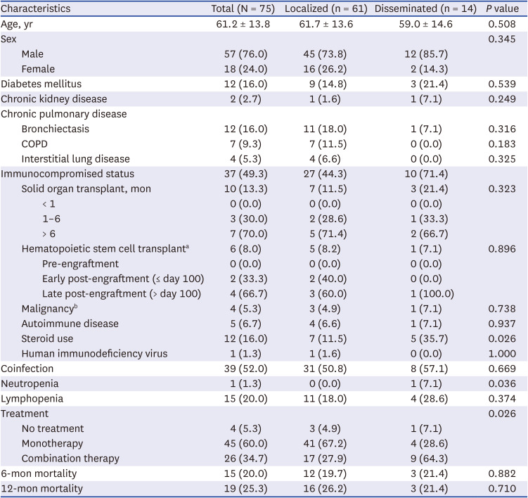
We initially identified 120 patients with positive Nocardia cultures in specimens or pathological results of Nocardia from PCNB. Among them, 25 patients exhibited positive cultures from sources other than respiratory samples, but did not demonstrate any signs of lung involvement. Six patients were categorized as colonization cases. Subsequently, 14 patients who did not undergo chest CT within 2 weeks after the diagnosis of nocardiosis were excluded. Finally, 75 patients were included in this study (Fig. 2). The average age was 61.2 ± 13.8 years, and 57 patients (76.0%) were male. Out of the total number of cases, 37 (49.3%) individuals had an immunocompromised state, including 10 (13.3%) with solid organ transplantations, 6 (8.0%) with hematopoietic cell transplantations, 4 (5.3%) with malignancy that were treated with chemotherapy or radiotherapy within the past 6 months, 5 (6.7%) with autoimmune disease, 12 (16.0%) with steroid use, and 1 (1.3%) with HIV infection (Table 1). Out of 10 patients who underwent solid organ transplantations, 7 (70.0%) developed Nocardia infection more than 6 months after transplantation. Among 6 patients who underwent hematopoietic cell transplantation, 4 (66.7%) had Nocardia infection during the late post-engraftment period. Among these, 4 out of 6 patients had graft-versus-host disease.
Fig. 2

Flow chart of included patients with pulmonary nocardiosis.

CT = computed tomography.
jkms-39-e107-g002
Table 1

Clinical characteristics of subjected patients

jkms-39-e107-i001
Characteristics Total (N = 75) Localized (n = 61) Disseminated (n = 14) P value
Age, yr 61.2 ± 13.8 61.7 ± 13.6 59.0 ± 14.6 0.508
Sex 0.345
Male 57 (76.0) 45 (73.8) 12 (85.7)
Female 18 (24.0) 16 (26.2) 2 (14.3)
Diabetes mellitus 12 (16.0) 9 (14.8) 3 (21.4) 0.539
Chronic kidney disease 2 (2.7) 1 (1.6) 1 (7.1) 0.249
Chronic pulmonary disease
Bronchiectasis 12 (16.0) 11 (18.0) 1 (7.1) 0.316
COPD 7 (9.3) 7 (11.5) 0 (0.0) 0.183
Interstitial lung disease 4 (5.3) 4 (6.6) 0 (0.0) 0.325
Immunocompromised status 37 (49.3) 27 (44.3) 10 (71.4)
Solid organ transplant, mon 10 (13.3) 7 (11.5) 3 (21.4) 0.323
< 1 0 (0.0) 0 (0.0) 0 (0.0)
1–6 3 (30.0) 2 (28.6) 1 (33.3)
> 6 7 (70.0) 5 (71.4) 2 (66.7)
Hematopoietic stem cell transplanta 6 (8.0) 5 (8.2) 1 (7.1) 0.896
Pre-engraftment 0 (0.0) 0 (0.0) 0 (0.0)
Early post-engraftment (≤ day 100) 2 (33.3) 2 (40.0) 0 (0.0)
Late post-engraftment (> day 100) 4 (66.7) 3 (60.0) 1 (100.0)
Malignancyb 4 (5.3) 3 (4.9) 1 (7.1) 0.738
Autoimmune disease 5 (6.7) 4 (6.6) 1 (7.1) 0.937
Steroid use 12 (16.0) 7 (11.5) 5 (35.7) 0.026
Human immunodeficiency virus 1 (1.3) 1 (1.6) 0 (0.0) 1.000
Coinfection 39 (52.0) 31 (50.8) 8 (57.1) 0.669
Neutropenia 1 (1.3) 0 (0.0) 1 (7.1) 0.036
Lymphopenia 15 (20.0) 11 (18.0) 4 (28.6) 0.374
Treatment 0.026
No treatment 4 (5.3) 3 (4.9) 1 (7.1)
Monotherapy 45 (60.0) 41 (67.2) 4 (28.6)
Combination therapy 26 (34.7) 17 (27.9) 9 (64.3)
6-mon mortality 15 (20.0) 12 (19.7) 3 (21.4) 0.882
12-mon mortality 19 (25.3) 16 (26.2) 3 (21.4) 0.710
Values are presented as number (%) or mean ± standard deviation.
COPD = chronic obstructive pulmonary disease.
aFour of 6 patients had graft-versus-host disease.
bPatients who had a malignancy and received chemotherapy or radiation therapy within the past 6 months.
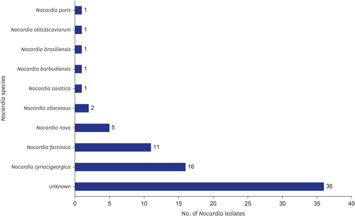
Positive cultures of Nocardia spp. were detected in various respiratory specimens: sputum specimens in 38 patients (50.7%), bronchoalveolar lavage in 17 patients (22.7%), and pleural fluid in 1 patient (1.3%). Among the 20 patients (26.7%) diagnosed through PCNB only, positive tissue culture was obtained in 8 patients, organisms were seen on histologic stains in 9 patients, and both identification of organisms and positive tissue cultures were found in 3 patients. The most common Nocardia species isolated were Nocardia cyriacigeorgica (n = 16) and Nocardia farcinica (n = 11). Thirty-six isolates’ species were unidentified (Fig. 3).
Fig. 3

Isolated Nocardia species.

jkms-39-e107-g003
Fourteen patients (18.7%) were found to have disseminated pulmonary nocardiosis. Among these cases, the brain was the most common site of dissemination in 9 patients (64.3%), followed by musculoskeletal involvement in 3 patients (21.4%), and positive blood culture in 3 patients (21.4%). One of the patients with disseminated infection showed involvement in multiple organs, including the lungs, brain, and skin with positive blood culture.
Coinfection was identified in 39 patients (52.0%), with nontuberculous mycobacteria (NTM) detected in 10 patients (13.3%), bacterial infection in 17 patients (22.7%), fungal infection in 12 patients (16.0%), and viral infection in 11 patients (14.7%).

Chest CT findings and comparison between localized and disseminated nocardiosis

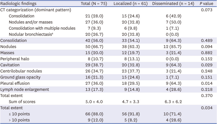
The most common CT findings were nodules (66.7%), followed by consolidation (56.0%) and cavitation (38.7%). Mediastinal lymph node enlargement was found in 13 out of 75 patients (17.3%). The most common CT pattern was solitary or multiple nodules and/or masses (36.0%), followed by consolidation (28.0%), nodular bronchiectasis (26.7%), and consolidation with multiple nodules (9.3%) (Table 2).
Table 2

Radiologic findings of patients with nocardiosis

jkms-39-e107-i002
Radiologic findings Total (N = 75) Localized (n = 61) Disseminated (n = 14) P value
CT categorization (dominant pattern) 0.073
Consolidation 21 (28.0) 15 (24.6) 6 (42.9)
Nodules and/or masses 27 (36.0) 20 (32.8) 7 (50.0)
Consolidation with multiple nodules 7 (9.3) 6 (9.8) 1 (7.1)
Nodular bronchiectasisa 20 (26.7) 20 (32.8) 0 (0.0)
Consolidation 42 (56.0) 33 (54.1) 9 (64.3) 0.489
Nodules 50 (66.7) 38 (62.3) 12 (85.7) 0.094
Masses 15 (20.0) 12 (19.7) 3 (21.4) 0.882
Peripheral halo 8 (10.7) 8 (13.1) 0 (0.0) 0.152
Cavitation 29 (38.7) 20 (32.8) 9 (64.3) 0.029
Centrilobular nodules 26 (34.7) 23 (37.7) 3 (21.4) 0.248
Ground glass opacity 16 (21.3) 15 (24.6) 1 (7.1) 0.151
Pleural effusion 27 (36.0) 18 (29.5) 9 (64.3) 0.014
Lymph node enlargement 13 (17.3) 9 (14.8) 4 (28.6) 0.218
Total extent 0.370
Sum of scores 5.0 ± 4.0 4.7 ± 3.3 6.3 ± 6.2
Total extent 0.034
≤ 10 points 66 (88.0) 56 (91.8) 10 (71.4)
> 10 points 9 (12.0) 5 (8.2) 4 (28.6)
Values are number (%) or mean ± standard deviation.
CT = computed tomography.
aThree of 20 patients had co-infection with nontuberculous mycobacteria.
When comparing CT findings between localized and disseminated infections, pulmonary nocardiosis with dissemination showed a higher frequency of cavitation (disseminated, 64.3% vs. localized, 32.8%, P = 0.029) and pleural effusion (disseminated, 64.3% vs. localized, 29.5%, P = 0.014). When divided into two groups based on a total extent score of 10, disseminated pulmonary nocardiosis showed a higher frequency of cases with a total extent score greater than 10 (disseminated, 28.6% vs. localized, 8.2%, P = 0.034); in contrast, the total extent scores were not significantly different between the two groups (disseminated, 6.3 ± 6.2 vs. localized, 4.7 ± 3.3, P = 0.370). Other image findings were not significantly different between the two groups.

Survival analysis

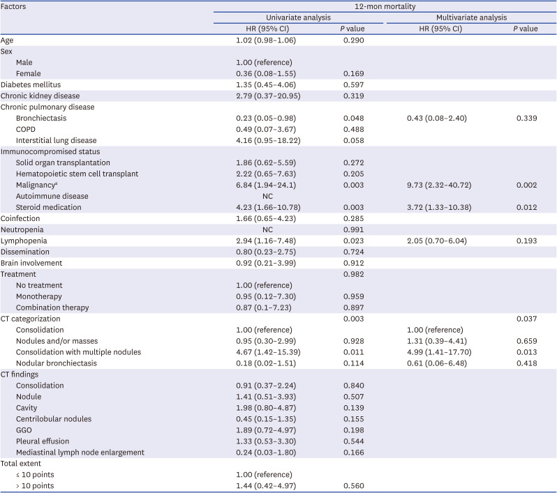
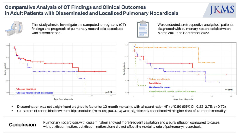
In total, 19 patients (25.3%) died within 12-month of diagnosis. The 12-month mortality rates were 21.4% (3/14) for pulmonary nocardiosis with dissemination and 22.2% (2/9) for pulmonary nocardiosis with brain involvement. The median time to death was 3.71 months (interquartile range [IQR], 1.05–5.54). Of the remaining 56 patients, 46 completed the 12-month follow-up and 10 patients were followed up for a median duration of 6.70 months (IQR, 5.24–9.30). Kaplan-Meier curves of survival are shown in Fig. 4. There was no significant difference in mortality between localized and disseminated pulmonary nocardiosis (P = 0.240, log-rank test). The presence of consolidation and multiple nodules and/or masses were found to be significantly associated with worse survival compared to the other CT categories (P < 0.001, log-rank test). Table 3 presents the factors associated with 12-month survival as determined by Cox regression analysis. In the univariate analysis, malignancy, steroid use, lymphopenia, and CT pattern of consolidation with multiple nodules were significantly associated with 12-month mortality, while disseminated infection did not show a significant association (hazard ratio [HR], 0.80; 95% CI, 0.23–2.75; P = 0.724). In multivariate analysis, malignancy (HR, 9.73; 95% CI, 2.32–40.72; P = 0.002), steroid use (HR, 3.72; 95% CI, 1.33–10.38; P = 0.012), and CT pattern of consolidation with nodules/masses (HR, 4.99; 95% CI, 1.41–17.70; P = 0.013) were identified as risk factors associated with 12-month mortality.
Fig. 4

Kaplan-Meier curves for overall survival. (A) Overall survival according to localized and disseminated pulmonary nocardiosis. (B) Overall survival according to computed tomography pattern.

jkms-39-e107-g004
Table 3

Univariable and multivariable Cox regression analysis of 12-month mortality after diagnosis of pulmonary nocardiosis

jkms-39-e107-i003
Factors 12-mon mortality
Univariate analysis Multivariate analysis
HR (95% CI) P value HR (95% CI) P value
Age 1.02 (0.98–1.06) 0.290
Sex
Male 1.00 (reference)
Female 0.36 (0.08–1.55) 0.169
Diabetes mellitus 1.35 (0.45–4.06) 0.597
Chronic kidney disease 2.79 (0.37–20.95) 0.319
Chronic pulmonary disease
Bronchiectasis 0.23 (0.05–0.98) 0.048 0.43 (0.08–2.40) 0.339
COPD 0.49 (0.07–3.67) 0.488
Interstitial lung disease 4.16 (0.95–18.22) 0.058
Immunocompromised status
Solid organ transplantation 1.86 (0.62–5.59) 0.272
Hematopoietic stem cell transplant 2.22 (0.65–7.63) 0.205
Malignancya 6.84 (1.94–24.1) 0.003 9.73 (2.32–40.72) 0.002
Autoimmune disease NC
Steroid medication 4.23 (1.66–10.78) 0.003 3.72 (1.33–10.38) 0.012
Coinfection 1.66 (0.65–4.23) 0.285
Neutropenia NC 0.991
Lymphopenia 2.94 (1.16–7.48) 0.023 2.05 (0.70–6.04) 0.193
Dissemination 0.80 (0.23–2.75) 0.724
Brain involvement 0.92 (0.21–3.99) 0.912
Treatment 0.982
No treatment 1.00 (reference)
Monotherapy 0.95 (0.12–7.30) 0.959
Combination therapy 0.87 (0.1–7.23) 0.897
CT categorization 0.003 0.037
Consolidation 1.00 (reference) 1.00 (reference)
Nodules and/or masses 0.95 (0.30–2.99) 0.928 1.31 (0.39–4.41) 0.659
Consolidation with multiple nodules 4.67 (1.42–15.39) 0.011 4.99 (1.41–17.70) 0.013
Nodular bronchiectasis 0.18 (0.02–1.51) 0.114 0.61 (0.06–6.48) 0.418
CT findings
Consolidation 0.91 (0.37–2.24) 0.840
Nodule 1.41 (0.51–3.93) 0.507
Cavity 1.98 (0.80–4.87) 0.139
Centrilobular nodules 0.45 (0.15–1.35) 0.155
GGO 1.89 (0.72–4.97) 0.198
Pleural effusion 1.33 (0.53–3.30) 0.544
Mediastinal lymph node enlargement 0.24 (0.03–1.80) 0.166
Total extent
≤ 10 points 1.00 (reference)
> 10 points 1.44 (0.42–4.97) 0.560
HR = hazard ratio, CI = confidential interval, COPD = chronic obstructive pulmonary disease, GGO = ground glass opacity, NC = not calculated.
aPatients who had a malignancy and received chemotherapy or radiation therapy within the past 6 months.

DISCUSSION

In this study, we compared CT findings between localized and disseminated pulmonary nocardiosis and identified the clinical and radiologic prognostic factors, including dissemination. We observed that the presence of a cavity and pleural effusion was associated with disseminated pulmonary nocardiosis. While dissemination itself was not associated with mortality for pulmonary nocardiosis. Patients with underlying malignancy and those taking steroid medication exhibited higher mortality rates. Additionally, CT findings of consolidation with multiple nodules were also associated with a poor outcome.
Previous studies have compared the clinical outcomes of localized and disseminated nocardiosis, and reported that disseminated infections have a worse prognosis compared to localized infection; however, these studies included various sites of Nocardia infection, including the skin, soft tissue, eyes, and as well as the lungs.418 Primary cutaneous infection of nocardiosis typically has a better outcome,119 while lung infection has a high mortality rate ranging from 16% to 25%, regardless of dissemination.20212223 Therefore, when considering the prognosis of pulmonary nocardiosis, it appears that the severity of the infection itself is more critical than its dissemination status. Our findings are consistent with another study, which stated that advanced infection, rather than dissemination alone, was a risk factor associated with worse 1-year mortality.23
The presence of cavitation has been associated with higher mortality in other studies,32021 but not in the present study. We thought that cavitation could serve as an indicator of the severity of the infection, such as disseminated infection, but it did not necessarily correlate with higher mortality. This discrepancy may be attributed to variations in the site of infection and the characteristics of the patients. Furthermore, it could be due to the improved control of pulmonary nocardiosis with cavitation, thanks to advances in medical care.
Whereas the presence of both consolidation and multiple nodules on CT scans was associated with a worse prognosis. This combination of findings likely indicates advanced pulmonary nocardiosis infection, with spread from one lung to another and increased disease involvement. In cases that exhibit both consolidation and multiple nodules, it is advisable to consider more aggressive treatment such as combination therapy and an extended treatment duration.124
Patients with underlying structural diseases such as bronchiectasis can show subacute manifestations of pulmonary nocardiosis. A few studies have reported that a nodular bronchiectatic pattern can be observed on chest CT scans, resembling NTM.2526 Although there are some cases of co-infection with NTM, it is worth noting that the nodular bronchiectatic pattern in pulmonary nocardiosis can occur independently of NTM co-infection, which can be resolved with medication specifically targeting nocardiosis. It suggests that this condition may not be solely attributed to NTM infection. In our study, some of the patients with a nodular bronchiectasis pattern may have represented subacute pulmonary nocardiosis, and only 3 of the 20 patients had co-infection with NTM. Further studies with larger sample sizes are needed for the diagnosis and treatment of subacute pulmonary nocardiosis.
PCNB in patients with a high suspicion of infection can occasionally provide useful information that can lead to significant changes in patient management.27 The role of PCNB was particularly critical in the diagnosis of pulmonary nocardiosis in this study. Patients diagnosed solely with PCNB accounted for as much as 26.7% (20/75) of the total patients. A previous study reported that 44% of cases of pulmonary nocardiosis required an invasive procedure for diagnosis in order to obtain adequate specimens.28 Pulmonary nocardiosis is often initially misdiagnosed as TB or cancer based on CT findings. Once pulmonary nocardiosis is diagnosed, specific antimicrobial therapy, such as trimethoprim-sulfamethoxazole, can be initiated,29 and additional procedures, such as a brain MRI, are often performed to confirm dissemination.30 Therefore, if an imaging finding indicates possible pulmonary nocardiosis without evidence of a positive culture, PCNB may be helpful for diagnosis and treatment.
Immunocompromised state is associated with higher mortality rate in patients with nocardiosis.1 Patients with malignancy have a mortality rate greater than 60%.31 In our study, three of four patients with underlying malignancy died within 12 months. Among them, two patients died due to nocardiosis, while one experienced mortality attributed to cancer aggravation. Our data set has limitations when it comes to interpreting mortality in the cancer population, as we analyzed all-cause mortality in a small sample. Therefore, further study is needed to examine whether pulmonary Nocardia infection affected mortality in cancer patients.
There are several limitations to this study. First, it was a retrospective study with a relatively small sample size. Second, patients received different management such as combination therapy or not, possibly based on disease severity, which may have influenced the clinical outcome. Third, not all patients had brain imaging, which may have resulted in an underdiagnosis of disseminated infection. Lastly, we analyzed all-cause mortality, which may include patients who died from other causes.
In conclusion, this study revealed that pulmonary nocardiosis with dissemination often showed more frequent cavitation and pleural effusion compared to cases without dissemination. However, dissemination alone did not affect the mortality rate of pulmonary nocardiosis. Instead, patients with underlying malignancy, steroid use, and CT findings of consolidation with multiple nodules were associated with a worse prognosis of pulmonary nocardiosis. Therefore, this study emphasizes the importance of considering these factors when managing pulmonary nocardiosis.

Notes

Disclosure: The authors have no potential conflicts of interest to disclose.

Author Contributions:

  • Conceptualization: Lee HN, Do KH.

  • Data curation: Lee HN, Do KH, Kim EY, Choe J, Sung H, Choi SH, Kim HJ.

  • Formal analysis: Lee HN, Kim EY, Kim HJ.

  • Investigation: Lee HN, Kim EY.

  • Writing - original draft: Lee HN.

  • Writing - review & editing: Lee HN, Do KH, Choe J, Sung H, Choi SH.

References

1. Traxler RM, Bell ME, Lasker B, Headd B, Shieh WJ, McQuiston JR. Updated review on Nocardia species: 2006-2021. Clin Microbiol Rev. 2022; 35(4):e0002721. PMID: 36314911.
2. Harpaz R, Dahl RM, Dooling KL. Prevalence of immunosuppression among US adults, 2013. JAMA. 2016; 316(23):2547–2548. PMID: 27792809.
3. Yetmar ZA, Khodadadi RB, Chesdachai S, McHugh JW, Challener DW, Wengenack NL, et al. Mortality after nocardiosis: risk factors and evaluation of disseminated infection. Open Forum Infect Dis. 2023; 10(8):ofad409. PMID: 37577117.
4. Soueges S, Bouiller K, Botelho-Nevers E, Gagneux-Brunon A, Chirouze C, Rodriguez-Nava V, et al. Prognosis and factors associated with disseminated nocardiosis: a ten-year multicenter study. J Infect. 2022; 85(2):130–136. PMID: 35654278.
5. Yetmar ZA, Challener DW, Seville MT, Bosch W, Beam E. Outcomes of nocardiosis and treatment of disseminated infection in solid organ transplant recipients. Transplantation. 2023; 107(3):782–791. PMID: 36303280.
6. Lebeaux D, Freund R, van Delden C, Guillot H, Marbus SD, Matignon M, et al. Outcome and treatment of nocardiosis after solid organ transplantation: new insights from a European study. Clin Infect Dis. 2017; 64(10):1396–1405. PMID: 28329348.
7. Chen YC, Lee CH, Chien CC, Chao TL, Lin WC, Liu JW. Pulmonary nocardiosis in southern Taiwan. J Microbiol Immunol Infect. 2013; 46(6):441–447. PMID: 23017691.
8. Presant CA, Wiernik PH, Serpick AA. Factors affecting survival in nocardiosis. Am Rev Respir Dis. 1973; 108(6):1444–1448. PMID: 4751735.
9. Restrepo A, Clark NM. Infectious Diseases Community of Practice of the American Society of Transplantation. Nocardia infections in solid organ transplantation: guidelines from the Infectious Diseases Community of Practice of the American Society of Transplantation. Clin Transplant. 2019; 33(9):e13509. PMID: 30817024.
10. Kanne JP, Yandow DR, Mohammed TL, Meyer CA. CT findings of pulmonary nocardiosis. AJR Am J Roentgenol. 2011; 197(2):W266–W272. PMID: 21785052.
11. Bhoil R. In response to the article “Computed Tomography Features of Pulmonary Nocardiosis in Immunocompromised and Immunocompetent Patients”; Pol J Radiol, 2015; 80: 13-17. Pol J Radiol. 2015; 80:281–282. PMID: 26082821.
12. Yoon HK, Im JG, Ahn JM, Han MC. Pulmonary nocardiosis: CT findings. J Comput Assist Tomogr. 1995; 19(1):52–55. PMID: 7822548.
13. Micek ST, Kollef KE, Reichley RM, Roubinian N, Kollef MH. Health care-associated pneumonia and community-acquired pneumonia: a single-center experience. Antimicrob Agents Chemother. 2007; 51(10):3568–3573. PMID: 17682100.
14. Su YB, Sohn S, Krown SE, Livingston PO, Wolchok JD, Quinn C, et al. Selective CD4+ lymphopenia in melanoma patients treated with temozolomide: a toxicity with therapeutic implications. J Clin Oncol. 2004; 22(4):610–616. PMID: 14726505.
15. Lee HN, Koo HJ, Kim SH, Choi SH, Sung H, Do KH. Human bocavirus infection in adults: clinical features and radiological findings. Korean J Radiol. 2019; 20(7):1226–1235. PMID: 31270986.
16. Hansell DM, Bankier AA, MacMahon H, McLoud TC, Müller NL, Remy J. Fleischner Society: glossary of terms for thoracic imaging. Radiology. 2008; 246(3):697–722. PMID: 18195376.
17. Wang Y, Dong C, Hu Y, Li C, Ren Q, Zhang X, et al. Temporal changes of CT findings in 90 patients with COVID-19 pneumonia: a longitudinal study. Radiology. 2020; 296(2):E55–E64. PMID: 32191587.
18. Takamatsu A, Yaguchi T, Tagashira Y, Watanabe A, Honda H. Japan Nocardia Study Group. Nocardiosis in Japan: a multicentric retrospective cohort study. Antimicrob Agents Chemother. 2022; 66(2):e0189021. PMID: 34902263.
19. Wang HK, Sheng WH, Hung CC, Chen YC, Lee MH, Lin WS, et al. Clinical characteristics, microbiology, and outcomes for patients with lung and disseminated nocardiosis in a tertiary hospital. J Formos Med Assoc. 2015; 114(8):742–749. PMID: 24008153.
20. Martínez Tomás R, Menéndez Villanueva R, Reyes Calzada S, Santos Durantez M, Vallés Tarazona JM, Modesto Alapont M, et al. Pulmonary nocardiosis: risk factors and outcomes. Respirology. 2007; 12(3):394–400. PMID: 17539844.
21. Chen J, Zhou H, Xu P, Zhang P, Ma S, Zhou J. Clinical and radiographic characteristics of pulmonary nocardiosis: clues to earlier diagnosis. PLoS One. 2014; 9(3):e90724. PMID: 24594890.
22. Takiguchi Y, Ishizaki S, Kobayashi T, Sato S, Hashimoto Y, Suruga Y, et al. Pulmonary nocardiosis: a clinical analysis of 30 cases. Intern Med. 2017; 56(12):1485–1490. PMID: 28626172.
23. Yetmar ZA, Thoendel MJ, Bosch W, Seville MT, Hogan WJ, Beam E. Risk factors and outcomes of nocardiosis in hematopoietic stem cell transplantation recipients. Transplant Cell Ther. 2023; 29(3):206.e1–206.e7.
24. Heo ST, Ko KS, Kwon KT, Ryu SY, Bae IG, Oh WS, et al. The first case of catheter-related bloodstream infection caused by Nocardia farcinica . J Korean Med Sci. 2010; 25(11):1665–1668. PMID: 21060759.
25. Nakagoshi K, Yaguchi T, Takahashi K, Morizumi S, Nishiyama M, Takahashi Y, et al. Pulmonary nocardiosis caused by Nocardia pneumoniae mimicking non-tuberculous mycobacterial disease. QJM. 2022; 115(9):625–626. PMID: 35587749.
26. Fujita T, Ikari J, Watanabe A, Tatsumi K. Clinical characteristics of pulmonary nocardiosis in immunocompetent patients. J Infect Chemother. 2016; 22(11):738–743. PMID: 27615155.
27. Haas BM, Clayton JD, Elicker BM, Ordovas KG, Naeger DM. CT-guided percutaneous lung biopsies in patients with suspicion for infection may yield clinically useful information. AJR Am J Roentgenol. 2017; 208(2):459–463. PMID: 27845850.
28. Georghiou PR, Blacklock ZM. Infection with Nocardia species in Queensland. A review of 102 clinical isolates. Med J Aust. 1992; 156(10):692–697. PMID: 1620016.
29. Wallace RJ Jr, Septimus EJ, Williams TW Jr, Conklin RH, Satterwhite TK, Bushby MB, et al. Use of trimethoprim-sulfamethoxazole for treatment of infections due to Nocardia. Rev Infect Dis. 1982; 4(2):315–325. PMID: 6981158.
30. Wang HL, Seo YH, LaSala PR, Tarrand JJ, Han XY. Nocardiosis in 132 patients with cancer: microbiological and clinical analyses. Am J Clin Pathol. 2014; 142(4):513–523. PMID: 25239419.
31. Torres HA, Reddy BT, Raad II, Tarrand J, Bodey GP, Hanna HA, et al. Nocardiosis in cancer patients. Medicine (Baltimore). 2002; 81(5):388–397. PMID: 12352633.
TOOLS
Similar articles