Journal List > J Stroke > v.26(1) > 1516086496

Ceulemans, Pinckaers, Postma, van Zwam, and van Oostenbrugge: Association Between Anemia and Clinical Outcome in Acute Ischemic Stroke Patients Treated With Endovascular Treatment

Abstract

Background and Purpose

Endovascular treatment (EVT) is the preferred treatment option in eligible acute ischemic stroke (AIS) patients with a large vessel occlusion of the anterior circulation. Several comorbidities have been identified that can affect clinical outcomes. Various studies have investigated the association between anemia and clinical outcome and found conflicting results. The aim is to investigate the association between pre-EVT anemia and clinical outcomes at different time points post-EVT, primarily focusing on the National Institutes of Health Stroke Scale (NIHSS) at 24–48 hours.

Methods

We prospectively included 560 AIS patients who received EVT in the Maastricht University Medical Center+. Hemoglobin levels (Hb; g/dL) were determined on admission. Hb levels were also categorized into two groups: anemia (male: Hb ≤12.9 g/dL; female: Hb ≤11.9 g/dL) and no anemia. Multiple imputation was used to handle missing data. Multivariable regression was used to investigate the association between anemia or Hb levels and clinical outcomes.

Results

Anemia was present in 26% of the patients. Multivariable regression did not show a significant association between anemia or Hb levels and NIHSS at 24–48 hours (adjusted β [aβ]anemia: 1.44, 95% confidence interval [CI]: -0.47 to 3.36; aβHb: -0.37, 95% CI: -0.88 to 0.13). However, multivariable regression showed significant associations with modified Rankin Scale (adjusted common odds ratio [acOR]anemia: 1.66, 95% CI: 1.12 to 2.48; acORHb: 0.83, 95% CI: 0.75 to 0.93) and poor functional outcome at 90 days (adjusted OR [aOR]anemia: 2.09, 95% CI: 1.21 to 3.63; aORHb: 0.80, 95% CI: 0.69 to 0.92).

Conclusion

Anemia was not independently associated with early neurological deficit (NIHSS) post-AIS, suggesting it is more suitable as a general frailty marker.

Introduction

Several studies have shown that acute ischemic stroke (AIS) patients with a large vessel occlusion (LVO) of the anterior circulation benefit from endovascular treatment (EVT) [1-5]. Based on these results, EVT has become the standard of care in this patient population [6].
The clinical outcome of EVT can be affected by various comorbidities in the patients [7,8]. As such, previous studies suggested that anemia, predominantly caused by iron deficiency, is associated with increased morbidity and mortality in AIS patients, especially in cases of LVO [8-10].
Several studies found anemia on admission to be an independent predictor for poor clinical outcome post-AIS when treated with intravenous thrombolysis, EVT, or a combination of both [11-20]. Conversely, other studies did not find this association [10,21-23]. Although a large meta-analysis concluded a positive association between anemia on admission and adverse clinical outcomes, heterogeneity between the included studies was substantial [9]. Notably, while previous studies used the modified Rankin Scale (mRS) at 30 days to 1-year post-AIS as a primary outcome measure, an association between anemia and earlier outcome measures has not yet been assessed. It has been suggested that early outcome assessment, e.g., by means of the National Institutes of Health Stroke Scale (NIHSS) during hospitalization, would be more appropriate, as it may be less influenced by other clinical factors compared to the mRS and is more useful in practice [24-26]. As up to 40% of AIS patients suffer from anemia, it is relevant to further study the relationship between anemia and clinical outcomes post-EVT.
Therefore, the aim of this retrospective study was to investigate the association between pre-EVT anemia and clinical outcomes at different time points post-EVT, with a primary focus on the NIHSS at 24–48 hours.

Methods

Ethics statement

The study was conducted in accordance with the Declaration of Helsinki. Ethical approval for this study was obtained from the ethics committee of the Maastricht University Medical Center, Maastricht, The Netherlands (MEC-2020-1456). The need for individual patient consent was waived owing to the retrospective nature of the study.

Design and study population

This retrospective study used data from the Maastricht University Medical Center+ (MUMC+). Patients who received EVT between 2010 and 2019 were screened. Patients with an occlusion of the internal carotid artery (ICA), ICA-terminus, or middle cerebral artery segment M1 or M2 were included. The exclusion criteria were as follows: (1) patients randomized into the MR CLEAN MED trial who received trial medication (acetylsalicylic acid and/or unfractionated heparin) [27], as this could affect the clinical outcome; and (2) patients who were included in the MR CLEAN LATE trial, as researchers were still blinded for endpoint data during the analysis of the current study.

Study parameters

Clinical and procedural parameters were collected from prospective stroke records. Because a large part of the included patient cohort participated in the MR CLEAN trial, MR CLEAN Registry, MR CLEAN NO-IV, or MR CLEAN MED, core lab imaging assessments were often readily available [1,27-29]. If imaging parameters were missing, an experienced neuroradiologist and core lab member (A.P.) scored the respective parameters.
Hemoglobin levels (Hb; in g/dL) determined on admission were not included in the prospective stroke records and were retrospectively collected from patient medical files. Hb levels were assessed upon arrival at the emergency department, either in the referring center or our center (treating center), before the onset of the EVT. If the Hb level was not determined on admission at the emergency department, Hb levels assessed in the prior 7 days were used. Notably, in the case of major bleeding, Hb levels were deemed missing and were therefore imputed. When multiple Hb levels were available, the level obtained closest to the EVT was used for analyses in the present study. Anemia was defined according to the World Health Organization criteria (Hbmale ≤12.9 g/dL; Hbfemale ≤11.9 g/dL) [30,31].
The primary outcome was the NIHSS score at 24–48 hours post-EVT. Secondary outcomes were the mRS score, poor functional outcome (mRS 3–6), and mortality at 90 days post-EVT. The NIHSS and mRS scores were prospectively assessed by trained personnel. Specifically, the mRS was assessed during a phone interview at 90 days post-AIS, which is the standard follow-up procedure for all stroke patients in the Netherlands. Missing follow-up NIHSS scores were, if possible, retrospectively reconstructed based on the patient’s medical file by a trained researcher (F.P.). The mRS scores were not retrospectively reconstructed. Hence, missing mRS scores were always imputed.

Missing data

In the included cohort, Hb levels were missing in 13% (73/560) of the patients. Considering our outcome measures, 5.7% of NIHSS scores at 24–48 hours and 4.1% of mRS scores at 90 days post-EVT were missing. The missing data percentage in all other baseline variables included in the analyses was 4.6%.
Multiple imputation by chained equations (MICE) using the mice package version 3.15.0 (https://cran.r-project.org/) was used to handle the missing data [32]. The imputation model included relevant covariates and outcome variables. The number of imputations was based on the fraction of missing information [33].

Statistical analysis

Owing to this study’s explorative and retrospective nature, a power calculation was not performed.
Statistical analysis was performed with R version 4.2.2 (R Foundation for Statistical Computing, Vienna, Austria). Crude data were used to describe baseline patient characteristics, laboratory parameters, and imaging and EVT characteristics of patients with and without anemia. In addition, a baseline table based on the presence/absence of Hb was provided to evaluate for potential imbalances. To test for significant differences between the anemia and no-anemia groups, the chi-square, Fisher’s exact, Student’s t, and Mann-Whitney U tests were used as appropriate.
Continuous data are presented as means±standard deviation or medians with interquartile range (IQR), based on the nature of the data. NIHSS scores were analyzed as a continuous variable.
Uni- and multivariable linear, binary logistic, and ordinal regression were used as appropriate to determine the association between Hb levels or anemia and outcome variables. Variables with P<0.10 in univariable regression analyses were included in the multivariable regression. To prevent overfitting of the regression models, the maximum number of confounders was restricted to 10% of the outcomes per outcome variable [34]. The effect estimates of linear regression are presented per 1 g/dL decrease in Hb.

Results

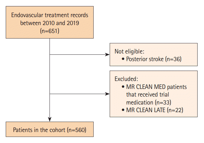
In total, 651 EVT records were screened for eligibility, of which 560 patients were included for statistical analysis. An inclusion flowchart is presented in Figure 1. Baseline characteristics are described in Table 1. Anemia was present in 26% of the patients. AIS patients suffering from anemia were slightly older with a median age of 77 years, compared to 70 years in non-anemic AIS patients (P<0.001). In addition, anemia was more common in female patient (56%) than in male patients, although not statistically significant (P=0.469). Median Hb levels of anemic patients were 11.3 g/dL and 12.1 g/dL for female and male anemic patients, respectively. A medical history of cardiovascular risk factors was more frequently seen in the anemia group (P=0.011). Consequently, patients suffering from anemia were more frequently on antihypertensive (P<0.001), cholesterol-lowering (P=0.048), antiplatelet (P=0.007), or anticoagulation medication (P=0.011). In addition, as shown in Supplementary Table 1, patients with a known versus an unknown Hb level were comparable in terms of stroke severity on admission and pre-morbid mRS, with only minimal differences in stroke history, the number of M1 occlusions, and total EVT-attempts.

Association with outcome measures

Effect estimates of the performed regression analyses are presented in Table 2. In the univariable analysis, both anemia and decreased Hb levels were significantly associated with an increase in NIHSS at 24–48 hours post-EVT (βanemia: 2.87, 95% confidence interval [CI]: 0.60 to 5.14; βHb: -0.80, 95% CI: -1.37 to -0.23). However, this was no longer significant in the multivariable regression analysis (adjusted [a]βanemia: 1.44, 95% CI: -0.47 to 3.36; and aβHb: -0.37, 95% CI:-0.88 to 0.13).
Considering the mRS at 90 days, both anemia and decreasing Hb levels were significantly associated with an mRS shift to poorer outcomes in the univariable regression analysis (common odds ratio [cOR]anemia: 2.01, 95% CI: 1.45 to 3.04; and cORHb: 0.78, 95% CI: 0.71 to 0.86). This effect remained significant in the multivariable regression analysis (adjusted cOR [acOR]anemia: 1.66, 95% CI: 1.12 to 2.48; and acORHb: 0.83, 95% CI: 0.75 to 0.93). In addition, relevant interaction terms were explored and, if significant, are reported in the supplemental materials (Supplementary Tables 2-9). As such, the interaction term of Hb*age is positively associated with mRS at 90 days (acORHb*age: 0.995, 95% CI: 0.991–0.999) (Supplementary Table 5).
Likewise, there was a significant association with a poor functional outcome in the univariable analysis (ORanemia: 2.49, 95% CI: 1.56 to 3.97; ORHb: 0.76, 95% CI: 0.68 to 0.86). This effect remained significant in the multivariable regression analysis (aORanemia: 2.09, 95% CI: 1.21 to 3.63; aORHb: 0.80, 95% CI: 0.69 to 0.92).
Anemia and a decrease in Hb levels were significantly associated with an increase in mortality at 90 days in the univariable regression analysis (ORanemia: 2.30, 95% CI: 1.48 to 3.58; ORHb: 0.77, 95% CI: 0.69 to 0.87). However, this effect was no longer significant in the multivariable regression analysis (aORanemia: 1.53, 95% CI: 0.88 to 2.66; aORHb: 0.86, 95% CI: 0.74 to 1.01).
Lastly, a sensitivity analysis with unimputed hemoglobin levels (i.e., a dataset containing 487/560 patients) is presented in Supplementary Table 10 and showed similar results as compared to the analyses with the imputed dataset.

Discussion

In this study, we aimed to identify an association between preEVT anemia and clinical outcomes in AIS patients with an LVO of the anterior circulation. Our main findings were that, in multivariable regression analysis, both anemia and Hb levels were significantly associated with the mRS and poor functional outcome at 90 days, but not with the NIHSS at 24–48 hours or mortality at 90 days.
Unlike other studies with a similar research question, we used the NIHSS at 24–48 hours as our primary outcome measure. As the presence of anemia is often accompanied by other comorbidities [35-39], the true effect of anemia on clinical outcome may be masked when using only long-term outcome measures [24,25]. Additionally, non-specific functional outcome measures (i.e., mRS or the Barthel Index), which are assessed after a typical recovery period of 90 days, might not accurately represent the relationship between anemia and neurological deficit immediately after AIS [24,25]. However, anemia and decreased Hb levels were not significantly associated with the NIHSS at 24–48 hours in our multivariable analyses, which may suggest that anemia is more suitable as a general frailty marker rather than as a marker for neurological deficit immediately post-AIS. This hypothesis is supported by the finding that the presence of anemia and lower Hb levels were significantly associated with advanced age and the presence of comorbidities in the present study. Additionally, Supplementary Table 5 shows that there is a significant effect of age on the association between Hb and mRS at 90 days. Noteworthy, the cOR of the interaction was 0.995, indicating that this effect decreased with increasing age, presumably due to the inverse relationship between age and Hb level. Nevertheless, this effect is minimal and has questionable clinical relevance and is therefore only shown in the supplemental materials (Supplementary Table 5). However, given the retrospective nature of the present study, we did not evaluate frailty in a specific way. Therefore, this hypothesis has to be interpreted with caution.
Previous literature has suggested that anemic patients, who likely also suffer from additional comorbidities, already have impaired cerebral autoregulation before stroke onset [40]. Additionally, a previous study proposes that during stroke, the cerebral oxygen uptake in the penumbral region progressively decreases with Hb levels below 10.0 g/dL. Consequently, one could reason that it might be more difficult to tolerate hypoxic/ischemic events in the presence of anemia with additional comorbidities compared to AIS patients without anemia and other comorbidities [10].
Besides the NIHSS at 24–48 hours, we investigated the association between anemia or lower Hb levels and poor functional outcome or mortality at 90 days. We found a significant association between the presence of anemia or decreased Hb levels and poor functional outcome, but not with mortality. These results are in line with previous studies [14,15]. Of note, one study only found this association in patients with moderate (Hb <10.0 g/dL for both sexes) to severe anemia (Hb <8.0 g/dL for both sexes)[ 10]. Our study population included only few patients suffering from moderate to severe anemia, rendering subgroups too small for statistical analysis with adequate power. Notably, approximately 55% of the patients included in the previously mentioned cohort suffered from mild anemia, while this proportion was roughly 67% in our cohort. Interestingly, since this previously mentioned study only included 90 anemic patients in total, it could be possible that there was not enough power to detect an association with mild anemia. Markedly, during the clinical follow-up, the aforementioned study performed restrictive red blood cell transfusion (RBCT), i.e., RBCT in patients with Hb levels <8.0 g/dL. Our center does not routinely provide RBCT in this patient population as the optimal RBCT strategy and threshold in AIS patients is still under debate [41,42].
While previous studies demonstrated a significant association between anemia and mortality at 90 days, our present study found no such effect [12,14,15]. Possible explanations for this unexpected result could be differences between study populations, i.e., mean population age, baseline NIHSS score, and correction for different confounders.
Several limitations have to be addressed. Firstly, the small sample sizes in the moderate to severe anemia subgroups did not allow for subgroup analysis on this subject. Secondly, although this study used prospectively collected data, Hb levels were collected retrospectively. In addition, it was not possible to determine the etiology (e.g., nutritional status) of anemia/low Hb. Consequently, we did not distinguish between different types of anemia (e.g., iron deficient anemia, vitamin deficient anemia, and hemolytic anemia) and had no information regarding the underlying cause of anemia, which can affect functional dependence and mortality [36]. In that line, anemic patients had significantly higher C-reactive protein levels than that in non-anemic patients (Table 1). Unfortunately, owing to the retrospective nature of the present study, we were unable to collect information regarding comorbidities other than cardiovascular risk factors and corresponding medication. Thirdly, though we had a very extensive clinical dataset it is possible that we did not adjust for all relevant confounders (e.g., computed tomography perfusion parameters).
Despite these limitations, there are several strengths of this study. First, our dataset was larger compared to that in other studies [8,14,23]. Second, while the majority of the studies on this topic use the mRS and/or mortality as a primary outcome measure, we used a neurological deficit score (NIHSS) early post-EVT as a primary outcome measure as it may be less influenced by other clinical factors [24,25]. Lastly, we used a multiple imputation strategy to handle missing data in both the dependent and independent variables. In addition, we performed a sensitivity analysis with unimputed hemoglobin data (i.e., data from 487 patients of whom a hemoglobin level was available upon hospital admission) and found no discrepancies as compared with the imputed data analysis, showing no biases introduced by the imputation strategy (Supplementary Table 10).

Conclusions

Anemia was found to be significantly associated with clinical outcomes at 90 days post-EVT in patients with an LVO of the anterior circulation. Given the fact we did not observe an association with the NIHSS at 24–28 hours post-EVT, it seems more likely that anemia is a marker for frailty rather than an early neurological outcome marker after AIS.

Supplementary materials

Supplementary materials related to this article can be found online at https://doi.org/10.5853/jos.2023.01669.
Supplementary Table 1.
Baseline patient characteristics and laboratory parameters based on the presence of hemoglobin levels (n=560)
jos-2023-01669-Supplementary-Table-1.pdf
Supplementary Table 2.
Model output of linear regression analysis for the association between anemia and NIHSS at 24-48 hours
jos-2023-01669-Supplementary-Table-2.pdf
Supplementary Table 3.
Model output of linear regression analysis for the association between hemoglobin level on admission and NIHSS at 24-48 hours
jos-2023-01669-Supplementary-Table-3.pdf
Supplementary Table 4.
Model output of ordinal logistic regression analysis for the association between anemia and mRS at 90 days
jos-2023-01669-Supplementary-Table-4.pdf
Supplementary Table 5.
Model output of ordinal logistic regression analysis for the association between hemoglobin level on admission and mRS at 90 days
jos-2023-01669-Supplementary-Table-5.pdf
Supplementary Table 6.
Model output of binary logistic regression analysis for the association between anemia and mRS 3–6 at 90 days
jos-2023-01669-Supplementary-Table-6.pdf
Supplementary Table 7.
Model output of binary logistic regression analysis for the association between hemoglobin level on admission and mRS 3–6 at 90 days
jos-2023-01669-Supplementary-Table-7.pdf
Supplementary Table 8.
Model output of binary logistic regression analysis for the association between anemia and mortality at 90 days
jos-2023-01669-Supplementary-Table-8.pdf
Supplementary Table 9.
Model output of binary logistic regression analysis for the association between hemoglobin level on admission and mortality at 90 days
jos-2023-01669-Supplementary-Table-9.pdf
Supplementary Table 10.
Sensitivity analyses without imputed hemoglobin values
jos-2023-01669-Supplementary-Table-10.pdf

Notes

Funding statement

None

Conflicts of interest

The authors have no financial conflicts of interest.

Author contribution

Conceptualization: RJvO, AC, FMEP. Study design: AC, FMEP. Methodology: AC, FMEP. Data collection: AC, FMEP. Investigation: AC, FMEP. Statistical analysis: AC. Writing—original draft: AC. Writing—review & editing: all authors. Approval of final manuscript: all authors.

ACKNOWLEDGMENTS

We thank the MR CLEAN trial, MR CLEAN Registry, MR CLEAN NO-IV, and MR CLEAN MED for providing part of the data.
Anonymized data from this study can be made available to other researchers, upon reasonable request to the corresponding author.

References

1. Berkhemer OA, Fransen PS, Beumer D, van den Berg LA, Lingsma HF, Yoo AJ, et al. A randomized trial of intraarterial treatment for acute ischemic stroke. N Engl J Med. 2015; 372:11–20.
2. Goyal M, Menon BK, van Zwam WH, Dippel DW, Mitchell PJ, Demchuk AM, et al. Endovascular thrombectomy after large-vessel ischaemic stroke: a meta-analysis of individual patient data from five randomised trials. Lancet. 2016; 387:1723–1731.
3. Olthuis SGH, Pirson FAV, Pinckaers FME, Hinsenveld WH, Nieboer D, Ceulemans A, et al. Endovascular treatment versus no endovascular treatment after 6-24 h in patients with ischaemic stroke and collateral flow on CT angiography (MR CLEANLATE) in the Netherlands: a multicentre, open-label, blinded-endpoint, randomised, controlled, phase 3 trial. Lancet. 2023; 401:1371–1380.
4. Albers GW, Marks MP, Kemp S, Christensen S, Tsai JP, OrtegaGutierrez S, et al. Thrombectomy for stroke at 6 to 16 hours with selection by perfusion imaging. N Engl J Med. 2018; 378:708–718.
5. Nogueira RG, Jadhav AP, Haussen DC, Bonafe A, Budzik RF, Bhuva P, et al. Thrombectomy 6 to 24 hours after stroke with a mismatch between deficit and infarct. N Engl J Med. 2018; 378:11–21.
6. Compagne KCJ, Kappelhof M, Hinsenveld WH, Brouwer J, Goldhoorn RB, Uyttenboogaart M, et al. Improvements in endovascular treatment for acute ischemic stroke: a longitudinal study in the MR CLEAN registry. Stroke. 2022; 53:1863–1872.
7. Simic´-Panic´ D, Boškovic´ K, Milic´evic´ M, Rabi Žikic´ T, Cvjetkovic´ Bošnjak M, Tomaševic´-Todorovic´ S, et al. The impact of comorbidity on rehabilitation outcome after ischemic stroke. Acta Clin Croat. 2018; 57:5–15.
8. Khan MF, Shamael I, Zaman Q, Mahmood A, Siddiqui M. Association of anemia with stroke severity in acute ischemic stroke patients. Cureus. 2018; 10:e2870.
9. Desai A, Oh D, Rao EM, Sahoo S, Mahajan UV, Labak CM, et al. Impact of anemia on acute ischemic stroke outcomes: a systematic review of the literature. PLoS One. 2023; 18:e0280025.
10. Akpinar CK, Gurkas E, Aytac E. Moderate to severe anemia is associated with poor functional outcome in acute stroke patients treated with mechanical thrombectomy. Interv Neurol. 2018; 7:12–18.
11. Altersberger VL, Kellert L, Al Sultan AS, Martinez-Majander N, Hametner C, Eskandari A, et al. Effect of haemoglobin levels on outcome in intravenous thrombolysis-treated stroke patients. Eur Stroke J. 2020; 5:138–147.
12. Barlas RS, Honney K, Loke YK, McCall SJ, Bettencourt-Silva JH, Clark AB, et al. Impact of hemoglobin levels and anemia on mortality in acute stroke: analysis of UK regional registry data, systematic review, and meta-analysis. J Am Heart Assoc. 2016; 5:e003019.
13. Park YH, Kim BJ, Kim JS, Yang MH, Jang MS, Kim N, et al. Impact of both ends of the hemoglobin range on clinical outcomes in acute ischemic stroke. Stroke. 2013; 44:3220–3222.
14. Nisar T, Shapouran S, Abu-Hadid O, Shaulov S, Tofade T, Patel J, et al. Association of anemia with functional outcomes in patients with mechanical thrombectomy. Clin Neurol Neurosurg. 2021; 211:107028.
15. Pienimäki JP, Protto S, Hakomäki E, Jolma P, Sillanpää N. Anemia predicts poor clinical outcome in mechanical thrombectomy patients with fair or good collateral circulation. Cerebrovasc Dis Extra. 2020; 10:139–147.
16. Bellwald S, Balasubramaniam R, Nagler M, Burri MS, Fischer SDA, Hakim A, et al. Association of anemia and hemoglobin decrease during acute stroke treatment with infarct growth and clinical outcome. PLoS One. 2018; 13:e0203535.
17. Nisar T, Lebioda K, Shaulov S, Shapouran S, Abu-Hadid O, Tofade T, et al. Interplay between anemia parameters and collateral status in patients who undergo mechanical thrombectomy. J Clin Neurosci. 2022; 104:34–41.
18. Li Z, Zhou T, Li Y, Chen P, Chen L. Anemia increases the mortality risk in patients with stroke: a meta-analysis of cohort studies. Sci Rep. 2016; 6:26636.
19. Hao Z, Wu B, Wang D, Lin S, Tao W, Liu M. A cohort study of patients with anemia on admission and fatality after acute ischemic stroke. J Clin Neurosci. 2013; 20:37–42.
20. Ganesh A, Ospel JM, Menon BK, Demchuk AM, McTaggart RA, Nogueira RG, et al. Assessment of discrepancies between follow-up infarct volume and 90-day outcomes among patients with ischemic stroke who received endovascular therapy. JAMA Netw Open. 2021; 4:e2132376.
21. Furlan JC, Fang J, Silver FL. Acute ischemic stroke and abnormal blood hemoglobin concentration. Acta Neurol Scand. 2016; 134:123–130.
22. Sharma K, Johnson DJ, Johnson B, Frank SM, Stevens RD. Hemoglobin concentration does not impact 3-month outcome following acute ischemic stroke. BMC Neurol. 2018; 18:78.
23. Chang TR, Boehme AK, Aysenne A, Albright KC, Burns C, Beasley TM, et al. Nadir hemoglobin is associated with poor outcome from intracerebral hemorrhage. Springerplus. 2013; 2:379.
24. Young FB, Weir CJ, Lees KR. Comparison of the National Institutes of Health Stroke Scale with disability outcome measures in acute stroke trials. Stroke. 2005; 36:2187–2192.
25. Taylor-Rowan M, Wilson A, Dawson J, Quinn TJ. Functional assessment for acute stroke trials: properties, analysis, and application. Front Neurol. 2018; 9:191.
26. Chalos V, van der Ende NAM, Lingsma HF, Mulder MJHL, Venema E, Dijkland SA, et al. National Institutes of Health Stroke Scale: an alternative primary outcome measure for trials of acute treatment for ischemic stroke. Stroke. 2020; 51:282–290.
27. van der Steen W, van de Graaf RA, Chalos V, Lingsma HF, van Doormaal PJ, Coutinho JM, et al. Safety and efficacy of aspirin, unfractionated heparin, both, or neither during endovascular stroke treatment (MR CLEAN-MED): an open-label, multicentre, randomised controlled trial. Lancet. 2022; 399:1059–1069.
28. Jansen IGH, Mulder MJHL, Goldhoorn RB; MR CLEAN Registry Investigators. Endovascular treatment for acute ischaemic stroke in routine clinical practice: prospective, observational cohort study (MR CLEAN registry). BMJ. 2018; 360:k949.
29. LeCouffe NE, Kappelhof M, Treurniet KM, Rinkel LA, Bruggeman AE, Berkhemer OA, et al. A randomized trial of intravenous alteplase before endovascular treatment for stroke. N Engl J Med. 2021; 385:1833–1844.
30. World Health Organization. Anaemia: how is it defined? Geneva: World Health Organization;2008. [cited Year Month Date]. Available from: https://www.who.int/data/nutrition/nlis/info/anaemia.
31. Cappellini MD, Motta I. Anemia in clinical practice-definition and classification: does hemoglobin change with aging? Semin Hematol. 2015; 52:261–269.
32. van Buuren S, Groothuis-Oudshoorn K. mice: Multivariate imputation by chained equations in R. J Stat Softw. 2011; 45:1–67.
33. von Hippel PT. How many imputations do you need? A two-stage calculation using a quadratic rule. Sociol Methods Res. 2020; 49:699–718.
34. Harrell FE Jr, Lee KL, Califf RM, Pryor DB, Rosati RA. Regression modelling strategies for improved prognostic prediction. Stat Med. 1984; 3:143–152.
35. Gandhi SJ, Hagans I, Nathan K, Hunter K, Roy S. Prevalence, comorbidity and investigation of anemia in the primary care office. J Clin Med Res. 2017; 9:970–980.
36. den Elzen WP, Willems JM, Westendorp RG, de Craen AJ, Assendelft WJ, Gussekloo J. Effect of anemia and comorbidity on functional status and mortality in old age: results from the Leiden 85-plus study. CMAJ. 2009; 181:151–157.
37. Salive ME, Cornoni-Huntley J, Guralnik JM, Phillips CL, Wallace RB, Ostfeld AM, et al. Anemia and hemoglobin levels in older persons: relationship with age, gender, and health status. J Am Geriatr Soc. 1992; 40:489–496.
38. Gaskell H, Derry S, Andrew Moore R, McQuay HJ. Prevalence of anaemia in older persons: systematic review. BMC Geriatr. 2008; 8:1.
39. Stauder R, Valent P, Theurl I. Anemia at older age: etiologies, clinical implications, and management. Blood. 2018; 131:505–514.
40. Gottesman RF, Sojkova J, Beason-Held LL, An Y, Longo DL, Ferrucci L, et al. Patterns of regional cerebral blood flow associated with low hemoglobin in the Baltimore Longitudinal Study of Aging. J Gerontol A Biol Sci Med Sci. 2012; 67:963–969.
41. Leal-Noval SR, Múñoz-Gómez M, Murillo-Cabezas F. Optimal hemoglobin concentration in patients with subarachnoid hemorrhage, acute ischemic stroke and traumatic brain injury. Curr Opin Crit Care. 2008; 14:156–162.
42. Ducrocq G, Gonzalez-Juanatey JR, Puymirat E, Lemesle G, Cachanado M, Durand-Zaleski I, et al. Effect of a restrictive vs liberal blood transfusion strategy on major cardiovascular events among patients with acute myocardial infarction and anemia: the REALITY randomized clinical trial. JAMA. 2021; 325:552–560.

Figure 1.
Inclusion flowchart.
jos-2023-01669f1.tif
Table 1.
Baseline patient characteristics and laboratory parameters based on the presence of anemia (n=487)
No anemia (n=361) Anemia (n=126) P
Baseline patient characteristics
 Age (yr) 70 (62–80) 77 (69–84) <0.001*
 Female sex 186 (52) 70 (56) 0.469
 Smoking 91 (32) 23 (26) 0.356
 History of cardiovascular risk factors 0.011*
  Previous hypertension 174 (48) 76 (60)
  Previous hypercholesterolemia 79 (22) 42 (33)
  Previous atrial fibrillation 71 (20) 28 (22)
 History of stroke 0.294
  Previous ischemic stroke 45 (13) 19 (16)
  Previous intracranial hemorrhage 3 (1) 2 (2)
 Antihypertensive medication 192 (53) 89 (70) <0.001*
 Cholesterol-lowering medication (statins) 119 (33) 54 (43) 0.048
 Antiplatelet medication 115 (32) 57 (45) 0.007*
 Anticoagulation medication 0.011*
  DOACs 18 (5) 8 (6)
  Coumarins 25 (7) 15 (13)
  Heparins 6 (2) 8 (6)
 Systolic blood pressure on admission (mm Hg) 150 (132–167) 150 (134–170) 0.486
 Stroke severity (NIHSS) on admission 15 (9–18) 15 (11–19) 0.280
 Modified Rankin Scale on admission ≥3 38 (11) 30 (24) <0.001*
 Intravenous thrombolysis 271 (75) 87 (69) 0.198
Baseline laboratory parameters
 Hb on admission (g/dL)
  Male 14.5 (13.9–15.5) 12.1 (11.3–12.6) <0.001*
  Female 13.5 (12.7–14.3) 11.3 (10.5–11.8) <0.001*
 Hematocrit (%) 0.42 (0.38–0.44) 0.35 (0.33–0.37) <0.001*
 Thrombocyte count (×103) 235 (198–282) 245 (195–304) 0.244
 Serum glucose on admission (mmol/L) 6.8 (6.0–8.5) 7.2 (6.1–8.5) 0.179
 Serum creatinine on admission (µmol/L) 84.0 (71.0–99.0) 88.0 (69.3–123.3) 0.058
 Serum CRP on admission (mg/L) 4.0 (2.0–9.0) 10.5 (3.8–47.3) <0.001*
Imaging and endovascular therapy characteristics
 ASPECTS on admission 9 (7–10) 9 (8–10) 0.143
 Poor collaterals (≤50%) on admission 112 (33) 36 (31) 0.732
 Occluded segment 0.786
  ICA-top 47 (13) 18 (14)
  ICA 28 (8) 10 (8)
  M1 221 (61) 71 (56)
  M2 65 (18) 27 (21)
 Total attempts 2 (1–3) 2 (1–4) 0.816
 Intervention complication(s) 0.817
  Spasm(s) 29 (8) 4 (3)
  Dissection 16 (4) 2 (1)
  Perforation 8 (2) 4 (3)
  Distal thrombus in the same vascular territory 52 (14) 22 (17)
  Embolus in a new vascular territory 18 (5) 6 (5)
 Recanalization (eTICI score 2B–3) 243 (68) 85 (67) 0.912
 Duration of EVT (min) 54 (30–83) 65 (28–87) 0.472

Values are presented as median (interquartile range) or n (%).

n, number; DOACs, direct oral anticoagulants; NIHSS, National Institutes of Health Stroke Scale; Hb, hemoglobin; CRP, C-reactive protein; ASPECTS, Alberta Stroke Program Early CT Score; ICA, internal carotid artery; eTICI, expanded treatment in cerebral ischemia; EVT, endovascular treatment.

* Statistical significance.

Table 2.
Results of multivariable regression analyses
Outcome measures EE Anemia
Hemoglobin (g/dL)
Unadjusted (95% CI) P Adjusted (95% CI) P Unadjusted (95% CI) P Adjusted (95% CI) P
NIHSS at 24-48 hours β 2.87 (0.60 to 5.14) 0.013* 1.44 (-0.47 to 3.36) 0.139 -0.80 (-1.37 to -0.23) 0.006* -0.37 (-0.88 to 0.13) 0.149
mRS at 90 days cOR 2.01 (1.45 to 3.04) <0.001* 1.66 (1.12 to 2.48) 0.012* 0.78 (0.71 to 0.86) <0.001* 0.83 (0.75 to 0.93) <0.001*
mRS 3–6 at 90 days OR 2.49 (1.56 to 3.97) <0.001* 2.09 (1.21 to 3.63) 0.009* 0.76 (0.68 to 0.86) <0.001* 0.80 (0.69 to 0.92) 0.001*
Mortality at 90 days§ OR 2.30 (1.48 to 3.58) <0.001* 1.53 (0.88 to 2.66) 0.130 0.77 (0.69 to 0.87) <0.001* 0.86 (0.74 to 1.01) 0.059

NIHSS, National Institutes of Health Stroke Scale; mRS, modified Ranking Scale; EE, effect estimate; CI, confidence interval; OR, odds ratio; cOR, common odds ratio; ASPECTS, Alberta Stroke Program Early CT Score; EVT, endovascular treatment; CRP, C-reactive protein.

* Statistical significance;

Adjustments: age, use of antithrombotic medication, stroke severity (NIHSS) on admission, intravenous thrombolysis, glucose level on admission, ASPECTS score at baseline, poor collaterals, occlusion segment, total EVT-attempts, intervention complications, recanalization, duration of EVT, presence of anemia or hemoglobin level;

Adjustments: age, history of cardiovascular risk factors, history of stroke, use of antithrombotic medication, systolic blood pressure, stroke severity (NIHSS) on admission, intravenous thrombolysis, glucose level on admission, ASPECTS score on admission, poor collaterals, occlusion segment, total EVT-attempts, recanalization, duration of EVT, presence of anemia or hemoglobin level;

§ Adjustments: age, history of cardiovascular risk factors, history of stroke, use of antithrombotic medication, stroke severity (NIHSS) on admission, glucose level on admission, creatinine level on admission, CRP level on admission, poor collaterals, occlusion segment, total EVT-attempts, recanalization, duration of EVT, presence of anemia or hemoglobin level.

TOOLS
Similar articles