

 PDF
PDF Citation
 Print
Print


I. Introduction
Calcium phosphate calculi or stones in human include dental calculus, salivary stone or sialolith, urinary tract stone or urolith, renal stone or nephrolith, rhinolith or nasal stone, antrolith or maxillary sinus stone, tonsillolith or tonsil stone, pancreatic calculus, uterine stone, gallstone, and other stones1. The oral cavity is susceptible to several calcifications such as the sialolith, dental calculus, and tonsillolith. Sialolith is the most common cause for salivary gland obstruction2. Sialoliths are composed of biphasic materials of organic and inorganic matrices, with a high amount of calcium phosphate, and contain a central core and a lamellar peripheral structure1,3. Sialoliths show a single well-defined core constituted by material with low mineralization that support the nucleation hypothesis of an initial organic nidus4. The submandibular gland is the most affected organ (85%) and parotid and sublingual glands are affected in 10% and 5% of cases, respectively5. Recent studies show that while sialoliths have complex structure, with a central core surrounded by concentric layers, calculus and tonsilloliths do not present distinctive architecture6. While some studies have been carried out, there are no comprehensive comparisons of morphology and composition of sialoliths, tonsilloliths, and antroliths.
Although many theories have been proposed, there is scarce evidence in the literature on the specific mechanism of lithogenesis, indicating a need for further research. The genesis of sialoliths may be related to factors such as hyposalivation, anatomical variation of the excretory salivary ducts7,8, dehydration, impaired crystalloid solubility9, agglomeration of sialomicroliths10, and decreased secretion rate and/or altered biochemical composition of saliva11. One study reported that the salivary concentration of phytate, citrate, and magnesium was decreased in patients with sialolithiasis12.
The oral cavity is susceptible to several calcifications such as the sialolith, dental calculus, and tonsillolith. Recent studies show that while sialoliths have complex structure, with a central core surrounded by concentric layers, calculus and tonsilloliths do not present distinctive architecture13. Sialoliths show a single well-defined core constituted by material with low mineralization that support the nucleation hypothesis of an initial organic nidus4. While some studies have been carried out, there are no comprehensive comparisons of morphology and composition of sialoliths, tonsilloliths, and antroliths.
Tonsilloliths are calcifications that form in the crypts of the palatal tonsils and are responsible for chronic infection. Tonsilloliths demonstrate similar architecture and physiological behavior to dental biofilms. Tonsilloliths are concentrations of aerobic and anaerobic bacteria that calcify over time from a soft gel to a “stone.” The center of tonsillolith is an oxygen-poor environment with depletion of surface sugar13,14. An adult palatine tonsil contains approximately 10-20 crypts, which become an anaerobic environment in which certain bacteria can accumulate and form biofilms. There have been no ultrastructural and elemental analyses of tonsilloliths.
Antroliths are calcified masses and deposited calcium salts that form through the deposition of inflammatory exudates within the maxillary sinus. Although a number of predisposing factors have been identified, such as chronic inflammation, odontogenic infection, inadequate sinus drainage, and the presence of foreign bodies in the sinus, the exact pathogenesis of antrolith formation has not been fully elucidated15. The foreign body at the core of the antrolith, such as tooth or bone fragments, mucus, blood, pus, or fungi, is usually of endogenous origin16.
The aim of this study was to investigate the lithogenesis of sialoliths compared with that of other calcifications such as tonsilloliths and antroliths. The null hypothesis of this study is that sialoliths, tonsilloliths, and antroliths show no differences in ultrastructure and chemical composition. These analyses may be the first step in developing intervention methods to prevent sialolith formation and its complications.
Go to : 
II. Materials and Methods
1. Sialolith, tonsillolith, and antrolith specimen collection
Sialolith, tonsillolith, and antrolith specimens were obtained from the Department of Oral Maxillofacial Surgery, School of Dentistry, Seoul National University. Samples were gathered between January 2017 and July 2022 and were maintained in three different fixations: without fixation, preserved in a 10% buffered formalin solution, and preserved in a 2.5% glutaraldehyde solution. Only stone specimens that remained in good condition and were fully intact underwent subsequent analysis.
This study and the access of patient records were approved by the Institutional Review Board of School of Dentistry, Seoul National University (S-D20220023). Written informed consent was obtained from all patients to publish the study.
After specimen screening, the patient records were reviewed to identify patients that fulfilled the following criteria:
• Patients with various pathologies that underwent sialolith, tonsillolith, and antrolith removal; these pathologies included sialolithiasis, tonsillitis, and chronic maxillary sinusitis.
• Patients with available clinical and radiogram data for all treatment periods and follow-up.
Finally, 19 specimens from 18 eligible patients (9 females, 9 males; mean age 39.0 years, ranging from 20 to 63 years) were included. The specimens were from patients with chronic sialadenitis with pain and swelling in the submandibular and parotid gland region, recurrent sore throat and odynophagia due to tonsillolith, and chronic odontogenic maxillary sinusitis.
2. Specimen selection and grouping
The 19 calcified stone samples were classified into three groups (Table 1): group A, sialolith (n=16); group B, tonsillolith (n=1); group C, antrolith (n=2).
Table 1
Patient demographic information
![]()
The specimens were named as Ai (i=1, …, 15).
3. Micro-computed tomography (micro-CT) analysis
Micro-CT scanning was performed using a Skyscan 1273 (Bruker) with a 136 μA source current, 110 kV source voltage, 18 μm resolution and 0.3 mm copper filter. The specimens were rotated over 360° with 0.3° steps. Calibration rod pairs composed of epoxy resin embedded with fine CaHA powder at concentrations of 0.25 g/cm3 and 0.75 g/cm3 and at a diameter of 8 mm were used as phantoms to evaluate the average attenuation.
Data were analyzed by NRecon 1.7.5.1 (Skyscan) with a ring artifact correction of 3 and beam hardening correction of 20%. Volumetric visualization was achieved with DataView software (Skyscan), which was used for raw dataset reconstruction. The resulting images were 1,536 pixels in width and height. Volume renderings were created with the CTVox program (Bruker) for three-dimensional (3D) visualization. Each specimen was analyzed with the 3D analysis tools in the CTAn program (Bruker).
4. Histopathological analysis
Sections were prepared by dehydration, clearing, and impregnation. First, samples were dehydrated by immersion in increasing concentrations of ethanol (EtOH): 70%→80%→90%→95%→100%. The dehydrating agent was then removed using Neo-clear (Aruimea, Madrid, Spain) in the following sequence: 100% (Neo-clear)→100% (Neo-clear)→100% (Neo-clear)→100% (paraffin)→100% (paraffin). The paraffin-embedded blocks were sliced into 5 µm sections and stained with H&E. The slides were scanned using a 3D scanner (PANNORAMIC 250 Flash III; 3DHISTECH) and examined with slide-viewing software (CaseViewer ver 2.0; 3DHISTECH).
5. Scanning electron microscopy (SEM) analysis
The samples underwent platinum (Pt) sputter coating to enhance the signal-to-noise ratio and prevent specimen charging. The samples were then analyzed by SEM (Apreo S; Thermo Fisher Scientific). For analysis of the surface’s fine structure, we used the secondary electron detection mode, while the backscattered electron detection mode was used to detect various phases and compositions based on differences in atomic number. We examined the specimen by progressing from the outer layer to the core, inspecting between 9 to 15 focal points. We analyzed the cross-sectional surface at 500× magnification and selected regions with representative features for subsequent micrograph and elemental analysis. Throughout the analysis, the SEM operated at 10 kV, capturing micrographs at magnifications of 65×, 500×, 1,000×, 2,500×, 5,000×, 10,000×, and 20,000×.
6. Energy dispersive X-ray spectroscopy (EDS) analysis
Elemental analysis was conducted using an EDS instrument (XFlash 6; Bruker), which was connected to a microscope detector. The analysis was facilitated by the ESPRIT software (Bruker). To perform the EDS element analysis, regions of interest were selected from the center, middle, and outer surfaces of each specimen. A representative point within each region was chosen for analysis, and the examination was carried out at a magnification of 10,000×. The EDS methodology included both qualitative and semi-quantitative microanalysis, which included the mapping of element distribution. The measurement of mass concentration (C) was categorized based on both weight percentage (wt%) and atomic weight (at%).
7. Transmission electron microscopy (TEM) analysis
The specimen was stripped into a 1 mm×1 mm×1 mm block, embedded in epoxy resin, and cut into ultrathin sections (70-80 nm). Next, 1 μm sections were stained with toluidine blue and examined microscopically (BX41 Light Microscope; Olympus Co.). The samples were then examined by TEM (JEM-1400 Flash; JEOL Ltd.) with 3,000×, 6,000×, and 10,000× magnifications.
8. Statistical analysis
For the chemical composition analysis by EDS, the means and standard deviations of the wt% and at% were calculated. Shapiro–Wilk test was used to check the normal distribution. The differences between groups were tested by one-way ANOVA using IBM SPSS Statistics (ver. 25.0; IBM Corp.). P<0.05 was considered statistically significant.
Go to :
III. Results
1. Patient characteristic and demographic data
This study included 18 patients that met the inclusion and the exclusion criteria, with a total of 19 specimens including sialoliths, tonsilloliths, and antroliths. The mean patient age was 39.0±15.8 years, ranging from 20 to 63 years. The patient group included 9 male patients (50.0%) and 9 female patients (50.0%).
2. Sialolith, tonsillolith, and antrolith specimen data
In 15 patients of group A, nine cases were presented on the hilum area of the submandibular salivary gland (60.0%), five presented on the Whartonʼs duct orifice (33.3%), and one case was on the Stensenʼs duct orifice (6.7%). Single stones were found in 13 cases (86.7%), and two stones were found in two cases (13.3%). In group B, the tonsillolith presented on the right tonsil; in group C, the antroliths were obtained from the right and left maxillary sinuses.
3. Micro-CT analysis sialolith, tonsillolith, and antrolith
Micro-CT was used for 3D observation of the specimens.(Fig. 1) In group A, 13 sialolith specimens had a single core (A2, A3, A6, A7, A8, A9, A10, A11, A12-1, A12-2, A13, A14, A15), one sialolith had multiple cores (A5), and one sialolith had two cores (A9). In group B, the tonsillolith showed a homogeneous structure without a concentric laminated pattern (B1). The specimen showed a higher degree of homogeneous mineralized structure compared with that of group A. The antrolith specimens in group C revealed a compact homogeneous structure that had more internal voids (C1, C2) compared with that of group A and group B.(Fig. 1) Table 2 shows the morphometric parameters of groups A, B, and C.
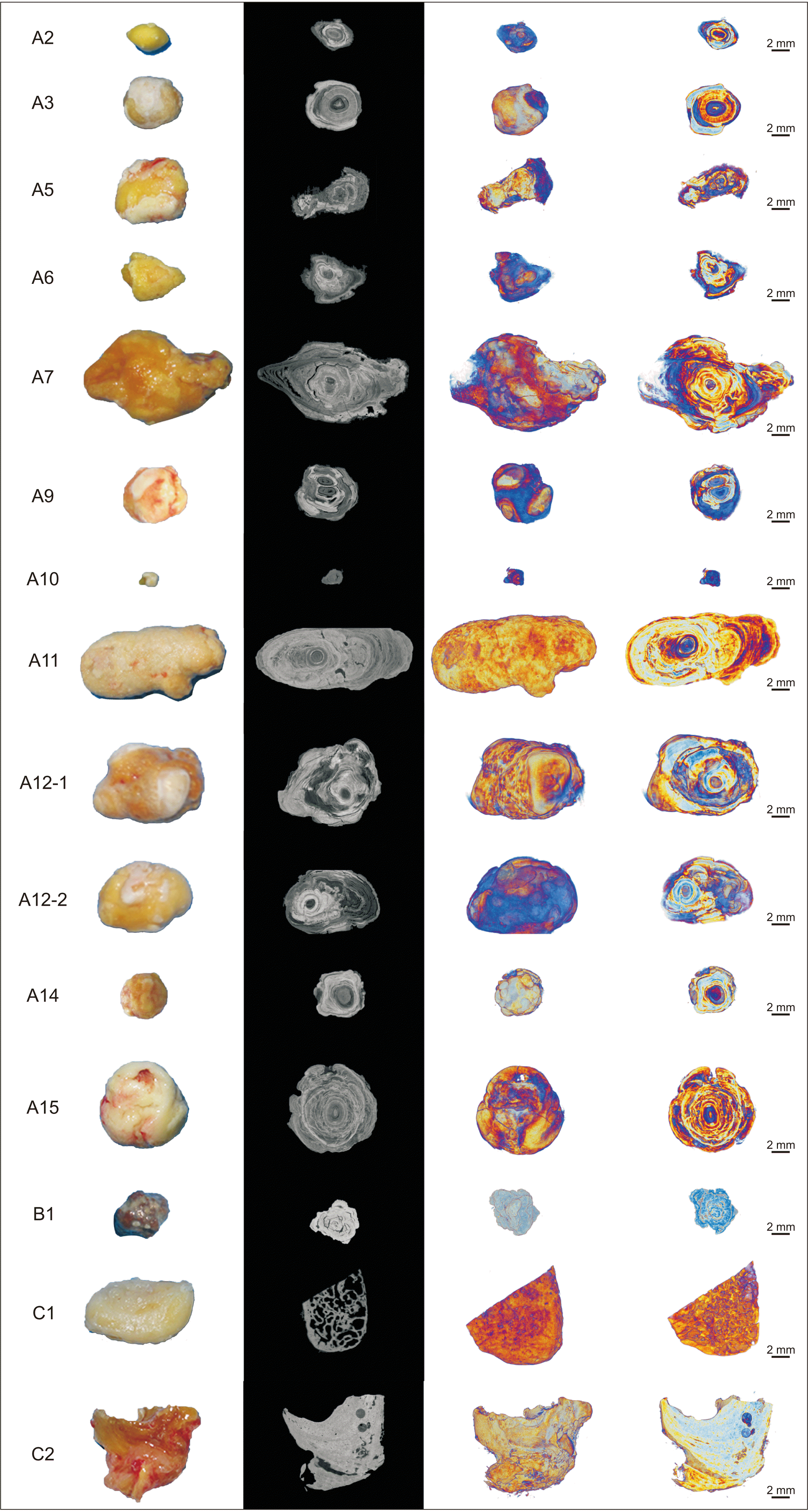 | Fig. 1Representative clinical, two-dimensional micro-computed tomography cross section and three-dimensional reconstructed images of specimen A2, A3, A5, A6, A7, A9, A10, A11, A12-1, A12-2, A14, A15, B1, C1 and C2. Sialoliths show an onion-like concentric lamellar structure. Brighter regions represent higher mineralization and dark regions represent organic substance. Scale bars=2 mm.
|
Table 2
Morphometric parameters analyzed with micro-computed tomography
(TV: total VOI [volume of interest] volume, Obj.V: object volume, Obj.V/TV: percent object volume, TS: total VOI surface, Obj.S: object surface, Obj.S/Obj.V: object surface/volume ratio, Obj.S/ TV: object surface density, St.Th: structure thickness, St.Sp: structure separation, Po(tot): total porosity)
![]()
4. Histopathological findings
1) Histological findings in group A
All sialolith specimens showed concentric lamellar structures with organic and inorganic substances. Eosinophilic characteristics were found at the core. A laminated pattern of a teardrop-shaped globular structure was seen at the periphery. A laminated concentric pattern of the basophilic zone indicating a highly mineralized area of the stone, which is mostly composed of inorganic material, was seen at the peripheral layers. Small amounts of bacteria were located at the outer shell of the sialolith containing salivary ductal epithelium.(Fig. 2. A-C)
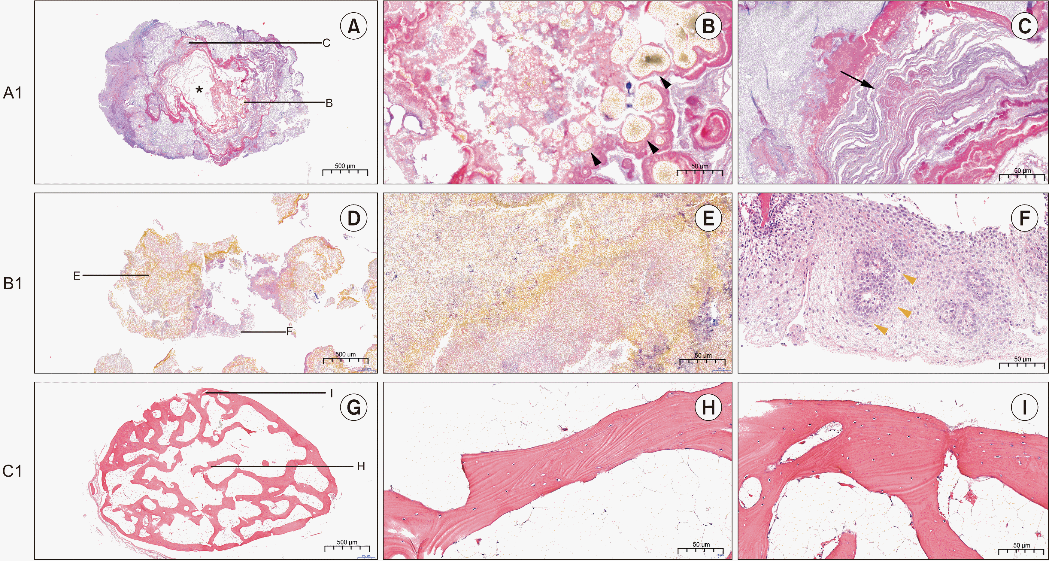 | Fig. 2Representative histological findings of specimen A1, B1, and C1. A. Sialolith A1 with a single organic core in which the core was lost during the histological slide preparation (H&E staining, 4×). Scale bar=500 μm. B. Globular lipid particles found near the core of the sialolith (black arrowheads), (H&E staining, 20×). Scale bar=50 μm. C. Mineralized nodules were found in the outer layers of the core showing irregular mineralization (black arrow) (H&E staining, 20×). Scale bar=50 μm. D. Tonsillolith specimen (B1) (H&E staining, 4×). Scale bar=500 μm. E. The overall specimen exhibits a crystalline structure of organic material at a mature stage of its development (H&E staining, 20×). Scale bar=50 μm. F. A duct-like structure was observed at the center of squamous epithelium indicating the minor salivary gland duct (yellow arrowheads) (H&E staining, 20×). Scale bar=50 μm. G. Antrolith specimen (C1) (H&E staining, 4×). Scale bar=500 μm. H. The lesion was not encapsulated and showed a homogeneous lamellar bone with fibrous marrow cavities (H&E staining, 4×). Scale bar=50 μm. I. The woven bone was replaced by lamellar bone with Haversian canals at the periphery (H&E staining, 4×). Scale bar=50 μm.
|
2) Histological findings in group B
The tonsillolith specimen stained with H&E demonstrated a mature stage of development; the tonsillolith was surrounded by fibrinous debris and inflammatory cells.(Fig. 2. D, 2. E) The tonsillolith showed degrees of immature calcification of organic and inorganic materials. A duct-like structure was observed at the center of squamous epithelium indicative of the minor salivary gland duct.(Fig. 2. F) Squamous epithelium on the surface of the tonsillolith was noted to extend toward the lining of the tonsillar crypt. High magnification revealed that the tonsillolith was composed of a dense matrix, and microbial colonies composed of rods and cocci were observed.
3) Histological findings in group C
The antrolith lesion was not encapsulated and showed a homogeneous lamellar bone with fibrous marrow cavities. The trabecular bone was replaced by lamellar bone with Haversian canals at the periphery.(Fig. 2. G-I) The lamellae of the bone tissue were organized longitudinally in a well-differentiated and interlacing trabecular pattern, which is characteristic of mature cancellous bone. Osteocytes were randomly and unevenly distributed with spindle-shaped lacunae. Abundant eosinophilic and finely granular cytoplasm, eccentrically placed oval nuclei, and absent nucleoli were present. Common characteristics of neoplastic bone, such as thickened trabeculae or architectural atypia, were absent. The attached soft tissue showed inflamed granulation tissue with bone sequestrum and mainly consisted of diffuse inflammatory cells including macrophages, lymphocytes, and plasma cells.
5. SEM findings of sialolith, tonsillolith, and antrolith specimens
1) Ultrastructural findings of group A
SEM findings of specimens from group A revealed a concentric lamellar structure with alternating mineralized and organic material. The sialoliths had distinct three layers: a peripheral multilayer zone, an intermediate compact zone and the central nidus area4,17. Sialolith A4, obtained from the submandibular salivary gland, showed a layered structure.(Fig. 3, A4) At the very outer layer of the sialolith, a long rod-shaped bacterial biofilm cave (yellow arrowheads) was seen with calcium nanoparticles on point 01. The SEM image shows the layered structure of the sialolith (white lines). At the intermediate layers on points 02, 03, and 04, dense hydroxyapatite aggregates were observed with bacterial empty casts (blue arrowheads) at 20,000× magnification. Point 05 showed a more porous structure than points 02, 03, and 04, but rudely hexagonal, fibrous, irregularly shaped hydroxyapatite crystals were observed. Fibrous and irregularly shaped hydroxyapatite crystals showed random orientation from point 05. Point 06 showed the irregularly shaped hydroxyapatite occurring in cluster masses or individual crystals. Points 07, 09, and 11 showed a coarser structure of hydroxyapatite crystal aggregation. Points 08 and 14 depicted a densely aggregated layer of microscopic mineral masses compatible with octacalcium phosphate. A filament pattern of the inorganic matter compatible with carbonate apatite arranged in different orientations was observed on point 10 (red arrowheads). Calcite-like crystals were seen on point 13. The irregular structure showed platy, roughly hexagonal and irregularly shaped hydroxyapatite crystals in random orientations.

2) Ultrastructural findings in group B
The group B specimen did not present with concentric laminated structures. SEM analysis of the tonsillolith showed hexahedral-shaped crystals on all points of interest.(Fig. 3, B1) The outer layer of the tonsillolith showed a filamentous structure that suggested bacterial accumulation on point 10.
3) Ultrastructural findings in group C
Group C showed a homogeneous structure without concentric laminated mineral deposits. The antrolith specimen from group C (C1) was analyzed at the cross-sectional surface with 14 focal points of interest. Non-crystalline and smooth surfaces were found in the peripheral and central area of the specimen at points 01-11. Coarse crystalline microaggregates were identified in the core regions.(Fig. 3, C1).
6. Chemical composition of sialolith, tonsillolith, and antrolith specimens
The chemical composition of the specimens was analyzed and recorded at the peripheral, middle, and core layers. The compositional analysis of specimens in group A, B, and C consistently demonstrated a high fraction of calcium (Ca), carbon (C), phosphate (P), and oxygen (O).(Table 3) However, the level of each element varied in each group. The wt% and at% of Ca in group A were higher (20.72±5.53 wt% and 9.55±2.77 at%) compared with group B (17.48±3.02 wt% and 8.23±1.51 at%) and group C (17.18±6.98 wt% and 7.28±3.31 at%). However, the differences were not significantly different.(Table 3) The C level wt% and at% were significantly higher in group C (32.86±17.60 wt% and 43.23±17.25 at%) compared with group A (21.36±4.50 wt% and 31.85±5.29 at%) and group B (15.33±4.50 wt% and 23.92±6.40 at%) (P=0.006; P=0.004, respectively). The wt% and at% of O was significantly higher in group B (38.31±2.70 wt% and 45.95±3.46 at%), compared with group C (29.24±3.21 wt% and 30.52±4.33 at%) (P=0.016; P=0.002, respectively). The wt% and at% Ca/P ratios were 1.19±0.85 and 2.20±2.33, respectively, in group A; group B showed a wt% of 1.27±0.62 and at% of 0.97±0.48, and group C showed a wt% of 3.59±1.54 and at% of 2.77±1.34.
Table 3
Comparison level of Ca, C, O, and P between groups
![]()
The comparative chemical composition analysis between the groups is shown in Table 4. In addition to the major three elements that constituted the sialolith, tonsillolith, and antrolith specimens, many other elements were detected, including fluorine (F), nitrogen (N), sodium (Na), silicon (Si), and magnesium (Mg); these were detected in all groups. Other elements such as copper (Cu) and zinc (Zn) were found only in group A. Traces of iron (Fe) were found only in group B (Fig. 4), while zirconium (Zr) was found in both groups A and B. The wt% of N was significantly higher in group A (14.68±5.63 wt%) compared with groups B and C (P=0.031).
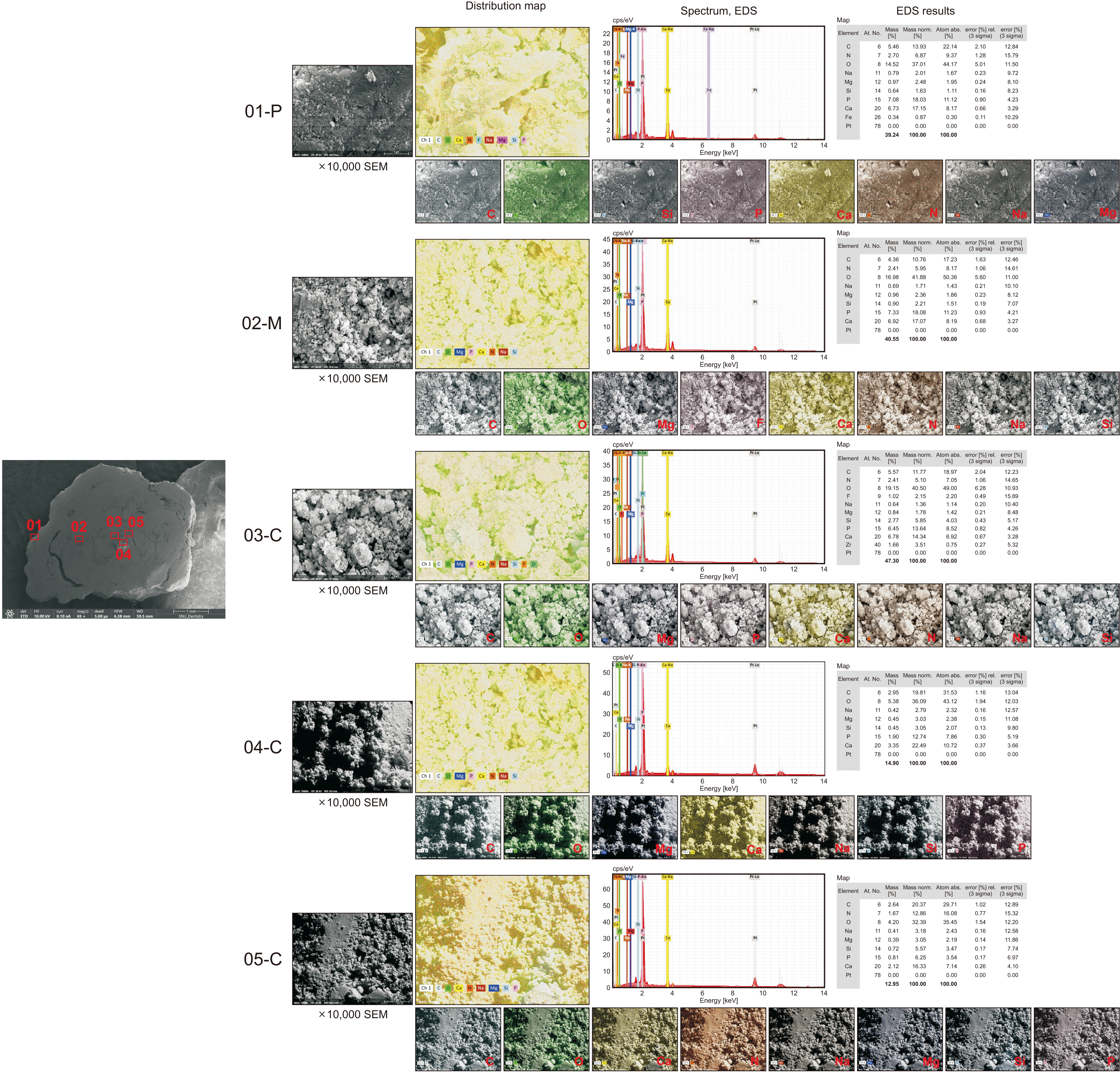 | Fig. 4Representative mapping of elemental distribution and a spectrum of the representative points with energy dispersive X-ray spectroscopy (EDS) results in tonsillolith (B1). Scanning electron microscopy (SEM) image, 10,000× magnification. EDS analysis was carried out at five representative points of interest on the peripheral, middle, and core layers.
|
Table 4
Elemental composition of the specimen in groups A, B, and C
![]()
7. TEM analysis
In TEM analysis, highly mineralized/crystalline regions appear dark in the bright field imaging due to staining and/or the diffraction of heavy elements. A layered appearance of sialolith A4 showing globular vesicular structure in the central core area with a heterogeneous crystalline needle-like pattern was observed18.(Fig. 5. A, 5. B) Large single crystals and deposition of inorganic material at the inner lamellae were observed in darker contrast compared with the surrounding structure. Needle-like filamentary crystals suggested hydroxyapatite structure.(Fig. 5. C, 5. D)
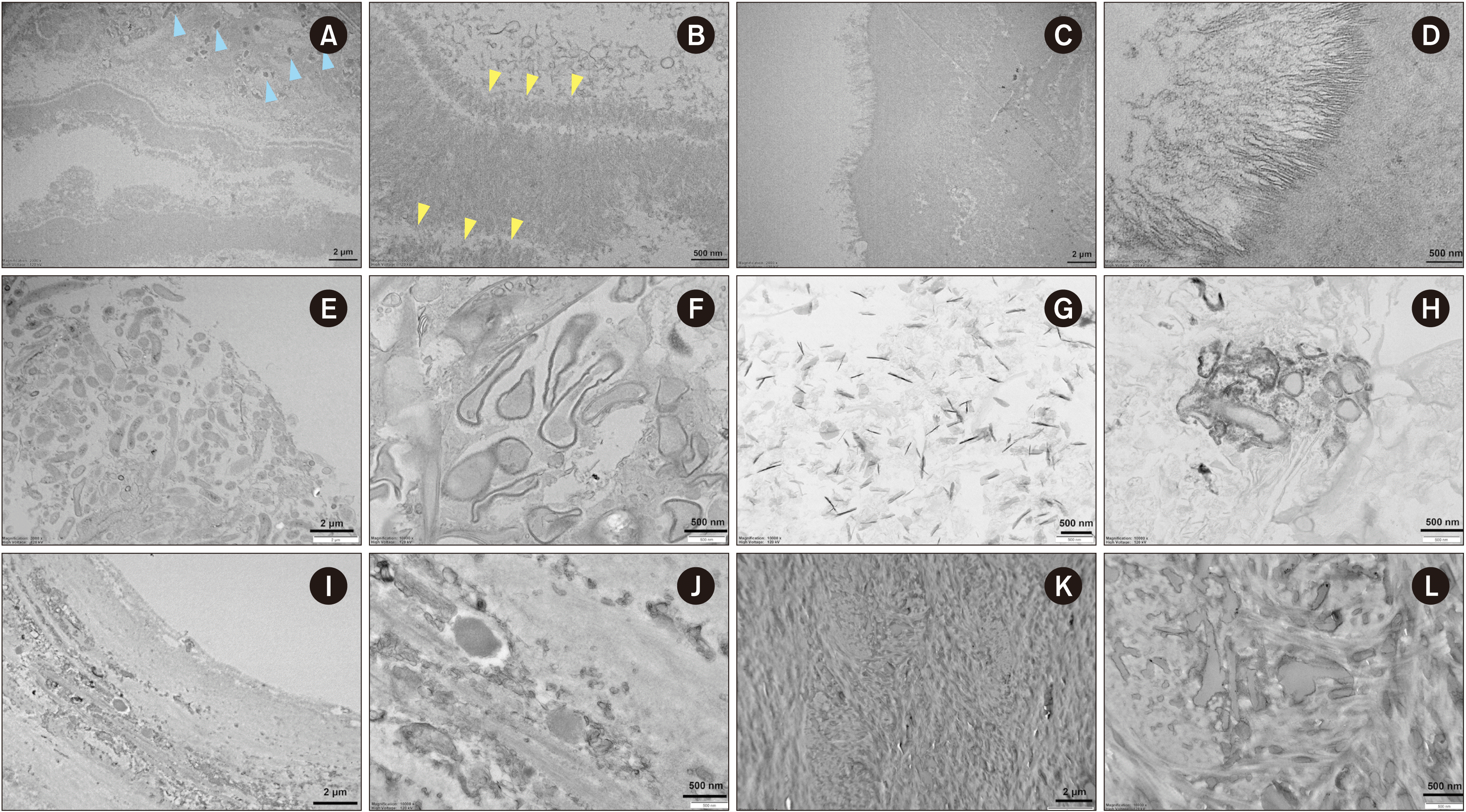 | Fig. 5Representative transmission electron microscopy (TEM) images of A4 sialolith (A-D), B1 tonsillolith (E-H), and C2 antrolth (I-L). Layered appearance of the sialolith showing the globular mineralized structure in internal lamella, (blue arrowheads), while the crystalline needle-like pattern was heterogeneous (yellow arrowheads), magnification 2,000×, 10,000× (A, B). Needle-like filamentary crystals, magnification 2,000×, 20,000× (C, D). Representative TEM images of B1 tonsillolith. The B1 tonsillolith had stratified squamous epithelium in its peripheral area, magnification 2,000×, 10,000× (E, F). Needle-like crystals in the core region and extra-vesicular deposition of inorganic material were also observed, magnification 10,000× (G, H). Representative TEM images of C2 antrolith. Outer layer showing osteoblastic rimming, magnification 2,000×, 10,000× (I, J). Dense, mature, predominantly lamellar bone in the middle area of the specimen, magnification 2,000×, 10,000× (K, L).
|
The B1 tonsillolith had stratified squamous epithelium in its peripheral area.(Fig. 5. E) Bacteria were present in the middle layer of the tonsillolith.(Fig. 5. F) Needle-like crystals in the core region and extra-vesicular deposition of inorganic material were also observed.(Fig. 5. G, 5. H) In the outer layer of the C2 antrolith, an osteoblastic rimming was observed.(Fig. 5. I, 5. J) The middle region of the specimen was mainly composed of dense, mature, and predominantly lamellar bone.(Fig. 5. K, 5. L) The core region had no Haversian canals and fibrous component.
Go to :
IV. Discussion
Kidney stones have been formally studied since 1802. Extensive study has been conducted on the pathophysiology, microstructure, chemistry, microbiome composition, prevention and treatment of kidney stone formation19-25. Calcifications in the articular cartilage, cardiovascular tissues, and kidneys are studied extensively due to their symptomatic nature. However, few studies have conducted ultrastructural and elemental analysis of calcifications in the head and neck region, including sialoliths, tonsilloliths, and antroliths.
Stoodley et al.14 showed that tonsilloliths exhibit a similar structure and chemical gradients to a dental biofilm. The physiological activity was analyzed by confocal microscopy and by microelectrodes that measured aerobic and anaerobic respirations and acid production14. In our study, we found filamentous bacteria at the peripheral layer of the specimen. The composition of the developing biofilm is determined by several factors such as the site of colonization and the type of the surface26.
Antroliths are a rare entity and are usually asymptomatic. In most cases, antroliths are discovered incidentally during routine examinations and are thought to have endogenous and exogenous origins16. In our literature review, there was a lack of information on ultrastructural and elemental analysis of antroliths. As the incidence of antroliths is rare and there is a lack of research on this field, the pathogenesis of antroliths is not completely understood. In our study, we found that the antrolith specimen had a different structure compared with sialoliths, which had a concentric laminated structure with a core.
The study of the core of lithiasis provides valuable information on the generation, precipitation, and aggregation of minerals27. The determination of the structural characteristics and the composition of all parts of the stone especially the core is essential for understanding the mechanism of stone genesis17. In human kidney stones, amorphous calcium phosphate was found to promote the aggregation of amorphous calcium oxalate complexes, which induce the nucleation and growth of urinary stones28. Several theories have been proposed to explain sialolith formation, and many have focused on the core or nucleus that may initiate the development. Takeda proposed a mechanism in which crystalloid present in a salivary gland may aggregate and form a core in which organic and inorganic materials deposit29. However, we found that the antrolith specimen had no core and consisted of a lamellar bone structure. This bone structure may originate from the surrounding bone of the maxillary sinus wall because of pathological conditions involving the sinus. Another study using SEM and TEM analysis reported similar qualities of sialoliths and calculus, such as a layered structure and presence of crystals. This correlation could clarify the mineralization process in sialoliths6. However, in the current study, we did not find similarities between sialoliths, tonsillolilths and antroliths.
The comparative micro-CT, SEM, and TEM analyses of groups A, B, and C revealed that the sialolith, tonsillolith, and antrolith specimens had different micromorphology, suggesting different pathways of formation. Sialoliths mostly had concentric laminated structure, in which the chronological order of development could be analyzed in detail. However, the tonsillolith and antrolith specimens showed homogeneous structures without any layered microstructure. Micro-CT analysis allowed nondestructive imaging of the major core of the sialolith. The cores are sometimes less mineralized and may be damaged or missed during the specimen preparation process for SEM and TEM30. From the micro-CT scanning, the cores of each sialolith could be classified as highly mineralized (A3, A15) and less mineralized (A5, A7, A9, A11, A12-1, A12-2, A13, A14) in comparison with the surrounding layers. These findings were confirmed with the histology findings showing a less mineralized core in sialolith A1 surrounded by contrasting higher mineralized middle and outer layers. In a previous study, no organic cores were observed in the central part of the sialolith31. The presence of concentric laminated structure threaded with globules could be explained by the Liesegang and Ostwald precipitation mechanism32,33. A combination of micro-CT and ultrastructural analysis with SEM and TEM can be effectively used to characterize specimens.
The main elements of sialolith are Ca, P and O. A study using Fourier transform infrared (FTIR), FT-Raman, and fluorescence spectroscopic techniques showed that the ratio of the major elements Ca and P was 7:334. In another study using X-ray microanalysis, the component elements of sialolith were Ca, O, S, and Na. The Ca and P ratio was calculated to be 1.60-1.8935,36. The chemical constitution of each specimen varied from one to another. In our study, we found Ca, Ca, C, O, P, F, N, Si, Na, and Mg in sialolith17, tonsillolith, and antrolith specimens. Only sialoliths had Cu, Zn, and Zr, while the tonsillolith had traces of Fe. The main inorganic components of sialoliths are reported to be apatite (Ca10(PO4)6(OH)2), whitlockite (Ca3(PO4)2), and brushite (CaHPO4 2H2O). Other components include weddellite and octacalcium phase.
Other human liths such as renal and kidney stones have been studied extensively due to their symptomatic nature, such as pain, infection, and obstruction, which can affect patient quality of life. Acute passage of kidney stones is the ninth most common cause for emergency visit37. In contrast, sialoliths are generally asymptomatic38. The mechanism/pathophysiology of kidney stone formation has been established and preventative methods along with minimally invasive treatment methods, such as dissolution therapy of kidney stones, have been developed19. In some cases, depending on the size, location and the accessibility of the sialolith, extirpation of the salivary gland may be performed, where the sialolith is located in the parenchyma of the salivary gland or the proximal part of the duct. Dissolution of a sialolith located in a difficult location as a minimally invasive method would be a desirable approach over the extensive extirpation of the whole salivary gland. Better understanding of sialolith growth can be achieved though gaining knowledge on the sialolith chemical composition, crystalline microstructure and the biomolecules derived from the host and the resident bacteria. In the current study, the main chemical composition of sialoliths were Ca, C, O, and P, which were found in all layers17. In the process of sialolith formation, the role of bacteria, the interaction of calcium nanoparticles, salivary exfoliated host cells, and protein coagulation by different crosslinking enzymes in saliva are important factors for sialolith formation. Therefore, the goal may be to reduce the risk of precipitation or aggregation of calcium nanoparticles in the lithogenesis of sialoliths.
Go to :
V. Conclusion
The main elemental components of sialoliths were O, C, Ca, N, Cu, P, Zn, Si, Zr, F, Na, and Mg. The peripheral layer had an expanded elemental composition that included Cu, Zn, and Zr compared with that of the middle and core regions, while the middle layer had a small amount of F. In the tonsillolith specimen, a small amount of Fe was found in the peripheral region. The antrolith had a shorter component list: Ca, C, O, P, F, N, Si, Na, and Mg. The sialoliths had distinct three layers: a peripheral multilayer zone, an intermediate compact zone and the central nidus area; the tonsillolith and antrolith specimens lacked distinct layers and a core. The antrolith showed a structure and composition similar to that of lamellar bone.
Go to :
 XML Download