Journal List > Ann Hepatobiliary Pancreat Surg > v.28(1) > 1516086292

Nguyen, Nguyen, Luong, Dang, Le, Tran, Do, Pham, Pham, Tran, Nguyen, Trinh, and Inoue: Pancreaticoduodenectomy with superior mesenteric artery first-approach combined total meso-pancreas excision for periampullary malignancies: A high-volume single-center experience with short-term outcomes

Abstract

Backgrounds/Aims

Pancreaticoduodenectomy (PD) is the only radical treatment for periampullary malignancies. Superior mesenteric artery (SMA) first approach combined with total meso-pancreas (MP) excision was conducted to improve the oncological results. There has not been any previous research of a technique that combines the SMA first approach and total MP excision with a detailed description of the MP macroscopical shape.

Methods

We prospectively assessed 77 patients with periampullary malignancies between October 2020 and March 2022 (18 months). All patients had undergone PD with SMA first approach combined total MP excision. The perioperative indications, clinical data, intra-operative index, R0 resection rate of postoperative pathological specimens (especially mesopancreatic margin), postoperative complications, and follow-up results were evaluated.

Results

The median operative time was 289.6 min (178−540 min), the median intraoperative blood loss was 209 mL (30−1,600 mL). Microscopically, there were 19 (24.7%) cases with metastatic MP, and five cases (6.5%) with R1-resection of the MP. The number of lymph nodes (LNs) harvested and metastatic LNs were 27.2 (maximum was 74) and 1.8 (maximum was 16), respectively. Some (46.8%) patients had pancreatic fistula, but mostly in grade A, with 7 patients (9.1%) who required re-operations. Some 18.2% of cases developed postoperative refractory diarrhea. The rate of in-hospital mortality was 1.3%.

Conclusions

The PD with SMA first approach combined TMpE for periampullary malignancies was effective in achieving superior oncological statistics (rate of MP R0-resection and number of total resected LNs) with non-inferior short-term outcomes. It is necessary to evaluate survival outcomes with long-term follow-up.

INTRODUCTION

Periampullary cancer is a widely used term of a heterogeneous malignant group from the periampullary area, which includes the pancreatic head, the distal common bile duct (CBD), the ampulla of Vater, and the duodenum [1]. Periampullary cancers are still aggressively lethal, and becoming the second most-common cancer-leading-death malignancies [2]. Radical surgery, or pancreaticoduodenectomy (PD), remains the only radical treatment. The indication of PD was extended due to the development of neoadjuvant therapies [3]. Moreover, several new methods have been developed in PD to accomplish the oncological outcomes. Among them, the most bullet points are the superior mesenteric artery (SMA) first-approach and total meso-pancreas (MP) excision. With up-to-date knowledge in the oncological process and the involvement of periampullary malignancies, especially pancreatic ductal adenocarcinoma (PDAC), many systematic reviews and meta-analysis have discussed and shown the superior aspects of the SMA first approach, as well as SMA circumferential lymphadenectomy, against a convenient approach [4,5].
As the positive posterior margin after PD in pancreatic head cancer has been reported as frequently as 82% of all cases, it is important to define retroperitoneal and retro-pancreatic soft tissues to achieve an adequate posterior clearance [6]. Gockel et al. [7] first established an anatomical concept named the MP in 2007, which is known in other terms, such as the meso-pancreato-duodenum, to represent the firm, well-vascularized, and peri-neural connective tissue between the posterior area between the pancreatic head and the celiac trunk, as well as the mesenteric vessels [8]. The application of total MP excision (TMpE) in benign, as well as borderline disease, is still discussed and not recommended. Otherwise, after over 15 years from its inception, several original articles, including a randomized cohort study, and a systematic review, had proven that TMpE raises the rate of R0-resections, and the number of harvested lymph nodes (LNs), as well as reducing the total and local recurrence rate, and improving the disease-free survival rate [9-12]. However, no previous research has analyzed the PD technique by combining the three procedures (SMA first approach, as well as SMA circumferential lymphadenectomy. and TMpE), in terms of its feasibility, safety, and efficacy in short-term operative and oncological outcomes. Therefore, this article 1. summarizes our experiences in conducting the PD technique that includes a combination of the SMA-first approach and total MP excision, 2. evaluates the feasibility, safety, and efficacy in short-term outcomes of this procedure, and 3. contributes to our understanding of the concept of MP and TMpE. In our study, we have focused on analyzing technical aspects, as well as the short-term operative and oncological outcomes of the three combined procedures (SMA first approach, SMA circumferential lymphadenectomy, and TMpE) in one procedure. In long-term outcomes, we are conducting a comparative study of each histological subgroup of patients who have undergone our procedure with a propensity score-matched analysis, and we anticipate publishing the results soon.

MATERIALS AND METHODS

Data collection

The Institutional Review Board of Bach Mai Hospital reviewed and approved the research protocol (IRB no. 00003121). Written informed consent for publication of their clinical details and clinical images was obtained from the patients’ family.
All patients with a histologically proven diagnosis of periampullary malignancies who underwent PD with TMpE via SMA first approach at the Department of Gastrointestinal and Hepato-Pancreato-Biliary Surgery from October 2020 to March 2022 (18 months) were prospectively enrolled.
Patients’ data were prospectively collected, including:
General and preoperative information: Demographic characteristics (sex, age, height, weight, body mass index [BMI], and medical history), tumor location (periampullary sites of origin: pancreatic head parenchyma, distal CBD, ampulla of Vater, and second portion of duodenum), the rate of preoperative biliary drainage (percutaneous transhepatic biliary drainage with cases where surgery was delayed at least 2 weeks due to severe obstructive jaundice (serum bilirubin ≥ 250 μmol/L), cholangitis, severe pruritus or other comorbidities, etc. [13,14]).
Intraoperative data and Short-term outcomes: intraoperative data (operative time, blood loss, intraoperative complications and vascular resections), Tumor dimension, types of SMA first approach and pancreatic reconstruction, postoperative statistics (length of hospital stay, postoperative complications including post pancreatectomy hemorrhage [PPH], in-hospital mortality) were collected, especially including the frequency of diarrhea (defined as a condition requiring opioid antidiarrheal drug for at least 6 months after surgery), and the rate and classification of postoperative lymphorrhea (PL) according to Besselink’s classification [15] and postoperative pancreatic fistula (POPF) according to the 2016 International Study Group (ISGPS) definition and grading updated version [16].
Histopathological data: The pancreaticoduodenal specimen, resected margins (including the MP margin), LNs, and the MP were marked and measured by the surgeon on the back-table. Tumor grading, number of LNs retrieved, number of metastatic LNs, metastatic status of LN No. 14 and MP, pathological TNM staging according to the eighth edition of the AJCC/UICC system of periampullary malignancies [17], and resected margins status were determined. The margin status was defined as follows: no cancer cells are identified microscopically at any of the resected margins (R0), microscopic tumor present at any margin or within 1 mm of a transection or circumferential margin according to the Royal College of Pathologists guidelines (R1), and macroscopically residual tumor as determined by the surgeon intraoperatively (R2) [18]. The MP size and dimension were recorded systematically with five indices.
The primary endpoint was LNs characteristics (retrieved/metastatic LN), metastatic status of LN no. 14, MP, and main complications (POPF, PPH, PL, and diarrhea).

Anatomic and histological concept of meso-pancreas

Histologically, MP, started by Gockel et al. [7] in 2007, is a well-vascularized and peri-neural connective tissue between the posterior pancreatic head and the celiac trunk, as well as the mesenteric vessels, including important structures: the first and second nerve plexuses of the pancreas head (plPh-I and plPh-II), the inferior pancreatoduodenal arteries (IPDAs), the first jejunal arteries (FJAs) and first jejunal veins (FJVs), and regional LNs [19]. In anatomic concept, it was a connective tissue between the pancreas head to the SMA and the celiac trunk. The anatomical landmarks were the posterior surface of the head, neck, and uncinate process of the pancreas, the right side of the SMA, superior mesenteric vein (SMV), and celiac artery (CA), and the anterior of the inferior vena cava, and abdominal aorta [20].

Surgical procedure

Our procedure can be conducted in laparotomy, as well as laparoscopically. In the laparotomic approach, an upper abdominal incision was performed to explore the peritoneum to exclude metastases. Meanwhile, in the laparoscopic approach, we use 5 trocars, which procedure was described in our technical article [21]. We use three approaches to first explore the SMA root:
With the left-posterior approach, which was our main approach, which was specified by removing the LNs groups 14th en bloc, we exposed the SMA from the anterior to the left posterior side from the caudal side to the origin (Fig. 1), including LN station No. 14 or the left side of SMA LNs, and the left-sided Kocher maneuver. The detailed technique has been described in our previous article (Fig. 2) [21].
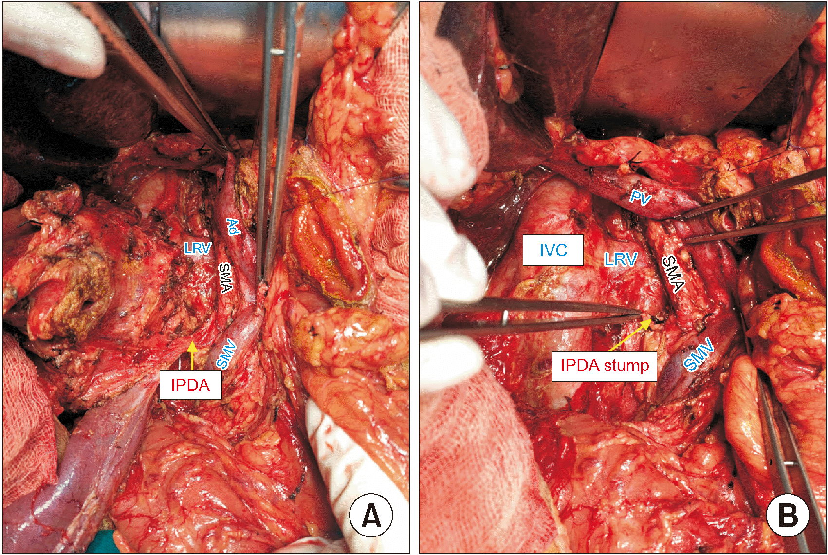
With the right or medial uncinate approach, which was applied in the cases of malignant tumors of uncinate process, we followed a procedure similar to the approach by Hackert et al. [22] (Fig. 3).
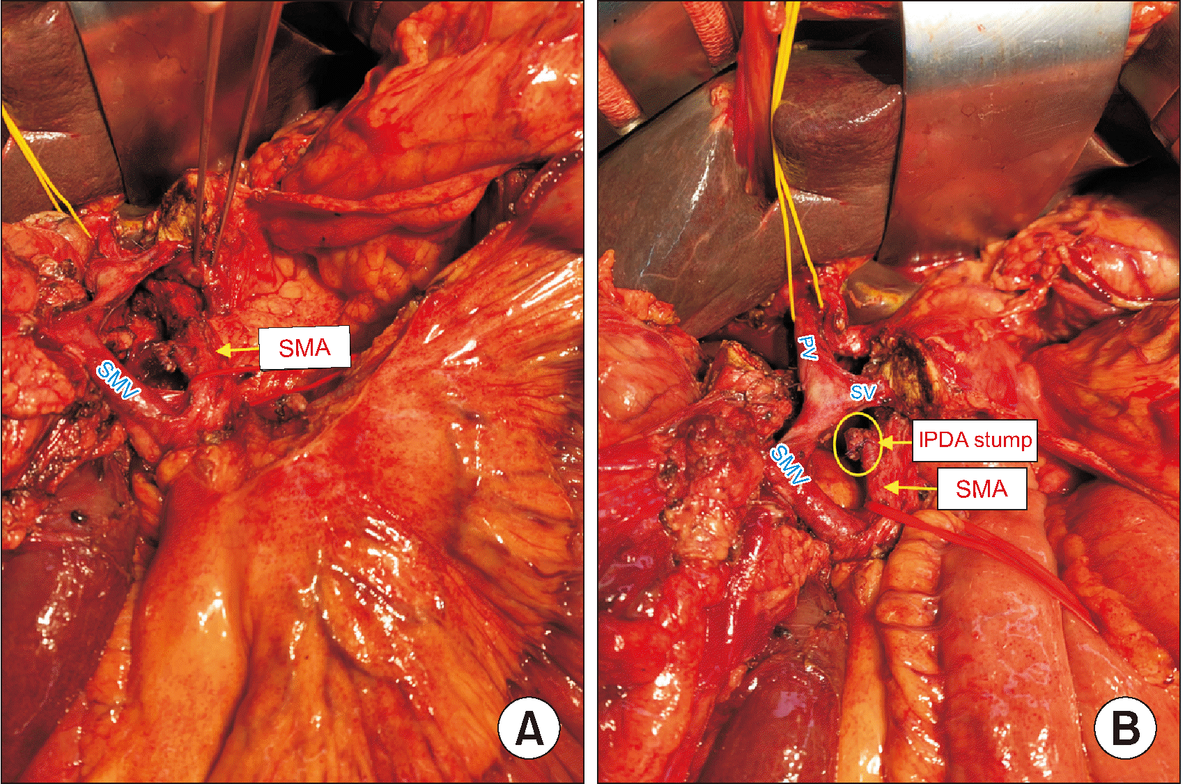
With the mesenteric or inferior infra-colic approach, which was applied in the cases of locally advanced tumors with susceptive infiltration of SMA origin, or malignant tumors of uncinate and ventral pancreas, we followed a procedure similar to the approach by Weitz et al. [23] (Fig. 4).
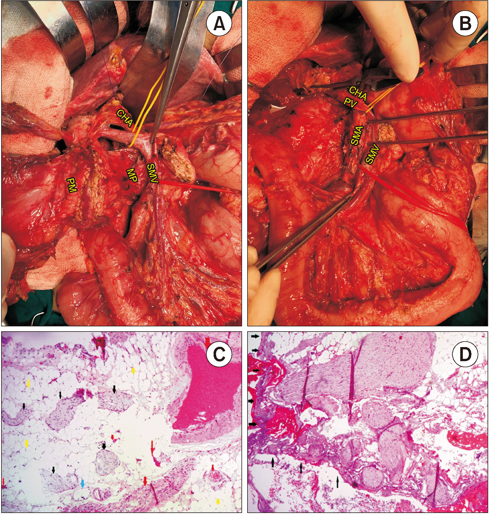
After continuous to mobilize of the entire pancreaticoduodenal mass by convenient Kocher maneuver and approaching the SMV/portal vein (PV) from the posterior side of the pancreas, as well as transecting the stomach and pancreatic neck, dissect the hepatoduodenal ligament and the celiac LNs; the total MP excision from the right side of the SMA and abdominal aorta will be easily and conveniently performed afterwards, including the right-side of SMA LNs (Fig. 5). The reconstruction phase includes pancreato-jejunostomy with double layers end-to-side, modified Blumgart-fashion, hepaticojejunostomy distal to the pancreato-jejunostomy about 10 cm with one layer fashion, and the Antecolic gastro-jejunostomy.
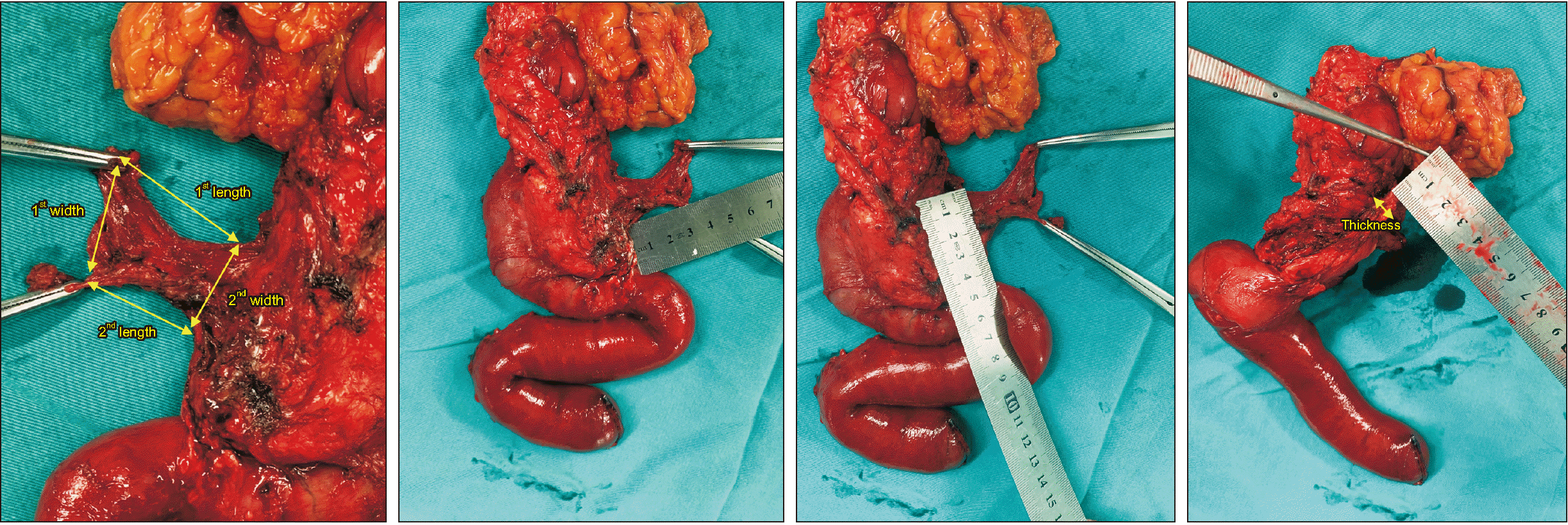
In our study, the MP was resected totally en bloc with entire pancreaticoduodenal mass. The MP size and margins in the specimen were recorded and marked in all cases on the back-table by the surgeon (Fig. 6). A extended lymphadenectomy was performed in all cases, which included LNs stations 5, 6, 7, 8a, 8p, 9, 12b1, 12b2, 12c, 12p, 13a, 13b, 14p, 14d, 17a, and 17b, according to the New Japanese Classification and the ISGPS consensus [11,19,24].

Statistical analysis

All variables were categorized into categorical, discrete, or continuous variables. Categorical variables contain a finite number of categories or distinct groups and are presented as a percentage of each value. Discrete variables are numeric variables that have a countable number of values between any two values. Continuous variables are numeric variables that have an infinite number of values between any two values, and are expressed as the mean ± standard deviation, or median (interquartile range) if without normal distribution. Continuous variables were compared by means of the t-test of the Wilcoxon rank sum test, when appropriate. Various nominal variables were analyzed using the chi-square test or Fisher’s exact test, when appropriate. All data were analyzed using SPSS for Windows, version 22.0 (SPSS Inc.).

RESULTS

From October 2020 to March 2022 (18 months), 105 cases of PD with SMA first approach and TMpE were conducted in our center (we have followed our procedure in all cases of resectable and/or borderline resectable periampullary neoplasms, because we have not taken preoperative pancreatic biopsy routinely, due to its high risk of pancreatic fistula and hemorrhage). Some 28 cases were excluded from the research due to non-malignancy in the final histological results. Finally, there were 77 cases of periampullary malignancies that underwent PD with SMA first approach and TMpE.

General and preoperative information

There were in toto 43 males and 34 females in our study. The median age was 58 years (yr) (range 12−83 yr). Height was 160 cm; weight at surgery was 54 kg, BMI was 21 kg/m2 (range 14−26 kg/m2). Preoperative biliary drainage was conducted before surgery in 11 patients. Some 26% of patients had a medical history of diabetes, as well as hypertension. Following the tumor location, 50.6% of patients had tumors arising from pancreatic parenchyma (Table 1).

Intraoperative data and short-term outcomes

PD was performed in 74 patients, while total pancreatectomy was performed on the remaining 3 patients (who had the locally advanced or multifocal pancreatic tumors [25]). There were 62 laparotomic PDs and 15 laparoscopic PDs. There were 9 cases in which resection of the PV and/or the SMV was performed. There were respectively (60, 9, and 8) patients for whom the left-posterior SMA first-approach, right approach, and mesenteric approach, respectively, was performed. TMpE was performed in all cases. Pancreatic reconstruction was performed using the binding pancreatico-enterostomy in 94.8% patients, while binding pancreaticogastrostomy was performed in 5.2% patients.
Median operative time was 289.6 ± 95.8 min (range 178−540 min). Median hospital stay was 23.2 ± 8.3 days (range 9−50 day). Mean intra-operative blood loss was 209 ± 273 mL (range 30−1,600 mL), and 10 patients needed intraoperative blood transfusion. Postoperative complications occurred in 37 cases (21 had grade-1-or-2 complications, while 16 had grade-3a-or-more complications, including hospital mortality according to the Clavien–Dindo classification). POPF occurred in 36 cases, with 34 cases of grade A or B, and 2 cases of grade C. PPH happened in 7 cases, with 3 cases requiring interventions, and 4 cases requiring re-operations (in total, 7 cases of re-operations; the other 3 cases were two cases due to complications of gastrojejunostomosis candidiasis [26], and one case of postoperative grade C pancreatic fistula). Some 14 (18.2%) cases developed postoperative refractory diarrhea. Some 15 (19.5%) cases were conditions of PL, with 10 cases of grade A or B, and 5 cases of Grade C requiring percutaneous hepatic lymphangiography (HLG) [27].
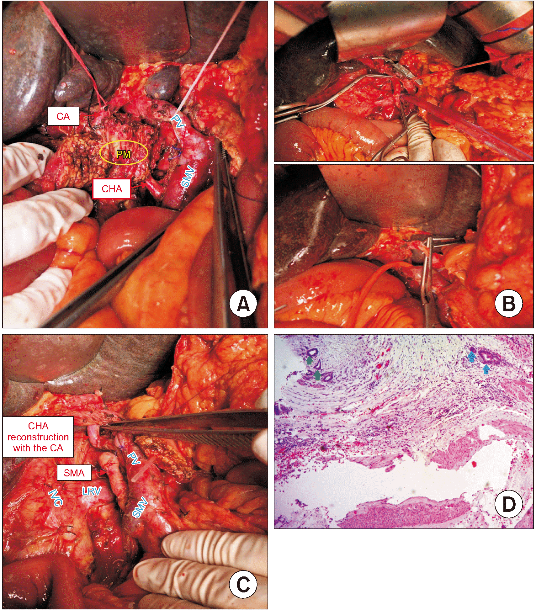
Hospital mortality occurred in 1 case (1.3%), secondary to grade C PPH: the patient was 52 years old with anatomical abnormality of the common hepatic artery (CHA) come from SMA, and the tumor had invaded to the CHA (preoperative negative biopsy did not allow neoadjuvant chemotherapy), so that an extended pancreatoduodenectomy with reconstruction of the CHA (vascular anastomosis between the CHA with the CA by the segment of inferior mesenteric vein) was performed; while in POD 10, the patient had an acute peritoneal hemorrhage due to vascular anastomosis rupture, probably from a pancreatic fistula, and died from cardiac arrest after reoperation (Table 2).

Histopathological data

All patients had a malignant tumor, including 39 of pancreatic parenchyma (adenocarcinoma, intraductal papillary mucinous carcinoma, neuroendocrine carcinoma…), 12 cases of distal CBD, 22 cases of ampulla of Vater, and 4 cases of duodenal second portion. The mean diameter of tumor was 33.3 ± 20.8 mm. Well, moderately, and poorly differentiated histopathology was obtained in 14, 47, and 16 cases (18.2%, 61.0%, and 20.8%, respectively).
Negative margin of pancreatic parenchyma was achieved in all cases. Eight in 9 cases of portal/SM vein resection had venous wall invasion. In general, MP was macroscopically trapezoid-liked shape with a size of 46.1 mm × 39.1 mm × 25.1 mm × 20.7 mm, and a thickness of 4.7 mm. Microscopically, there were 19 (24.7%) cases with metastatic MP, and five cases (6.5%) with R1-resection of the MP (Fig. 7, 8). The number of LNs harvested and metastatic LNs was 27.23 (maximum was 74) and 1.79 (maximum was 16), respectively. The rate of metastatic LN No. 14 was 17.5% (Table 3).

Logistics regression for primary endpoint

For short-term complications, in the logistics regression, abdominal approach (open vs. laparoscopic) is the only predictor for POPF complications (odds ratio [OR], 3.558; 95% confidence interval [CI], 1.006−12.585; p = 0.041), metastatic status of MP is the only predictor for PL complications (OR, 3.646; 95% CI, 1.105−12.034; p = 0.028) (Table 4).

DISCUSSION

The SMA, or artery, first-approach was first established in 2006 by Pessaux et al. [28]. There were some reasons for applying this technique as a standard approach for the treatment of periampullary malignancies. Firstly, following the updated Guideline of the National Comprehensive Cancer Network, the accurate SMA invasion was decided at the preoperative stage and resectability of the periampullary tumor, for which SMA circumferential interface > 180° is an unresectable sign, while SMA circumferential interface < 180° is borderline resectable [29]. So that, it is required and necessary to determine the tumoral invasion of the celiac trunk, as well as the superior mesenteric vessels, especially before the point of no return (the step of dissection of the pancreatic neck or division of the bile duct). Secondly, a majority of postoperative morbidity and mortality was associated with the complexity of anatomical variation of superior mesenteric vessel branches and tributaries [4]. The advantages of the SMA first-approach in the R0-resection rate, postoperative complications (including pancreatic fistula and bleeding), as well as overall survival (OS), were proven in recent meta-analyses [4,5]. With this importance, the “SMA-first approach” concept has been developed in many studies, in open PD, as well as total or assisted laparoscopic PD. To date, there are six methods in the “SMA-first approach” concept in open PD that have been summarized with the advantages, disadvantages, and indications of each approach by Sanjay et al. [30]. In laparoscopic PD, a recent systematic review has categorized the modified “SMA-first approach” methods into four approaches that include: anterior, posterior, right, and left approaches [31]. Our technique, which was developed initially in laparoscopic PD, and was then applied successfully in open PD, was called the “left posterior SMA first approach,” being based on dividing the FJA and FJV in their origin from the left-side after the removal of circumferential SMA LNs from the left anterior side to the posterior side by pulling the proximal jejunum to the left, then dissecting the first jejunal loop.
The high frequency of LN metastasis is one of the most influential, as well as an independent prognostic factor for the survival outcomes of resectable PDAC tumor [32]. According to Okada’s study [33], the rate of left-sided SMA LNs metastasis in PDAC was 12%. Moreover, right-sided SMA LNs are conventionally resected in PD with total MP dissection [34, 35]. Following the most recent Japanese LN station system, the LN No. 14 is divided into LN No. 14p and 14d, which was located surrounding the SMA, as well as in an anatomical–surgical layer of the “mesopancreatoduodenum,” which are not included in standard lymphadenectomy [7,36]. Hence, the term of SMA LNs circumferential dissection was a form of extended lymphadenectomy. Otherwise, there were some preliminary evidence that proved that SMA LNs circumferential dissection has superior survival outcomes without increasing postoperative complications, especially postoperative diarrhea [34]. In our research, the rate of metastatic left-sided LN No. 14 was 17.5%, which was quite similar to the result of Okada’s study [33]. This can be explained by our patients often detecting the disease at an advanced stage when the tumor has appeared with LN metastases.
In our study, there were 19 (24.7%) cases with metastatic MP. Several recent research reports have revealed that the MP was represented as an anatomical area of 66.6% cases of positive (R1 and R2) resection margins [37,38]. From a pathological aspect, the MP was a retroperitoneal, well-vascularized, and nerve-rich structure surrounding the SMA, including important structures: the first and second nerve plexuses (pl-) of the pancreas head (plPh-I and plPh-II), the IPDAs, the FJAs and FJVs, and LNs [8,19]. Otherwise, until now, there has not been any consensus yet in anatomical landmarks of the MP, though basically, the MP connects the pancreatic head to the SMA and right celiac ganglion [38]. Following the results of recent retrospective studies and a systematic review, TMpE has increased the R0-resection rate, so that the local tumor control and long-term outcomes in periampullary malignancies have been improved [7, 39]. The term “TMpE” was classified by Inoue et al. [38] into 3 levels according to the type and extent of periampullary tumors, and the level-2 TMpE includes en bloc SMA LNs circumferential and the MP dissection with the entire preservation of the pl–SMA. In all our cases, the TMpE has been conducted systematically at level 2 at least. Although the technique had contributed to the high rate of POPF (46.8%), most cases had mild Pancreatic fistula (80.1% of cases were Grade A), so this condition could be accepted. In our research, 18.2% of cases developed postoperative refractory diarrhea, which was comparable with results in the previous studies of Kuroki et al. [40], as well as Inoue et al. [41] (rate of diarrhea in level-2 TMpE in two groups of patients were 12% and 21%, respectively. Fifteen (19.5%) cases have been conditions of PL, with 10 cases of grade A or B, and 5 cases of grade C, according to Besselink’s classification [15]. Percutaneous HLG is a minor-invasive intervention for the treatment of lymphatic fistula with optimistic results [27]. Due to limited resources, we could not form a group of patients with conventional PD. All our patients with resectable and/or borderline resectable Periampullary Malignancies were undergoing PD with SMA first approach, as well as SMA circumferential lymphadenectomy and TMpE. However, the results have shown that our procedure was comparable in safety outcomes, compared to other authors with similar approaches (TMpE, SMA first-approach, etc.). So, we have concluded that our procedure was non-inferior in terms of safety (morbidity and mortality), compared to the conventional PD.
In our series, five cases (6.5%) had R1-resection of the MP, and 11 cases required major vascular resection. In these cases, it is necessary to perform a more radical resection, which was TRIANGLE operation, including level-3 TMpE, with right hemi-circumferential pl-SMA, pl-CA, and mesenteric–PV dissection [38,42] (Fig. 7, 8). In our procedure, we have restored the circumferential plexus nerve of SMA and CA (pl-SMA, pl-CA), excepting cases with invasive pancreatic head tumor. The concept of circumferential dissection of the SMA nerve plexus along with SMA LNs circumferential dissection, to accomplish R0 resection with the potential for nerve plexus invasion, showed no significant difference in oncological outcome, as well as significantly increasing the rate of postoperative diarrhea requiring opioids, according to recent studies and trials [34,41]. In our research, the MP size and shape were recorded in cases (Fig. 6). To the best of our knowledge, there has been no previous research that described the macroscopic shape with detailed indices of the MP. In general, the MP was macroscopically trapezoid-liked shape, with a size of 46.1 mm × 39.1 mm × 25.1 mm × 20.7 mm, and a thickness of 4.7 mm (Fig. 6). Our main approach, the left-posterior SMA first approach, allowed us to easily isolate and resect important structures of the MP, such as the IPDAs, and the FJAs and FJVs from the left-side (Fig. 1). Then, after transection of the distal stomach, first jejunal loop, and pancreatic gland, the TMpE was conducted based on the MP boundaries that were clearly identified from the right-side (Fig. 5). Moreover, the total number of LNs harvested and metastatic LNs were 27.2 (maximum was 74) and 1.8 (maximum was 16), respectively, which was comparable and quite superior, compared to the results from other studies with the application of TMpE alone (the mean of total number LNs harvested in the studies of Kawabata et al. [8] and Xu et al. [11] were 23 and 16, respectively. With that superior histopathological result, it was necessary to evaluate survival outcomes with long-term follow-up.
We analyzed the predictors (age, sex, type of anastomosis, etc.) for short-term complications (POPF, PPH, and PL). The abdominal approach (open vs. laparoscopic) is the only predictor for POPF complications (OR, 3.558; 95% CI, 1.006−12.585; p = 0.041); otherwise, the metastatic status of the MP is the only predictor for PL complications (OR, 3.646; 95% CI, 1.105−12.034; p = 0.028). The results were quite different from the studies by Nguyen et al. [43] and Torres et al. [44], which pointed out vessel resection and ASA classification, respectively, were the only associated predictors for short-term outcomes. These differences came from different study design and sample collection, and could not rule out biases. Secondly, the sample size was relatively small with short study duration, with the limitations of a single-center experience. To evaluate the long-term outcomes of our technique, further investigations and follow-up with a larger sample and control group or multi-center design must be conducted.
In conclusion, the PD with SMA first approach (especially the left-posterior first approach) combined TMpE for periampullary malignancies was safe and effective to achieve R0-resection with favorable short-term outcomes. This technique can be used widely in clinical practice. With the development of surgical support instruments and tools, our technique could be more standardized in the laparoscopic or robotic field. It is necessary to evaluate the survival outcomes with long-term follow-up.

ACKNOWLEDGEMENTS

The authors wish to thank to all the colleagues of the Department of Gastrointestinal and Hepato-pancreato-biliary surgery, Bach Mai Hospital, Hanoi, Vietnam for their assistance during the time of our patients’ in-hospital observation.

Notes

CONFLICT OF INTEREST

No potential conflict of interest relevant to this article was reported.

AUTHOR CONTRIBUTIONS

Conceptualization: TKN, HHN. Data curation: THL, KKD, VDL, VMD, TLT. Methodology: TKN, HHN, THL. Visualization: DDT, THL. Writing - original draft: THL. Writing - review & editing: TKN, HMP, HQP, CTN, HST, YI.

REFERENCES

1. Fernandez-Cruz L. Holzheimer RG, Mannick JA, editors. 2001. Periampullary carcinoma. Surgical Treatment: Evidence-Based and Problem-Oriented. Zuckschwerdt;p. 1–9.
2. Rahib L, Smith BD, Aizenberg R, Rosenzweig AB, Fleshman JM, Matrisian LM. 2014; Projecting cancer incidence and deaths to 2030: the unexpected burden of thyroid, liver, and pancreas cancers in the United States. Cancer Res. 74:2913–2921. DOI: 10.1158/0008-5472.CAN-14-0155. PMID: 24840647.
crossref
3. Hackert T, Sachsenmaier M, Hinz U, Schneider L, Michalski CW, Springfeld C, et al. 2016; Locally advanced pancreatic cancer: neoadjuvant therapy with folfirinox results in resectability in 60% of the patients. Ann Surg. 264:457–463. DOI: 10.1097/SLA.0000000000001850. PMID: 27355262.
4. Ironside N, Barreto SG, Loveday B, Shrikhande SV, Windsor JA, Pandanaboyana S. 2018; Meta-analysis of an artery-first approach versus standard pancreatoduodenectomy on perioperative outcomes and survival. Br J Surg. 105:628–636. DOI: 10.1002/bjs.10832. PMID: 29652079.
crossref
5. Jiang X, Yu Z, Ma Z, Deng H, Ren W, Shi W, et al. 2020; Superior mesenteric artery first approach can improve the clinical outcomes of pancreaticoduodenectomy: a meta-analysis. Int J Surg. 73:14–24. DOI: 10.1016/j.ijsu.2019.11.007. PMID: 31751791.
crossref
6. Menon KV, Gomez D, Smith AM, Anthoney A, Verbeke CS. 2009; Impact of margin status on survival following pancreatoduodenectomy for cancer: the Leeds Pathology Protocol (LEEPP). HPB (Oxford). 11:18–24. DOI: 10.1111/j.1477-2574.2008.00013.x. PMID: 19590619. PMCID: PMC2697870.
crossref
7. Gockel I, Domeyer M, Wolloscheck T, Konerding MA, Junginger T. 2007; Resection of the mesopancreas (RMP): a new surgical classification of a known anatomical space. World J Surg Oncol. 5:44. DOI: 10.1186/1477-7819-5-44. PMID: 17459163. PMCID: PMC1865381.
crossref
8. Kawabata Y, Hayashi H, Ishikawa N, Tajima Y. 2016; Total meso-pancreatoduodenum excision with pancreaticoduodenectomy in lower biliary tract cancer. Langenbecks Arch Surg. 401:463–469. DOI: 10.1007/s00423-016-1435-y. PMID: 27102325.
crossref
9. Adham M, Singhirunnusorn J. 2012; Surgical technique and results of total mesopancreas excision (TMpE) in pancreatic tumors. Eur J Surg Oncol. 38:340–345. DOI: 10.1016/j.ejso.2011.12.015. PMID: 22264964.
crossref
10. Wu W, Wang X, Wu X, Li M, Weng H, Cao Y, et al. 2016; Total mesopancreas excision for pancreatic head cancer: analysis of 120 cases. Chin J Cancer Res. 28:423–428. DOI: 10.21147/j.issn.1000-9604.2016.04.05. PMID: 27647970. PMCID: PMC5018537.
crossref
11. Xu J, Tian X, Chen Y, Ma Y, Liu C, Tian L, et al. 2017; Total mesopancreas excision for the treatment of pancreatic head cancer. J Cancer. 8:3575–3584. DOI: 10.7150/jca.21341. PMID: 29151943. PMCID: PMC5687173.
crossref
12. Ramia JM, De-la-Plaza R, Manuel-Vazquez A, Lopez-Marcano A, Morales R. 2018; Systematic review of the mesopancreas: concept and clinical implications. Clin Transl Oncol. 20:1385–1391. DOI: 10.1007/s12094-018-1869-5. PMID: 29675778.
crossref
13. Nehme F, Lee JH. 2022; Preoperative biliary drainage for pancreatic cancer. Dig Endosc. 34:428–438. DOI: 10.1111/den.14081. PMID: 34275165.
crossref
14. S , Lahiri RP, Phillips M, Pinn G, Pencavel TD, Kumar R, et al. 2021; Which patients benefit from preoperative biliary drainage in resectable pancreatic cancer? Expert Rev Gastroenterol Hepatol. 15:855–863. DOI: 10.1080/17474124.2021.1915127. PMID: 34036856.
crossref
15. MG , van Rijssen LB, Bassi C, Dervenis C, Montorsi M, Adham M, et al. 2017; Definition and classification of chyle leak after pancreatic operation: a consensus statement by the International Study Group on Pancreatic Surgery. Surgery. 161:365–372. DOI: 10.1016/j.surg.2016.06.058. PMID: 27692778.
crossref
16. Bassi C, Marchegiani G, Dervenis C, Sarr M, Abu Hilal M, Adham M, et al. 2017; The 2016 update of the International Study Group (ISGPS) definition and grading of postoperative pancreatic fistula: 11 years after. Surgery. 161:584–591. DOI: 10.1016/j.surg.2016.11.014. PMID: 28040257.
crossref
17. Amin MB, Greene FL, Edge SB, Compton CC, Gershenwald JE, Brookland RK, et al. 2017; The Eighth Edition AJCC Cancer Staging Manual: continuing to build a bridge from a population-based to a more "personalized" approach to cancer staging. CA Cancer J Clin. 67:93–99. DOI: 10.3322/caac.21388. PMID: 28094848.
crossref
18. Campbell F, Foulis A, Verbeke C. 2010. Dataset for the histopathological reporting of carcinomas of the pancreas, ampulla of Vater and common bile duct [Internet]. The Royal College of Pathologists;Available from: https://www.rcpath.org/static/34910231-c106-4629-a2de9e9ae6f87ac1/g091-pancreasdataset-mar17.pdf. cited 2023 May 22.
19. Isaji S, Murata Y, Kishiwada M. Neoptolemos JP, Urrutia R, Abbruzzese JL, Büchler MW, editors. 2018. New Japanese Classification of Pancreatic Cancer. Pancreatic Cancer. Springer New York;p. 1021–1037. DOI: 10.1007/978-1-4939-7193-0_84.
crossref
20. Peparini N. 2015; Mesopancreas: a boundless structure, namely the rationale for dissection of the paraaortic area in pancreaticoduodenectomy for pancreatic head carcinoma. World J Gastroenterol. 21:2865–2870. DOI: 10.3748/wjg.v21.i10.2865. PMID: 25780282. PMCID: PMC4356904.
crossref
21. Khiem T, Hoi H, Hiep T, Khue K, Duy V, Inoue Y, et al. 2022; Total laparoscopic pancreaticoduodenectomy with left posterior superior mesenteric artery first-approach and plexus-preserving circumferential lymphadenectomy: step-by-step technique with a surgical case report (with video). World J Surg Oncol. 20:269. DOI: 10.1186/s12957-022-02730-y. PMID: 36028841. PMCID: PMC9419321.
crossref
22. Hackert T, Werner J, Weitz J, Schmidt J, Büchler MW. 2010; Uncinate process first--a novel approach for pancreatic head resection. Langenbecks Arch Surg. 395:1161–1164. DOI: 10.1007/s00423-010-0663-9. PMID: 20582600.
crossref
23. Weitz J, Rahbari N, Koch M, Büchler MW. 2010; The "artery first" approach for resection of pancreatic head cancer. J Am Coll Surg. 210:e1–e4. DOI: 10.1016/j.jamcollsurg.2009.10.019. PMID: 20113929.
crossref
24. Tol JA, Gouma DJ, Bassi C, Dervenis C, Montorsi M, Adham M, et al. 2014; Definition of a standard lymphadenectomy in surgery for pancreatic ductal adenocarcinoma: a consensus statement by the International Study Group on Pancreatic Surgery (ISGPS). Surgery. 156:591–600. DOI: 10.1016/j.surg.2014.06.016. PMID: 25061003.
crossref
25. Coco D, Leanza S, Guerra F. 2019; Total pancreatectomy: indications, advantages and disadvantages - a review. Maedica (Bucur). 14:391–396.
26. Nguyen TK, Nguyen HH, Nguyen CL, Luong TH, Dinh LD, Le VD, et al. 2022; Case report: Candidiasis of gastrojejunostomosis after pancreaticoduodenectomy: preliminary experience from two cases. Front Oncol. 12:888927. DOI: 10.3389/fonc.2022.888927. PMID: 36091142. PMCID: PMC9449841.
crossref
27. Nguyen TK, Luong TH, Nguyen NC, Nguyen HH, Le VK, Trinh HS, et al. 2021; Hepatic lymphorrhea following pancreaticoduodenectomy: preliminary diagnosis and treatment experience from case series of four patients. Ann Med Surg (Lond). 68:102648. DOI: 10.1016/j.amsu.2021.102648. PMID: 34386232. PMCID: PMC8346360.
crossref
28. Pessaux P, Varma D, Arnaud JP. 2006; Pancreaticoduodenectomy: superior mesenteric artery first approach. J Gastrointest Surg. 10:607–611. DOI: 10.1016/j.gassur.2005.05.001. PMID: 16627229.
crossref
29. Tempero MA, Malafa MP, Al-Hawary M, Behrman SW, Benson AB, Cardin DB, et al. 2021; Pancreatic Adenocarcinoma, Version 2.2021, NCCN Clinical Practice Guidelines in Oncology. J Natl Compr Canc Netw. 19:439–457. DOI: 10.6004/jnccn.2021.0017. PMID: 33845462.
crossref
30. Sanjay P, Takaori K, Govil S, Shrikhande SV, Windsor JA. 2012; 'Artery- first' approaches to pancreatoduodenectomy. Br J Surg. 99:1027–1035. DOI: 10.1002/bjs.8763. PMID: 22569924.
31. Nagakawa Y, Watanabe Y, Kozono S, Boggi U, Palanivelu C, Liu R, et al. 2021; Surgical approaches to the superior mesenteric artery during minimally invasive pancreaticoduodenectomy: a systematic review. J Hepatobiliary Pancreat Sci. 29:114–123. DOI: 10.1002/jhbp.905. PMID: 33523604.
crossref
32. Einama T, Takihata Y, Aosasa S, Konno F, Kobayashi K, Yonamine N, et al. 2023; Prognosis of pancreatic cancer based on resectability: a single center experience. Cancers (Basel). 15:1101. DOI: 10.3390/cancers15041101. PMID: 36831444. PMCID: PMC9954753.
crossref
33. Okada K, Murakami Y, Kondo N, Uemura K, Nakagawa N, Seo S, et al. 2019; prognostic significance of lymph node metastasis and micrometastasis along the left side of superior mesenteric artery in pancreatic head cancer. J Gastrointest Surg. 23:2100–2109. DOI: 10.1007/s11605-019-04359-x. PMID: 31410820.
crossref
34. Kondo N, Uemura K, Nakagawa N, Okada K, Seo S, Takahashi S, et al. 2020; Superior mesenteric artery plexus-preserving pancreatoduodenectomy with circumferential dissection of lymph nodes. J Gastrointest Surg. 24:1712–1719. DOI: 10.1007/s11605-020-04629-z. PMID: 32410173.
crossref
35. Kim JR, Kim H, Kwon W, Jang JY, Kim SW. 2021; Pattern of local recurrence after curative resection in pancreatic ductal adenocarcinoma according to the initial location of the tumor. J Hepatobiliary Pancreat Sci. 28:105–114. DOI: 10.1002/jhbp.854. PMID: 33084211.
crossref
36. Kang MJ, Kim SW. 2020; En bloc proximal peri-mesenteric clearance for pancreatic head cancer surgery. Ann Hepatobiliary Pancreat Surg. 24:389–395. DOI: 10.14701/ahbps.2020.24.4.389. PMID: 33234741. PMCID: PMC7691194.
crossref
37. Gaedcke J, Gunawan B, Grade M, Szöke R, Liersch T, Becker H, et al. 2010; The mesopancreas is the primary site for R1 resection in pancreatic head cancer: relevance for clinical trials. Langenbecks Arch Surg. 395:451–458. DOI: 10.1007/s00423-009-0494-8. PMID: 19418067. PMCID: PMC2848727.
crossref
38. Inoue Y, Saiura A, Yoshioka R, Ono Y, Takahashi M, Arita J, et al. 2015; Pancreatoduodenectomy with systematic mesopancreas dissection using a supracolic anterior artery-first approach. Ann Surg. 262:1092–1101. DOI: 10.1097/SLA.0000000000001065. PMID: 25587814.
crossref
39. Ramia JM, De-la-Plaza R, Manuel-Vazquez A, Lopez-Marcano A, Morales R. 2018; Systematic review of the mesopancreas: concept and clinical implications. Clin Transl Oncol. 20:1385–1391. DOI: 10.1007/s12094-018-1869-5. PMID: 29675778.
crossref
40. Kuroki N, Ono Y, Sato T, Inoue Y, Oba A, Ito H, et al. 2022; Long-term outcome of patients with postoperative refractory diarrhea after tailored nerve plexus dissection around the major visceral arteries during pancreatoduodenectomy for pancreatic cancer. World J Surg. 46:1172–1182. DOI: 10.1007/s00268-022-06457-5. PMID: 35119513.
crossref
41. Inoue Y, Saiura A, Oba A, Kawakatsu S, Ono Y, Sato T, et al. 2019; Optimal extent of superior mesenteric artery dissection during pancreaticoduodenectomy for pancreatic cancer: balancing surgical and oncological safety. J Gastrointest Surg. 23:1373–1383. DOI: 10.1007/s11605-018-3995-3. PMID: 30306451.
crossref
42. Hackert T, Strobel O, Michalski CW, Mihaljevic AL, Mehrabi A, Müller-Stich B, et al. 2017; The TRIANGLE operation - radical surgery after neoadjuvant treatment for advanced pancreatic cancer: a single arm observational study. HPB (Oxford). 19:1001–1007. DOI: 10.1016/j.hpb.2017.07.007. PMID: 28838632.
crossref
43. Nguyen LT, Do DH, Van Nguyen H, Nguyen KT, Nguyen CD. 2022; Lymph node characteristics and short-term outcomes for resectable pancreatic ductal adenocarcinoma in vietnam: a retrospective single-center study. Indian J Surg. 84:458–463. DOI: 10.1007/s12262-022-03344-0.
crossref
44. Torres SM, Vaz da Silva DG, Ribeiro HSC, Diniz AL, Lobo MM, de Godoy AL, et al. 2020; Short-term outcomes after vascular resection for pancreatic tumors: lessons learned from 72 cases from a single Brazilian Cancer Center. J Surg Oncol. 121:857–862. DOI: 10.1002/jso.25799. PMID: 31808559.
crossref

Fig. 1
Left posterior SMA-first approach: the root of SMA was explored from the left anterior side to the posterior side by pulling the proximal jejunum to the left. (A) Laparoscopic. (B) Laparotomic. SMA, superior mesenteric artery; SMV, superior mesenteric vein; IMV, inferior mesenteric vein; FJA, first jejunum artery; FJV, first jejunum vein; IPDA, inferior pancreatoduodenal artery.
ahbps-28-1-59-f1.tif
Fig. 2
SMA circumferential lymphadenectomy with preservation of the neural plexus. (A) Left side. (B) Right side. SMA, superior mesenteric artery; SMV, superior mesenteric vein; IMV, inferior mesenteric vein; PV, portal vein; IVC, inferior vena cava; FJA, first jejunum artery; FJV, first jejunum vein; LRV, left renal vein.
ahbps-28-1-59-f2.tif
Fig. 3
Right or medial uncinate approach with IPDA resection in final step (A) before, (B) after, which was applied in cases of malignant tumors of the uncinate process. CHA, common hepatic artery; SMA, superior mesenteric artery; SMV, superior mesenteric vein; PV, portal vein; IVC, inferior vena cava; LRV, left renal vein; IPDA, inferior pancreatoduodenal artery.
ahbps-28-1-59-f3.tif
Fig. 4
(A) Mesenteric or inferior infra-colic approach, which was applied in cases of locally advanced tumors with susceptive infiltration of SMA origin, or malignant tumors of uncinate and ventral pancreas. (B) A case of tumor involving the meso-pancreas and SMV. MP, meso-pancreas; SMA, superior mesenteric artery; SMV, superior mesenteric vein; SV, splenic vein; PV, portal vein; IPDA, inferior pancreatoduodenal artery.
ahbps-28-1-59-f4.tif
Fig. 5
The total MP excision (A) before, (B) after en bloc with the entire pancreaticoduodenal mass from the right side of the SMA and abdominal aorta will be easily and conveniently done afterwards, including the right-side of SMA lymph nodes. Microscopically, (C) the MP contains blood vessels and capillaries (red arrows), adipose tissue (yellow arrows), peripheral nerves and nerve plexus (black arrows), and lymphatic vessels (blue arrow), and (D) fibrous sheath and fascia were not found around these structures (black arrows, resection margin). CHA, common hepatic artery; SMA, superior mesenteric artery; SMV, superior mesenteric vein; PV, portal vein; MP, meso-pancreas; PM, pancreaticoduodenal mass.
ahbps-28-1-59-f5.tif
Fig. 6
The meso-pancreas (MP) was resected systematically en bloc with the entire pancreaticoduodenal mass, and the MP size was recorded in all pancreaticoduodenectomy cases we conducted.
ahbps-28-1-59-f6.tif
Fig. 7
(A) A case of CHA come from SMA and the tumor infiltrating to the CHA and the MP. (B, C) An extended PD with TMpE level 3 and reconstruction of CHA (vascular anastomosis between the CHA with the CA by the segment of inferior mesenteric vein). (D) Histologically, tumor cells invade directly into the MP (blue arrows) and nerve fiber in the MP (green arrows). CA, celiac trunk; CHA, common hepatic artery; SMA, superior mesenteric artery; SMV, superior mesenteric vein; IVC, inferior vena cava; LRV, left renal vein; PV, portal vein; MP, meso-pancreas; PM, pancreaticoduodenal mass.
ahbps-28-1-59-f7.tif
Fig. 8
(A) A case of tumor involving the MP and MPA (yellow circle). (B, C) An extended PD with total MP excision level 3 with mesenteric–portal axis dissection and construction was performed. (D) Specimen showed tumor invading directly into the mesopancreas (yellow circle). SMA, superior mesenteric artery; SMV, superior mesenteric vein; MPA, mesenteric–portal axis; PV, portal vein; MP, meso-pancreas.
ahbps-28-1-59-f8.tif
Table 1
General and preoperative information
Index Value (n = 77)
Sex
Male 43 (55.8)
Female 34 (44.2)
Age (yr) 58.21 ± 12.21 (12–83)
Height (cm) 159.86 ± 7.75
Weight (kg) 53.59 ± 9.07
BMI (kg/m2) 20.86 ± 2.48 (14.22–26.06)
Medical history
Diabetes 20 (26.0)
Hypertension 20 (26.0)
Chronic pancreatitis 7 (9.1)
Hepatitis B or/and C 4 (5.2)
Smoking 6 (7.8)
ASA score
ASA II 66 (85.7)
ASA III 11 (14.3)
Tumor location
Pancreatic parenchyma (adenocarcinoma, IPMC, NEC…) 39 (50.6)
Distal CBD 12 (15.6)
Ampulla of Vater 22 (28.6)
Duodenal second portion 4 (5.2)
Preoperative biliary drainage 11 (14.3)

Values are presented as number (%), mean ± standard deviation (range) or mean ± standard deviation.

BMI, body mass index; IPMC, intraductal papillary mucinous carcinoma; NEC, neuroendocrine carcinoma; CBD, common bile duct.

Table 2
Intraoperative data with technical details and short-term outcomes
Index Value (n = 77)
Operative time (min) 289.6 ± 95.8 (178–540)
Abdominal approach
Laparotomy 62 (80.5)
Laparoscopic 15 (19.5)
SMA first approach
Left posterior 60 (77.9)
Mesenteric 8 (10.4)
Medial uncinate 9 (11.7)
Tumor dimension (mm) 33.3 ± 20.8
Type of operation
Pancreatoduodenectomy 74 (96.1)
Total pancreatectomy 3 (3.9)
Type of pancreatic reconstruction
Pancreatico-enterostomy 70 (94.6)
Pancreatico-gastrostomy 4 (5.4)
Vascular resection
PV/SMV 9 (11.7)
CHA/SMAa) 2 (2.6)
Estimated blood loss (mL) 209 ± 273 (30–1,600)
Type of postoperative complications
Pancreatic fistula 36 (46.8)
Bleeding (GI bleeding, peritoneal hemorrhage) 7 (9.1)
Lymphatic leakage 15 (19.5)
Grade A/B 10 (13.0)
Grade C 5 (6.5)
Pancreatic fistula
Grade A 29 (37.7)
Grade B 5 (6.5)
Grade C 2 (2.6)
Re-operationsb) 7 (9.1)
Diarrhea
Absent 63 (81.8)
Present 14 (18.2)
Non-opioid antidiarrheal 5 (6.5)
Opioid antidiarrheal 9 (11.7)
Length of hospital stay (day) 23.2 ± 8.3 (9–50)
In-hospital mortality 1 (1.3)

Values are presented as number (%), mean ± standard deviation, or mean ± standard deviation (range).

SMA, superior mesenteric artery; PV, portal vein; SMV, superior mesenteric vein; CHA, common hepatic artery; CA, celiac axis.

a)One case of anatomical abnormality of CHA come from SMA and the tumor had invaded to the CHA (preoperative negative biopsy did not allow neoadjuvant chemotherapy), so that an extended PD with reconstruction of CHA (vascular anastomosis between the CHA with the CA by the segment of inferior mesenteric vein) and one case of CA stenosis required collateral pathways management to avoid hepatic ischemic complications. b)4 cases of post-pancreatectomy hemorrhage (one cases from vascular anastomosis between the CHA with the CA, one case from CHA, one case from right colic artery and one case from SMV), 2 cases due to complications of gastrojejunostomosis candidiasis and 1 case of postoperative grade C pancreatic fistula.

Table 3
Histopathological data
Index Value
Pathological TNM stagea)
Stage I 15 (19.5)
Stage II 51 (66.3)
Stage III 11 (14.2)
Grading
G1 (Well-differentiated) 14 (18.2)
G2 (Moderate-differentiated) 47 (61.0)
G3 (Poor-differentiated) 16 (20.8)
LNs harvested 27.23 ± 13.10 (11–74)
Metastatic LNs 1.79 ± 3.05 (0–16)
Metastatic LNs No. 14 15 (19.5)
Metastatic MP 19 (24.7)
MP dimension (mm)
1st length 46.1 ± 11.2
2nd length 39.1 ± 8.5
1st width 25.1 ± 9.1
2nd width 20.7 ± 9.9
Thickness 4.7 ± 2.8
MP margin
R0 72 (93.5)
R1/R2 5/0 (6.5/0)

Values are presented as number (%), mean ± standard deviation, or mean ± standard deviation (range). R0: complete resection, no microscopic residual tumor.

R1: Microscopic residual tumor. R2: Macroscopic residual tumor.

TNM, tumor–node–metastatic; LN, lymph node; MP, meso-pancreas; No.14, along the superior mesenteric artery; R0, microscopically curative resection; R1, microscopically positive resection margin; R2, macroscopically positive resection margin.

a)Pathological TNM staging according to the eighth edition of the AJCC/UICC system of periampullary (pancreatic head, distal bile duct, and ampullary and the second portion of duodenum) malignancies.

Table 4
Logistics regression for primary endpoints
Primary endpoints Simple logistics regression

OR 95% CI p
POPF
Sex (M, F) 1.937 0.778–4.823 0.153
Age (< 60, ≥ 60) 0.462 0.185–1.152 0.095
Retrieved LNs (< 15, ≥ 15) 0.667 0.164–2.707 0.726
Abdominal approach (open, laparoscopic) 3.558 1.006–12.585 0.041
Vascular resection (no, yes) 1.408 0.426–4.658 0.574
SMA first approach (left-posterior, other) 1.833 0.601–5.597 0.283
Metastatic status of meso-pancreas (no, yes) 0.779 0.274–2.218 0.640
PPH
Sex (M, F) 0.187 0.021–1.634 0.126
Age (< 60, ≥ 60) 0.596 0.124–2.864 0.695
Retrieved LNs (< 15, ≥ 15) 1.119 1.029–1.216 0.305
Abdominal approach (open, laparoscopic) 1.933 0.335–11.167 0.455
Vascular resection (no, yes) 2.145 0.369–12.491 0.387
SMA first approach (left-posterior, other) 0.682 0.120–3.870 0.664
Metastatic status of meso-pancreas (no, yes) 1.247 0.221–7.023 0.802
PL
Sex (M, F) 0.388 0.111–1.351 0.128
Age (< 60, ≥ 60) 1.318 0.419–4.151 0.636
Retrieved LNs (< 15, ≥ 15) 1.294 1.135–1.475 0.110
Abdominal approach (open, laparoscopic) 0.641 0.127–3.228 0.587
Vascular resection (no, yes) 3.375 0.915–12.449 0.058
SMA first approach (left-posterior, other) 1.167 0.288–4.723 0.829
Metastatic status of meso-pancreas (no, yes) 3.646 1.105–12.034 0.028
Number of retrieved LNs
Abdominal approach (open, laparoscopic) 0.597
SMA first approach (left-posterior, other) 0.516

M, male; F, female; POPF, postoperative pancreatic fistula; LN, lymph node; SMA, superior mesenteric artery; PPH, post pancreatectomy hemorrhage; PL, postoperative lymphorrhea; OR, odds ratio; CI, confidence interval.

TOOLS
Similar articles