Abstract
Backgrounds/Aims
Hepatolithiasis and choledocholithiasis are frequent pathologies and unfortunately, with the current treatment strategies, the recurrence incidence is still high. This study aimed to assess the outcomes of laparoscopic choledochotomy using cholangioscopy via the percutaneous-choledochal tube for the treatment of hepatolithiasis and choledocholithiasis in Vietnamese patients.
Methods
A cross-sectional study of patients with hepatolithiasis and/or choledocholithiasis who underwent laparoscopic choledochotomy using intraoperative cholangioscopy via percutaneous-choledochal tube at the Department of Hepatopancreatobiliary Surgery, 108 Military Central Hospital, from June 2017 to March 2020.
Results
A total of 84 patients were analyzed. Most patients were females (56.0%) with a median age of 55.56 years. Among them, 41.8% of patients had previous abdominal operations, with 33.4% having choledochotomy. All patients underwent successful laparoscopic common bile duct exploration followed by T-tube drainage without needing to convert to open surgery. Most patients (64.3%) had both intrahepatic and extrahepatic stones. The rate of stones ≥ 10 mm in diameter was 64.3%. Biliary strictures were observed in 19.1% of patients during cholangioscopy. Complete removal of stones was achieved in 54.8% of patients. Intraoperative complications were encountered in two patients, but there was no need to change the strategy. The mean operating time was 121.85 ± 30.47 minutes. The early postoperative complication rate was 9.6%, and all patients were managed conservatively. The residual stones were removed through the T-tube tract by subsequent choledochoscopy in 34/38 patients, so the total success rate was 95.2%.
Hepatolithiasis (HL) and choledocholithiasis (CL) are a burden in East Asia, with a prevalence as high as 30%–50% [1]. In addition, bile duct stones in this population tend to be primary within the bile ducts rather than secondary from the gallbladder, like in Western patients [2]. Current treatment strategies focus on removing all stones and restoring bile duct circulation, which helps to reduce the chances of stone retention and recurrence and prevent and manage complications. However, because the etiology and pathogenesis of HL and CL are not fully understood [3], patients’ outcomes are still far from satisfactory. Biliary stone may become a chronic disease associated with relatively high stone recurrence rates (up to 22%–50%), reoperation, and complications [4-6].
Endoscopic therapy or transcystic approach is less invasive but challenging in patients with primary bile duct stones due to the large size, the number of stones, and the change in the anatomy of the biliary system [7]. Laparoscopic choledochotomy is more advanced in intervening for intrahepatic and extrahepatic stones; however, it faces challenges in the case of extensive bile duct stones because the location of stones will determine the site of choledochotomy. Therefore, in this study, we applied a self-made percutaneous-choledochal tube (PCT) and assessed the outcomes of laparoscopic choledochotomy combined with cholangioscopy through percutaneous-choledochal channels for HL and CL in Vietnamese patients.
A retrospective study was performed on 84 consecutive patients at the Department of Hepatopancreatobiliary Surgery, 108 Military Central Hospital, from June 2017 to March 2020. The inclusion criteria were: 1) patients diagnosed with HL and/or CL; and 2) common bile duct (CBD) diameter ≥ 8 mm on magnetic resonance cholangiography. Patients with contraindications for laparoscopic surgery or patients with an indication of hepatectomy or biliary bypass procedure due to complications of intrahepatic stones or CBD stones were excluded. The study was approved by the Ethics Committee of the 108 Military Central Hospital (Reference: 886/QĐ-BV, dated 08 February 2017). Each patient signed an informed consent form at the beginning of the study.
The following data were recorded for analysis: age, sex, signs and symptoms, previous abdominal surgery, operative time, length of stay, perioperative complications, the rate of residual stones, and subsequent interventions for removing residual stones.
All the patients underwent laparoscopic choledochotomy combined with intraoperative cholangioscopy via PCT. The patient was supine, using 4 or 5 ports depending on intra-abdominal adhesion. The camera port was placed at the umbilical. Two working ports (5 mm) were placed at the left and right of the hypochondriac regions. Working port 4 (10 mm) was placed 2 to 3 cm inferior to the right subcostal margin on the midclavicular line, and it was used for placing the choledochoscopy.
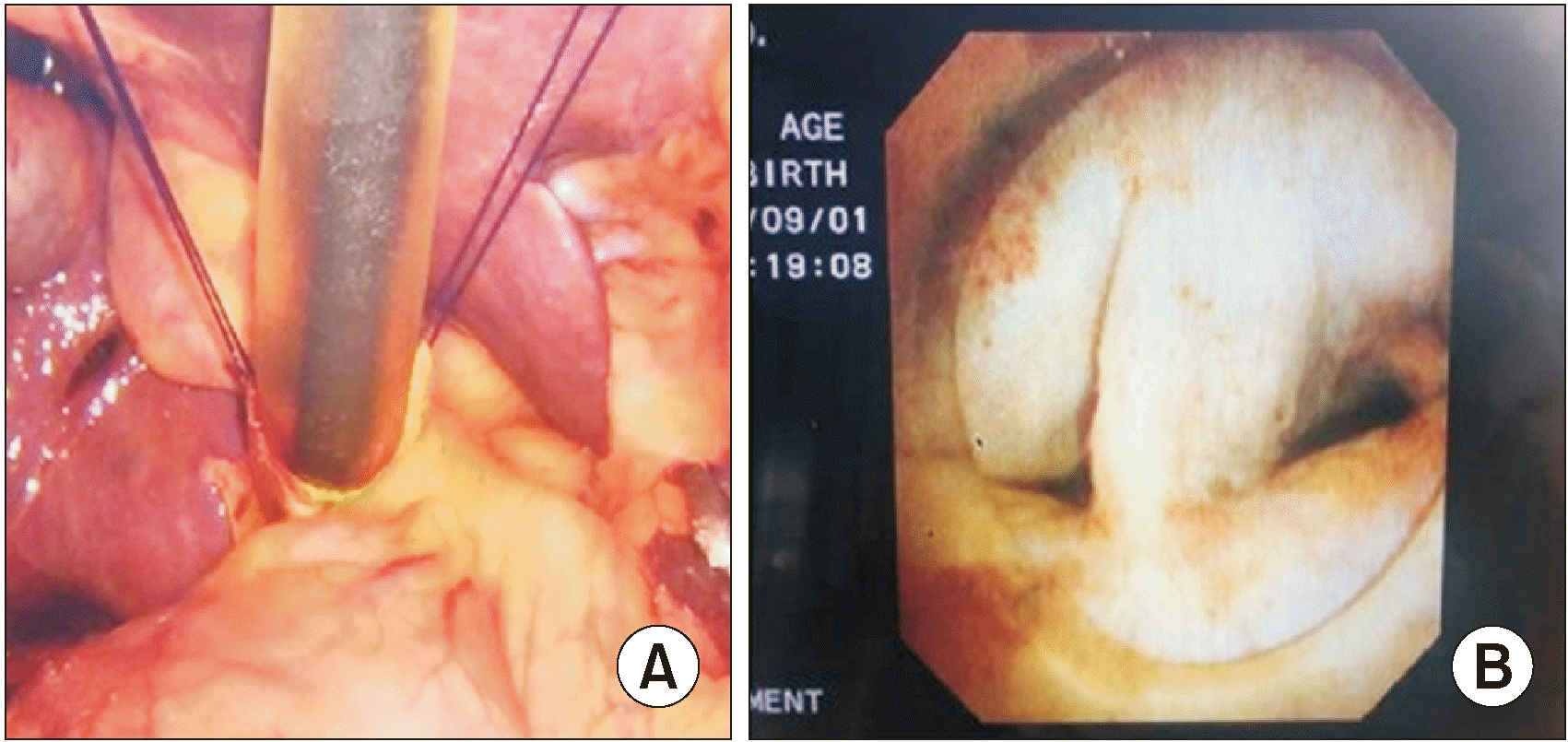
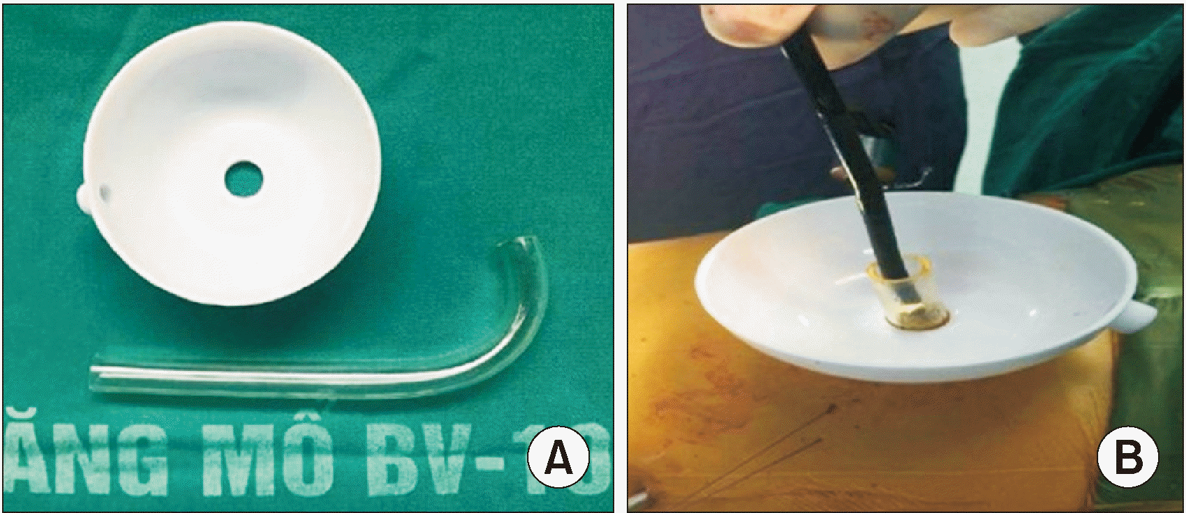
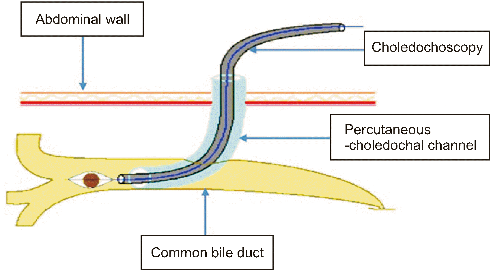
Next, we exposed the CBD and performed choledochotomy through a 10–12 mm longitudinal incision on the CBD's anterior wall, starting 1cm inferior to the hilum. We placed two stay sutures between the abdominal wall and edges of the CBD (Fig. 1A) to make it convenient when putting the self-made PCT (Fig. 2). Then, we inserted the PCT via the fourth working port. The stones and water came out from the outer end of the tube, collected into a small bowl, and continuously suctioned (Fig. 3). A 5-mm flexible choledochoscope (CHF-V; Olympus) was inserted through the PCT. The tip of the PCT can be turned down to explore the CBD or turned up to explore the intrahepatic bile duct; we can also rotate the tip to the left or right for left or right bile ducts (Fig. 1B). Stones were usually extracted using normal saline flushing and/or the Dormia basket. When the stones were large or impacted, an electric hydraulic lithotriptor (EHL) (Lithotron EL 27 Compact; WALZ Elektronik GMBH) was used to fragment them. We routinely inserted a T-tube into the CBD incision, and watertight closure of the CBD incision using 4-0 Vicryl (Ethicon; Johnson & Johnson).
An abdominal ultrasound was performed on postoperative day (POD) 6 to confirm the stone removal from the biliary system. The T-tube was withdrawn about one week later if it was confirmed that there was no remnant stones or bile duct stenosis. If residual stones were confirmed, we instructed patients to self-flush the T-tube daily at home or the local hospital. A subsequent choledochoscopy via the T-tube tract was performed four weeks after surgery if the stones were still there.
Descriptive statistics were presented as the mean and standard deviation. Categorical variables were expressed as proportions. Differences between groups were evaluated using Chi-squared tests. Univariable and multivariable analyses with logistic regression were used to assess the association between covariates. A p < 0.05 was considered statistically significant. All statistical analyses were performed using SPSS (version 22.0; SPSS Inc.).
From June 2017 to March 2020, a total of 84 consecutive patients (37 males, 47 females) aged 22 to 91 years (mean, 55.56 ± 14.66 years) were enrolled in our research study. All the patients had right upper quadrant pain and 12/84 (14.3%) patients had Charcot’s triad at admission. Nearly half of the patients (41.8%) had previous abdominal surgery, 33.4% of the patients had choledochotomy before, and 21.5% of the patients had their gallbladders removed. Among the patients, 28.6%, of the patients had CBD ≤ 10 mm in diameter and the remaining patients had CBD > 10 mm in diameter.
All the patients, 84/84 (100%), successfully underwent laparoscopic cholodochotomy combined with intraoperative cholangioscopy via PCT without the need to convert to laparotomy or other strategies. Due to extensive adhesion from previous surgery, we used five trocars in 4 patients (4.8%). Most patients (64.3%) had both intra- and extrahepatic stones, and only 19/84 (22.6%) patients had simultaneous gallstones—the distribution of biliary stones is shown in more detail in Table 1. The rate of stones ≥ 10 mm in diameter in CBD, left intrahepatic stones (IHS), and right IHS were 64.3%, 38.1%, and 27.4%, respectively.
The mean time to place the PCT was 5.05 ± 2.47 minutes (2–15 minutes). There were no stones or fluid spilled out into the intra-abdominal cavity. Up to 92.5% of patients needed EHL to fragment the stones because the stones were impacted or too large to be entrapped by the Dormia basket. The mean operative time was 121.85 ± 30.47 minutes (70–200 minutes). The time was more in patients with previous abdominal surgery, 137.71 ± 30.23 minutes compared with 110.51 ± 25.38 minutes, but without statistical significance (p = 0.114). During choledochoscopy, we found biliary strictures (could not pass through the scope) in 16 patients (19.1%). They were intrahepatic strictures and caused incomplete stone removal in 15/16 patients. We encountered 2 (2.4%) intraoperative complications. One patient got a hemorrhage when dilating the biliary tract, he was managed by irrigated warm normal saline to coagulate, and we continued the procedure when the bleeding stopped. The other patient had a transverse colonic serosa injury, which was repaired with laparoscopic suturing.
In 46 patients (54.8%), total removal of stones was achieved by combining intraoperative cholangioscopy, postoperative abdominal ultrasound, and T-tube cholangiogram. The mean length of stay after the primary surgery was 9.48 ± 3.60 days (4–24 days).
Overall, 5 patients (6.0%) experienced postoperative complications; all were classified Clavien-Dindo Grade I or II (Table 2). In one patient, transient biliary leakage developed on POD5, and it sealed spontaneously one week later. We encountered one case of an intestinal leak on POD4 without signs of peritonitis, so we managed conservatively, and the leak was sealed after nearly two weeks.
At re-examination at the clinic (the mean duration 31.77 ± 11.23 days), the T-tube was removed in 58 patients after confirming total stones removal.
After performing binary logistic regression, we found that stone residuals were associated with biliary stricture (Table 3). Meanwhile, the characteristics of stones (location, shape, and number of stones) and history of previous abdominal surgery had no effect on the rate of incomplete stone removal. For patients with residual stones, we successfully conducted choledochoscopy via the T-tube tract to clear residual stones in 22/26 patients, so complete stone removal was achieved in a total of 80/84 patients (95.2%).
In the past, the standard procedure for biliary stones removal was choledochotomy through a laparotomy [8]. From the 1970s, endoscopic sphincterotomy (EST) gradually superseded open CBD exploration in treating recurrent CBD stones; however, EST is associated with complications, such as pancreatitis, biliary infection, and bleeding. Moreover, EST has the intrinsic drawback of destroying the Oddi sphincter and the incapability to treat intrahepatic stones [9]. With the constant evolution of laparoscopic techniques, laparoscopic choledochotomy is preferred in primary intervention and reoperation [10-13]. Nevertheless, there are a few reports included patients with a history of biliary surgery, a familiar entity in Vietnamese patients. In this study, 33.4% of patients had a history of choledochotomy, which is higher than in the Western population, for instance, the rate of previous upper abdominal surgery was 14.2% in Khaled et al.’s report [13]. This retrospective study evaluated the clinical data of laparoscopic choledochotomy for HL and CL to explore the feasibility of the laparoscopic approach in patients with and without previous abdominal surgery.
Up to 39.3% of patients had previous biliary tract surgery, and most of the patients had choledochotomy. Up to 64.3% of patients had intra- and extrahepatic stones and only 22.6% had simultaneous gallstones (Table 1). These findings were consistent with the characteristics of primary bile duct stones—a common etiology of biliary stones in the Asian population [3]. In addition, Vietnamese patients rarely have periodic health examinations; they usually come to the hospital in the late stages, when they experience bile duct stones symptoms or even complications such as cholangitis or biliary strictures. In this study, all patients had abdominal pain, and up to 14.3% of patients had already developed Charcot’s triad, which is higher compared with Khaled et al. [13] report where the rates of cholangitis and pancreatitis were 2.5% and 3.3%, respectively. The later the patients seek medical attention, the more complexity of the bile duct stone. Over a quarter of our patients (28.6%) had an extensive distribution of both CBD and bilateral intrahepatic stones, which is higher than that reported by Liu et al. [10], only 8.7% in the laparoscopy group and 13.9% in total. Most patients had more than three stones. The rate of stones ≥ 10 mm in diameter, which needed to be fragmented by EHL, in CBD, left IHS, and right IHS were 64.3%, 38.1%, and 27.4%, respectively. Notably, these features make it more challenging to remove all the stones.
Finding CBD was the essential step of the procedure. In the group of patients without a history of abdominal surgery, we quickly exposed the dilated CBD; however, in the reoperation group, the adhesion made it more challenging. From our experience, with a focus on surgical requirements, excessive adhesiolysis should be avoided. If it is challenging to find the CBD, the puncture needle should be used to find the bile overflow to confirm the position of the CBD. Although the laparoscopic approach was relatively complicated in the reoperation group, there was no need to convert to an open approach, and the mean operative time in the two groups was not significantly different. This was primarily due to the surgeon’s experience in laparoscopic surgery and the priority of exposing the CBD rather than extensive adhesiolysis.
We chose to open the CBD at 1cm inferior to the hilum to maximize the range and explore the intrahepatic duct. With the PCT, we easily turned the tip up or down to approach the intrahepatic duct or CBD without pulling out the PCT, while not damaging the CBD wall. The two stay sutures make this process more accessible, and the PCT pull-out only occurred in three patients, but we quickly reinserted without the bile pill. During choledochoscopy, the normal saline solution must continuously flow to create a transparent environment, dilate the biliary tract, and flush the stone out [14]. We could infuse unlimited water because water and stones follow the PCT to the outside of the abdominal wall without flowing into the abdomen.
Our study’s rate of residual stones after surgery was 45.2%, which was higher than that reported in recent studies (Table 4) [10,12,13,15]. This could be because our patients mainly had more extensive primary bile duct stones, both in CBD and bilateral intrahepatic stones, and most of them had previous choledochotomy, which causes complications in the biliary structure. This is also the reason we routinely left a T-tube, although the primary closure of the choledochotomy was proved to be convincing in many reports [15,16]. Due to the complexity of extensive stones, complete stone removal was infeasible and unnecessary. This could prolong the procedure and cause more harm to the patients when put in general anesthesia for a long time. In addition, 16 patients (19.1%) had biliary strictures, which makes thorough cholechoscopy impossible, and it would be more reasonable to leave the T-tube and instruct the patient to self-flush the tube at home to gradually dilate the biliary tree as well as flush out residual stones [17]. In our patients, the T-tube was proven safe and feasible; only one patient had a biliary leak at the T-tube insertion site and was managed conservatively. Self-flush helped clear all residual stones in 12 patients (14.3%). In addition, the T-tube tract was successfully used for subsequent choledochoscopy to clear residual stones in 22/26 patients. Overall, complete stone removal was achieved in 95.2% of patients, which is comparable with the reports by Khaled et al. [13], Liu et al. [10], and Lien et al. [12]. In our study, retained stones were associated with biliary strictures (odds ratio, 16.00; 95% confidence interval, 4.01–63.86; p < 0.001) which is consistent with results of multiple previous studies [18,19]. These lesions may need to be intervened with biliary stenting or bypass procedures such as Roux-en-Y hepaticojejunostomy [20].
During follow-up, only one patient developed a biliary leak on POD5 and it spontaneously sealed after one week. There was no biliary stricture secondary to CBD exploration. Previously, it was recommended that choledochotomy should be avoided if the CBD is less than 10 mm in diameter [21]. However, our experience demonstrated that choledochotomy could be safely performed if the CBD is ≥ 8 mm in diameter, which is consistent with the report by Lien et al. [12].
Although our study showed satisfactory results, there are still several limitations. First, this was a retrospective study without a control group, so we could not determine the advantages of our approach using the PTC compared to conventional choledochoscopy and endoscopic removal or cystic approaches. Furthermore, the mean duration of follow-up was short, 31.77 ± 11.23 days, so we could not assess the late recurrence.
In conclusion, laparoscopic surgery was safe and effective in treating HL and CL using cholangioscopy via percutaneous-choledochal channels, even in patients with previous choledochotomy. It has the advantage of being less invasive and repeatable via the T-tube tract. Our promising findings warrant further generalization and practical application, particularly in the Asian population. This study was a retrospective analysis with a small sample size, which might lead to selection bias and require validation by prospective studies with large sample sizes.
REFERENCES
1. Feng X, Zheng S, Xia F, Ma K, Wang S, Bie P, et al. 2012; Classification and management of hepatolithiasis: a high-volume, single-center's experience. Intractable Rare Dis Res. 1:151–156. DOI: 10.5582/irdr.2012.v1.4.151. PMID: 25343089. PMCID: PMC4204570.
2. Molvar C, Glaenzer B. 2016; Choledocholithiasis: evaluation, treatment, and outcomes. Semin Intervent Radiol. 33:268–276. DOI: 10.1055/s-0036-1592329. PMID: 27904245. PMCID: PMC5088099.
3. Pausawasdi A, Watanapa P. 1997; Hepatolithiasis: epidemiology and classification. Hepatogastroenterology. 44:314–316. DOI: 10.3138/9781442672789-053.
4. European Association for the Study of the Liver (EASL). 2016; EASL Clinical Practice Guidelines on the prevention, diagnosis and treatment of gallstones. J Hepatol. 65:146–181. DOI: 10.1016/j.jhep.2016.03.005. PMID: 27085810.
5. Kim HJ, Kim JS, Suh SJ, Lee BJ, Park JJ, Lee HS, et al. 2015; Cholangiocarcinoma risk as long-term outcome after hepatic resection in the hepatolithiasis patients. World J Surg. 39:1537–1542. DOI: 10.1007/s00268-015-2965-0. PMID: 25648078.
6. Xia H, Zhang H, Xin X, Liang B, Yang T, Liu Y, et al. 2023; Surgical management of recurrence of primary intrahepatic bile duct stones. Can J Gastroenterol Hepatol. 2023:5158580. DOI: 10.1155/2023/5158580. PMID: 36726399. PMCID: PMC9886471.
7. Wu Y, Xu CJ, Xu SF. 2021; Advances in risk factors for recurrence of common bile duct stones. Int J Med Sci. 18:1067–1074. DOI: 10.7150/ijms.52974. PMID: 33456365. PMCID: PMC7807200.

8. Csendes A, Burdiles P, Diaz JC. 1998; Present role of classic open choledochostomy in the surgical treatment of patients with common bile duct stones. World J Surg. 22:1167–1170. DOI: 10.1007/s002689900537. PMID: 9828726.

9. Andriulli A, Loperfido S, Napolitano G, Niro G, Valvano MR, Spirito F, et al. 2007; Incidence rates of post-ERCP complications: a systematic survey of prospective studies. Am J Gastroenterol. 102:1781–1788. DOI: 10.1111/j.1572-0241.2007.01279.x. PMID: 17509029.

10. Liu YY, Li TY, Wu SD, Fan Y. 2022; The safety and feasibility of laparoscopic approach for the management of intrahepatic and extrahepatic bile duct stones in patients with prior biliary tract surgical interventions. Sci Rep. 12:14487. DOI: 10.1038/s41598-022-18930-1. PMID: 36008517. PMCID: PMC9411189.

11. Wang Y, Huang Y, Shi C, Wang L, Liu S, Zhang J, et al. 2022; Efficacy and safety of laparoscopic common bile duct exploration via choledochotomy with primary closure for the management of acute cholangitis caused by common bile duct stones. Surg Endosc. 36:4869–4877. DOI: 10.1007/s00464-021-08838-8. PMID: 34724579. PMCID: PMC9160116.

12. Lien HH, Huang CC, Huang CS, Shi MY, Chen DF, Wang NY, et al. 2005; Laparoscopic common bile duct exploration with T-tube choledochotomy for the management of choledocholithiasis. J Laparoendosc Adv Surg Tech A. 15:298–302. DOI: 10.1089/lap.2005.15.298. PMID: 15954833.

13. Khaled YS, Malde DJ, de Souza C, Kalia A, Ammori BJ. 2013; Laparoscopic bile duct exploration via choledochotomy followed by primary duct closure is feasible and safe for the treatment of choledocholithiasis. Surg Endosc. 27:4164–4170. DOI: 10.1007/s00464-013-3015-3. PMID: 23719974.

14. Zhang WL, Fang ZP, Shi BY, Chen T, Lv SD, Wang C, et al. 2021; Low-pressure pulse flushing choledochoscopy combined with neodymium laser lithotripsy for the treatment of intrahepatic bile duct stones. Hepatobiliary Pancreat Dis Int. 20:383–386. DOI: 10.1016/j.hbpd.2021.04.002. PMID: 33931315.

15. Zhou Y, Wu XD, Fan RG, Zhou GJ, Mu XM, Zha WZ, et al. 2014; Laparoscopic common bile duct exploration and primary closure of choledochotomy after failed endoscopic sphincterotomy. Int J Surg. 12:645–648. DOI: 10.1016/j.ijsu.2014.05.059. PMID: 24879343.

16. Gómez DA, Mendoza Zuchini A, Pedraza M, Salcedo Miranda DF, Mantilla-Sylvain F, Pérez Rivera CJ, et al. 2023; Long-term outcomes of laparoscopic common bile duct exploration through diathermy, choledochotomy, and primary closure: a 6-year retrospective cohort study. J Laparoendosc Adv Surg Tech A. 33:281–286. DOI: 10.1089/lap.2022.0453. PMID: 36576507.

17. Al-Qudah G, Tuma F. T Tube [Internet]. 2022. Available from: https://www.ncbi.nlm.nih.gov/books/NBK532867/. cited 2023 Jun 15.
18. Li S, Su B, Chen P, Hao J. 2018; Risk factors for recurrence of common bile duct stones after endoscopic biliary sphincterotomy. J Int Med Res. 46:2595–2605. DOI: 10.1177/0300060518765605. PMID: 29865913. PMCID: PMC6124257.

19. Deng F, Zhou M, Liu PP, Hong JB, Li GH, Zhou XJ, et al. 2019; Causes associated with recurrent choledocholithiasis following therapeutic endoscopic retrograde cholangiopancreatography: a large sample sized retrospective study. World J Clin Cases. 7:1028–1037. DOI: 10.12998/wjcc.v7.i9.1028. PMID: 31123675. PMCID: PMC6511924.

20. Dilek ON, Atasever A, Acar N, Karasu Ş, Özlem Gür E, Özşay O, et al. 2020; Hepatolithiasis: clinical series, review and current management strategy. Turk J Surg. 36:382–392. DOI: 10.47717/turkjsurg.2020.4551. PMID: 33778398. PMCID: PMC7963303.

21. Jutric Z, Hammill CW, Hansen PD. Jarnagin WR, editor. 2017; Chapter 36B - Stones in the bile duct: Minimally invasive surgical approaches. Blumgart's Surgery of the Liver, Biliary Tract and Pancreas, 2-Volume Set. Sixth Edition. Elsevier;2017; 604–610.e1. DOI: 10.1016/B978-0-323-34062-5.00143-6.
Fig. 1
Intraoperative findings: (A) the percutaneous-choledochal tube insert into the common bile duct easier with the stay sutures; (B) the tip of the percutaneous-choledochal tube can be rotated to the left or right to explore the left or right intrahepatic bile duct.

Table 1
Distribution of biliary stones
Table 2
Postoperative complications
Table 3
Factors related to stone residuals
Table 4
Characteristics of the studies about laparoscopic choledochotomy
| Study | Khaled et al. [13] | Liu et al. [10] | Lien et al. [12] | Zhou et al. [15] | 108 Hospital |
|---|---|---|---|---|---|
| Year | 2013 | 2022 | 2005 | 2014 | 2023 |
| Patient (n) | 120 | 22 | 82 | 78 | 84 |
| Stone distribution | CL | HL + CL | CL | CL | HL + CL |
| Cholangitis | 2.5% | Not mention | Not mention | 15% | 14.3% |
| Previous abdominal surgery | 14.2% | 100% | Not mention | 0% | 41.8% |
| Biliary stricture | Excluded | Excluded | Not mention | Excluded | 19% |
| CBD diameter (mm) | 9.4 (3–30) | 14.65 ± 4.79 | ≥ 8 | ≥ 11 | ≥ 8 |
| Residual stones | 2.5% | 8.7% | 17.1% | 0% | 45.2% |


 PDF
PDF Citation
 Print
Print


 XML Download