Abstract
A 27-year-old male patient, previously diagnosed with Hodgkin lymphoma (HL), presented with gait disturbance. Brain MRI showed a 4.5 cm mass lesion in the right occipital lobe, suggesting either intracranial involvement of HL or a potential meningioma. Despite high-dose methotrexate and steroid treatment, the patient’s symptoms persisted, and imaging showed an enlarging mass, leading to surgical intervention. Histopathological examination confirmed central nervous system (CNS) involvement of HL. Postoperatively, the patient underwent whole-brain radiotherapy and demonstrated marked clinical improvement. Our literature review from 1980 to 2023 identified only 46 cases of intracranial HL (IC-HL), underscoring its rarity. Lymphomas represent 2.2% of brain tumors, with 90%–95% being diffuse large B-cell lymphoma (DLBCL). In contrast, the incidence of CNS-HL patients is a mere 0.02%. Notably, IC-HL and intracranial DLBCL have differences in their typical locations and treatment strategies. Unlike DLBCL, which predominantly appears in the supratentorial region (87%), IC-HL is found there in 61.5% of cases. Additionally, 33.3% of IC-HL cases occur in the cerebellum, with 43.5% associated with posterior circulation regions. Furthermore, while biopsy followed by chemotherapy induction is a common strategy for DLBCL, 81.8% of IC-HL cases underwent surgical resection, and only 18.1% had a biopsy alone. The distinct characteristics of IC-HL tumors, including their larger size, attachment to the dura, and fibrotic nature with clear boundaries, might account for the preference for surgical intervention. The unique features of IC-HL compared to DLBCL highlight the need for distinct considerations in diagnosis and management.
Hodgkin lymphoma (HL), a hematopoietic malignancy, affects lymph nodes throughout the body [123]. While it is a significant malignancy, its progression to the central nervous system (CNS) is remarkably rare, with an incidence of only 0.02% [456]. Primary or secondary lymphomas account for 2.2%–4.0% of all brain tumors [578910]. In the spectrum of CNS lymphomas, diffuse large B-cell lymphoma (DLBCL) is the predominant histological subtype, comprising an estimated 90%–95% of cases [7810]. Burkitt lymphoma, T-cell lymphoma, and HL each have frequencies that are comparable to each other but are significantly rarer than DLBCL [11]. Of these, our case specifically focuses on HL. To gain deeper insight into these cases, we conducted a detailed literature review spanning from 1980 to 2023, focusing on intracranial HL (IC-HL) cases [91213141516171819202122232425262728293031323334353637383940414243444546]. Through our literature review and understanding of our case, our goal is to emphasize the key differences between DLBCL and HL.
A 27-year-old male patient, with a known history of HL, presented with gait disturbance. A brief summary of his HL history is as follows. Three years ago, he presented with persistent high fever and weight loss. Imaging revealed systemic lymphadenopathies in the axillary, neck, and mediastinal regions, as well as multifocal osseous lesions. A diagnostic biopsy from a cervical lymph node, coupled with a bone marrow assessment, confirmed a diagnosis of classic HL, lymphocyte-rich variant, at stage IVXB with documented bone marrow involvement. After confirming the diagnosis, the patient underwent serial chemotherapy regimens according to the National Comprehensive Cancer Network (NCCN) guideline [4748]. Initially, the patient underwent two cycles of ABVD (adriamycin, bleomycin, vinblastin, dacarbazine), which resulted in a partial response. This was followed by four cycles of BEACOPP (bleomycin, etoposide, adriamycin, cyclophosphamide, vincristine, procarbazine, prednisone). Although this regimen also led to a partial response, disease progression subsequently occurred. The treatment strategy was augmented by two cycles of ICE (ifosfamide, carboplatin, etoposide), but this approach resulted in further disease progression. The patient’s therapy was then adjusted to include four cycles of brentuximab vedotin, which successfully led to the absence of disease in the bone marrow. Further management involved six cycles of GDP (gemcitabine, dexamethasone, cisplatin), achieving a metabolic complete response. The treatment course was concluded with autologous stem cell transplantation.
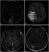
During follow-up, the patient complained of headaches and nausea, but other than increased size of small lymph nodes at four locations in the neck, no other sites of disease were identified. This prompted an MRI examination. Upon his presentation, a brain MRI revealed a 4.5 cm sized mass lesion in the right occipital lobe with a clear and well-defined margin, showing strong contrast enhancement, with attachment to the dura mater and the superior sagittal sinus (Fig. 1A). Accompanying peritumoral edema was prominent in the right cerebral hemisphere, extending to the thalamus and corpus callosum. This edematous change precipitated both subfalcine and transtentorial herniations (Fig. 1B). Initially, two cycles of high-dose methotrexate combined with steroids were given, assuming CNS involvement of lymphoma in HL. However, both symptoms and follow-up imaging results continued to worsen. Upon further review with MRI, and given the lack of therapeutic response, it became apparent that the possibility of another type of brain tumor different from HL, especially meningioma, had to be considered. Consequently, a craniotomy was scheduled both to reduce the ongoing mass effect and to obtain a biopsy. Before surgery, considering the possibility of a meningioma, an external carotid angiography was conducted. During the angiography, a tumor blush was observed from the distal branch of the occipital artery, leading to feeding artery embolization.
A craniotomy was performed for tumor resection. Upon dissecting the dura mater, which showed intense adhesion (Fig. 2A), an extensive fibrotic lesion with a well-defined margin, resembling fibrous meningioma, was identified (Fig. 2B). The tumor was encapsulated, and there was almost no bleeding tendency due to feeding artery embolization. The intraoperative findings were markedly different from the typical characteristics observed during DLBCL surgery, which often presents with soft tissue, unclear boundaries, and a severe bleeding tendency. An intraoperative frozen biopsy revealed an abundance of small, round cells. Macroscopically complete removal of tumor and dural coagulation were done (Fig. 2C and D).
The histopathological examination of the resected tissue revealed mononucleated characteristic Reed-Sternberg cells (Fig. 3A). Immunohistochemical staining demonstrated the cells to be positive for CD30 and CD15 (Fig. 3B and C), while negative for CD20 (Fig. 3D), confirming classic HL. These findings confirmed the diagnosis of systemic classic HL with intracranial involvement [123449].
Postoperatively, the patient underwent whole-brain radiotherapy, receiving a total dose of 40 Gy. The latest follow-up revealed MRI indicating no remnant tumor in the previous tumor bed (Fig. 1C and D). Clinically, he demonstrated significant improvement in his gait disturbances and regained his ability to walk independently. Currently, the patient has been in remission for 8 months and remains under close monitoring.
Between 1980 and 1999, only eight cases of intracranial involvement in HL were reported [1213141516171819]. From 2000 to the time of our current case, 39 additional cases have surfaced [9202122232425262728293031323334353637383940414243444546]. The increasing number of case reports underscores the importance of focusing on IC-HL. While sporadic case reports on IC-HL have emerged over time, a few articles have collected, organized, and published these reports [192021293334354246]. In fact, some cases were missed even within these limited publications. To ensure a complete collection of all cases, we conducted detailed searches on PubMed, Scopus, and Google Scholar. We carefully cross-referenced each case report and attempted to make sure that no cases were missed. Through this process, we gathered all the published literature on IC-HL from 1980 to 2023 and summarized it in Table 1 [91213141516171819202122232425262728293031323334353637383940414243444546]. Based on this table, we aim to highlight some differences between DLBCL and IC-HL.
First, examining the age distribution, while primary CNS lymphoma has a significantly higher incidence between the ages of 60 and 80 compared to other age groups [781011], IC-HL also has a higher incidence in the 60s, but not markedly more than in other age ranges (Fig. 4). This is consistent with the observation that HL displays a bimodal incidence peak in both younger and older populations [148].
Of a total of 47 cases, including ours, 39 had clearly described locations. Among these, 24 cases (61.5%) of the total, had tumors located in the supratentorium. This rate is significantly lower compared to DLBCL, where tumors are located in the supratentorium in approximately 87% of cases. Furthermore, IC-HL was found in the cerebellum in 13 cases (33.3%). Including the 2 cases from the occipital lobe and 2 from the brain stem, 43.5% of cases are associated with posterior circulation regions, such as the brainstem, cerebellum, and occipital lobe. Within the supratentorium, while DLBCL predominantly affects the frontal lobe and periventricular white matter, IC-HL often arises adjacent to the dura mater, presenting in varied locations. This can lead to potential misdiagnoses of IC-HL as other tumors, particularly meningiomas, or as ischemic or inflammatory lesions during the initial radiologic diagnosis. Our case also displayed unique morphological characteristics, from well-defined borders to intense enhancement patterns, extending into the dura and sinus. These features are similar to those seen in meningiomas (Fig. 1A). These characteristics distinguish IC-HL from DLBCL and are beneficial for its diagnosis. There were 19 cases (40.4%) accompanied by systemic HL, while 28 cases (60.6%) were not, indicating that the frequency of isolated IC-HL is relatively high. As previously mentioned, it is hard to diagnose HL using just radiologic findings, so a biopsy is essential to differentiate it from other tumors like meningiomas. Pathologically, the presence of monucleated Reed-Sternburg cells and immunohistochemical staining for CD30 and CD15 are key markers, making the biopsy-based diagnosis of classic HL definitive [1248].
While the primary treatment strategy for DLBCL involves chemotherapy induction after a biopsy, out of 44 cases where surgical details were clearly recorded, 36 (81.8%) of IC-HL underwent surgical resection, and only eight cases (18.1%) had just a biopsy. Among the 44 cases where postoperative treatment details were clearly described, 21 cases (47.7%) underwent concurrent chemotherapy and radiotherapy, 15 cases (34.1%) had radiotherapy alone, and 4 cases (9.1%) had chemotherapy alone. The majority of the cases received radiotherapy. The follow-up period for “no evidence of disease” varied. Only one case reported a death soon after surgery, with the patient passing away 10 days postoperation due to complications from pulmonary edema [38].
In the radiologic findings unique to IC-HL cases, the majority of the literature suggests that maximal safety resection is achievable. This is largely attributed to the clear demarcation between the tumor and normal tissue, and the common presentation of IC-HL on the brain’s surface. These characteristics notably distinguish IC-HL from DLBCL. Furthermore, the minimal bleeding risk simplified the surgical approach. In our case too, these advantageous conditions allowed for an uncomplicated piecemeal resection (Fig. 2). Hence, when IC-HL is suspected, pursuing maximal safety resection combined with chemotherapy and radiotherapy appears to be advantageous. Currently, a universally accepted treatment for IC-HL remains elusive, emphasizing the need for further research to determine the optimal treatment strategy.
In conclusion, through our case report and literature review, we have examined the distinct differences in IC-HL from radiological, surgical, and etiological findings. We suggest that our insights be helpful and hope they contribute to further research and advancements in the treatment of IC-HL.
Notes
Author Contributions:
Conceptualization: Seung Heon Cha, Won Ho Cho.
Data curation: Hwanhee Lee, Sangjun Ahn, Won Ho Cho.
Funding acquisition: Seung Heon Cha, Won Ho Cho.
Investigation: Seung Heon Cha, Won Ho Cho.
Software: Hwanhee Lee, Sangjun Ahn.
Supervision: Seung Heon Cha, Won Ho Cho.
Validation: Hwanhee Lee, Sangjun Ahn, Won Ho Cho.
Visualization: Sangjun Ahn.
Writing—original draft: Hwanhee Lee, Sangjun Ahn.
Writing—review & editing: Sangjun Ahn, Won Ho Cho.
Availability of Data and Material
The datasets generated or analyzed during the study are available from the corresponding author on reasonable request.
References
1. Brice P, de Kerviler E, Friedberg JW. Classical Hodgkin lymphoma. Lancet. 2021; 398:1518–1527. PMID: 33493434.
2. Wang HW, Balakrishna JP, Pittaluga S, Jaffe ES. Diagnosis of Hodgkin lymphoma in the modern era. Br J Haematol. 2019; 184:45–59. PMID: 30407610.
3. Shanbhag S, Ambinder RF. Hodgkin lymphoma: a review and update on recent progress. CA Cancer J Clin. 2018; 68:116–132. PMID: 29194581.
4. Grimm S, Chamberlain M. Hodgkin’s lymphoma: a review of neurologic complications. Adv Hematol. 2011; 2011:624578. PMID: 20975772.
5. Kaji FA, Martinez-Calle N, Sovani V, Fox CP. Rare central nervous system lymphomas. Br J Haematol. 2022; 197:662–678. PMID: 35292959.
6. Re D, Fuchs M, Schober T, Engert A, Diehl V. CNS involvement in Hodgkin’s lymphoma. J Clin Oncol. 2007; 25:3182.
8. Phillips EH, Fox CP, Cwynarski K. Primary CNS lymphoma. Curr Hematol Malig Rep. 2014; 9:243–253. PMID: 24969265.
9. Gerstner ER, Abrey LE, Schiff D, Ferreri AJ, Lister A, Montoto S, et al. CNS Hodgkin lymphoma. Blood. 2008; 112:1658–1661. PMID: 18591379.
10. Hochberg FH, Baehring JM, Hochberg EP. Primary CNS lymphoma. Nat Clin Pract Neurol. 2007; 3:24–35. PMID: 17205072.
11. Camilleri-Broët S, Martin A, Moreau A, Angonin R, Hénin D, Gontier MF, et al. Primary central nervous system lymphomas in 72 immunocompetent patients: pathologic findings and clinical correlations. Am J Clin Pathol. 1998; 110:607–612. PMID: 9802345.
12. Nagashima K, Mori S, Yoshimasu N, Takahashi K. Primary Hodgkin’s disease of the falx cerebri. Acta Neuropathol. 1980; 51:161–163. PMID: 7435148.
13. Bender BL, Mayernik DG. Hodgkin’s disease presenting with isolated craniospinal involvement. Cancer. 1986; 58:1745–1748. PMID: 3756795.
14. Doorly TP, Farrell MA, Phillips J. Primary intracerebral Hodgkin’s lymphoma. J Neurol Neurosurg Psychiatry. 1987; 50:1048–1050. PMID: 3655810.
15. Ashby MA, Barber PC, Holmes AE, Freer CE, Collins RD. Primary intracranial Hodgkin’s disease. A case report and discussion. Am J Surg Pathol. 1988; 12:294–299. PMID: 3354756.
16. Sickler GK, Hanson SK, Hsu SM, Papasozomenos SC. Primary intracerebral Hodgkin’s disease: a case report. Clin Neuropathol. 1990; 9:143–147. PMID: 2364594.
17. Clark WC, Callihan T, Schwartzberg L, Fontanesi J. Primary intracranial Hodgkin’s lymphoma without dural attachment: case report. J Neurosurg. 1992; 76:692–695. PMID: 1545264.
18. Deckert-Schlüter M, Marek J, Setlík M, Marková J, Pakos E, Fischer R, et al. Primary manifestation of Hodgkin’s disease in the central nervous system. Virchows Arch. 1998; 432:477–481. PMID: 9645450.
19. Klein R, Müllges W, Bendszus M, Woydt M, Kreipe H, Roggendorf W. Primary intracerebral Hodgkin’s disease: report of a case with Epstein-Barr virus association and review of the literature. Am J Surg Pathol. 1999; 23:477–481. PMID: 10199479.
20. Biagi J, MacKenzie RG, Lim MS, Sapp M, Berinstein N. Primary Hodgkin’s disease of the CNS in an immunocompetent patient: a case study and review of the literature. Neuro Oncol. 2000; 2:239–243. PMID: 11265233.
21. Herrlinger U, Klingel K, Meyermann R, Kandolf R, Kaiserling E, Kortmann RD, et al. Central nervous system Hodgkin’s lymphoma without systemic manifestation: case report and review of the literature. Acta Neuropathol. 2000; 99:709–714. PMID: 10867808.
22. Johnson MD, Kinney MC, Scheithauer BW, Briley RJ, Hamilton K, McPherson WF, et al. Primary intracerebral Hodgkin’s disease mimicking meningioma: case report. Neurosurgery. 2000; 47:454–456. discussion 456-7. PMID: 10942021.
23. Figueroa BE, Brown JR, Nascimento A, Fisher DC, Tuli S. Unusual sites of Hodgkin’s lymphoma: case 2. Hodgkin’s lymphoma of the CNS masquerading as meningioma. J Clin Oncol. 2004; 22:4228–4230. PMID: 15483035.
24. Hirmiz K, Foyle A, Wilke D, Burrell S, Brownstone R, Ago C, et al. Intracranial presentation of systemic Hodgkin’s disease. Leuk Lymphoma. 2004; 45:1667–1671. PMID: 15370222.
25. de Castro AF, Júnior AS, de Lins e Horta H, Neuenschwander LC, Fonseca RP, Lima SS, et al. Primary intracerebral Hodgkin lymphoma. Br J Haematol. 2007; 138:562. PMID: 17686050.
26. Hwang CY, Song YJ, Kim DC, Choi SS, Choi YM, Kim KU. Primary cerebellar Hodgkin’s lymphoma. J Korean Neurosurg Soc. 2007; 42:149–152.
27. Morawa E, Ragam A, Sirota R, Nabhan C. Hodgkin’s lymphoma involving the CNS. J Clin Oncol. 2007; 25:1437–1438. PMID: 17416864.
28. Subklewe M, Anagnostopoulos I. Radiologic and pathologic features of a posttransplantation primary central nervous system lymphoma demonstrating Epstein-Barr virus-positive Hodgkin lymphoma. Clin Lymphoma Myeloma. 2007; 7:535–537. PMID: 18021471.
29. Apollonsky N, Edelman M, Johnson A, Bhuiya T, Karayalcin G. Intracerebral presentation of Hodgkin disease mimicking meningioma in a young woman: case presentation with literature review. J Pediatr Hematol Oncol. 2008; 30:369–372. PMID: 18458571.
30. Almhanna K, Wongchaowart N, Sweetenham J. Intracerebral Hodgkin’s lymphoma in a patient with chronic lymphocytic leukemia/small lymphocytic lymphoma: a case report and literature review. Cancer Invest. 2009; 27:215–220. PMID: 19235595.
31. Foo WC, Desjardins A, Cummings TJ. Primary intracerebral Hodgkin lymphoma with recurrence. Clin Neuropathol. 2011; 30:75–79. PMID: 21329616.
32. Torgerson S, Olteanu H, Tinguely M, Fenske TS. Central nervous system Hodgkin lymphoma: case report and review of the literature. J Neurooncol. 2011; 102:329–334. PMID: 20676729.
33. Gessi M, Kuchelmeister K, Kellner U, Ritter M, Morgner A, Urbach H, et al. Unusual clinico-pathological features in primary Hodgkin’s lymphomas of the central nervous system. Acta Neurochir (Wien). 2013; 155:19–24. PMID: 23139103.
34. Kresak JL, Nguyen J, Wong K, Davis R. Primary Hodgkin lymphoma of the central nervous system: two case reports and review of the literature. Neuropathology. 2013; 33:658–662. PMID: 23530967.
35. Henkenberens C, Franzke A, Raab P, Oschlies I, Klapper W, Christiansen H. Primary EBV-positive Hodgkin’s lymphoma of the CNS under azathioprine treatment: case report and review of the literature. Strahlenther Onkol. 2014; 190:847–852. PMID: 24823896.
36. Martinez DL, Gujrati M, Geoffroy F, Tsung AJ. Isolated CNS Hodgkin’s lymphoma: implications for tissue diagnosis. CNS Oncol. 2014; 3:383–387. PMID: 25438809.
37. Sharaf N, Lobo B, Lee J, Prayson RA. Primary Hodgkin lymphoma of the central nervous system. J Clin Neurosci. 2014; 21:1271–1273. PMID: 24589557.
38. van Blydenstein SA, Patel M, Philip V, Lakha A, Pather S, Westgarth-Taylor T, et al. Classical Hodgkin lymphoma involving the central nervous system (brain) - an unusual presentation. Clin Case Rep. 2014; 2:88–92. PMID: 25356257.
39. Shivane A, Smith ME, Lewis D, Berei T. A rare case of primary intracerebral Hodgkin lymphoma. Clin Neuropathol. 2016; 35:389–392. PMID: 27719749.
40. Alfaseh A, Rajeh MN, Hamed G. Primary central nervous system Hodgkin lymphoma: a case discussion and a hypothesis on the etiology. Avicenna J Med. 2019; 9:28–31. PMID: 30697523.
41. Azriel A, Towner JE, Gaillard F, Box G, Rogers T, Morokoff A. Solitary intraventricular Hodgkin lymphoma post-transplant lymphoproliferative disease (HL-PTLD): case report. J Clin Neurosci. 2019; 69:269–272. PMID: 31451379.
42. Riccioni L, Cremonini AM, Gessaroli M. Primary intracranial Hodgkin’s lymphoma after a blunt trauma: a case report. J Neurosci Neurol Disord. 2020; 4:79–83.
43. Szczepanek D, Szumiło J, Stoma F, Szymczyk A, Jarosz B, Szczepanek A, et al. A case report of a female patient with Hodgkin lymphoma localized in the central nervous system and with concomitant pulmonary lymphomatoid granulomatosis. Front Neurol. 2020; 11:963. PMID: 33013640.
44. Ahmed S, Irfan B, Raza M, Haider G. Atypical involvement of central nervous system in classic Hodgkin lymphoma: a case report. J Med Case Rep. 2021; 15:532. PMID: 34711281.
45. Fu H, Shi S, Chen L, Xu B, Huang W, Chen Y, et al. Primary central nervous system Hodgkin’s lymphoma: a case report. J Int Med Res. 2021; 49:300060521999533. PMID: 33874776.
46. Kanagalingam T, Velker V, Pejhan S, Zhang Q, Mangel J, Young S. Isolated Hodgkin lymphoma of the intracranial dura: a case report and review of the literature. Clin Case Rep. 2023; 11:e7562. PMID: 37361649.
47. Ajani JA, D’Amico TA, Bentrem DJ, Chao J, Cooke D, Corvera C, et al. Gastric cancer, version 2.2022, NCCN clinical practice guidelines in oncology. J Natl Compr Canc Netw. 2022; 20:167–192. PMID: 35130500.
48. Hoppe RT, Advani RH, Ai WZ, Ambinder RF, Armand P, Bello CM, et al. Hodgkin lymphoma, version 2.2020, NCCN clinical practice guidelines in oncology. J Natl Compr Canc Netw. 2020; 18:755–781. PMID: 32502987.
49. Cheah CY, Bröckelmann PJ, Chihara D, Moskowitz AJ, Engert A, Jerkeman M, et al. Clinical characteristics and outcomes of patients with Hodgkin lymphoma with central nervous system involvement: an international multicenter collaboration. Am J Hematol. 2016; 91:894–899. PMID: 27222367.
Table 1
Study | Yrs | Age (yr) | Sex | Location | HL | Surg | Tx | Follow-up |
---|---|---|---|---|---|---|---|---|
Nagashima et al. [12] | 1980 | 60 | M | Falx cerebri | No | R | Cx+Rx | 11 mo, DOD |
Bender and Mayernik [13] | 1986 | 34 | M | Frontal lobe and dura, right | Yes | B | Cx+Rx | N/A |
Doorly et al. [14] | 1987 | 51 | M | Cerebellum | No | R | Cx+Rx | 12 mo, NED |
Ashby et al. [15] | 1988 | 62 | M | Frontoparietal lobe, right | No | R | Cx+Rx | 14 mo, NED |
Sickler et al. [16] | 1990 | 84 | F | Parietooccipital lobe, right | No | R | Rx | 8 mo, NED |
Clark et al. [17] | 1992 | 53 | F | Cerebellum | No | R | Rx | 6 mo, NED |
Deckert-Schlüter [18] | 1998 | 62 | F | Fronto-parietal lobe, left | Yes | R | Cx+Rx | 13 mo, NED |
Klein et al. [19] | 1999 | 54 | M | Occipital lobe, left | No | R | Cx+Rx | 12 mo, NED |
Biagi et al. [20] | 2000 | 52 | M | Tempoparietal lobe | No | R | Rx | 29 mo, NED |
Herrlinger et al. [21] | 2000 | 66 | W | Fronto-parietal lobe, left | No | R | Cx+Rx | 18 m, NED |
Johnson et al. [22] | 2000 | 55 | F | Cerebellum | No | R | Rx | 8 mo, NED |
Figueroa et al. [23] | 2004 | 23 | F | Posterior fossa | Yes | R | Cx+Rx | N/A |
Hirmiz et al. [24] | 2004 | 38 | M | Parietal lobe, right | Yes | R | Cx+Rx | 12 mo, NED |
de Castro et al. [25] | 2007 | 63 | M | Frontotemporal lobe, left and cerebellum | No | N/A | N/A | N/A |
Hwang et al. [26] | 2007 | 64 | F | Cerebellum | No | R | Rx | 16 mo, NED |
Morawa et al. [27] | 2007 | 51 | M | Frontal lobe, right | Yes | R | Cx+Rx | N/A |
Subklewe and Anagnostopoulos [28] | 2007 | 65 | F | Temporal lobe, right | No | N/A | N/A | 18 mo, NED |
Apollonsky et al. [29] | 2008 | 22 | F | Frontal lobe, left | Yes | R | Cx+Rx | 48 mo, NED |
Gerstner et al. [9] | 2008 | 58 | F | Not specified | No | R | Rx | 90 mo, NED |
2008 | 60 | F | Not specified | No | R | Rx | 1 mo, NED | |
2008 | 46 | M | Not specified | Yes | B | N/A | N/A | |
2008 | 72 | M | Not specified | Yes | R | No | 5 mo, NED | |
2008 | 37 | F | Not specified | Yes | B | Cx+Rx | 15 mo, DOD | |
2008 | 19 | M | Not specified | Yes | B | Cx+Rx | 128 mo, NED | |
2008 | 44 | M | Not specified | Yes | No | Cx+Rx | 4 mo, DOD | |
2008 | 23 | M | Not specified | Yes | R | Cx+Rx | 53 mo, NED | |
Almhanna et al. [30] | 2009 | 65 | M | Temporal lobe, left | Yes | R | Cx+Rx | N/A, DOD |
Foo et al. [31] | 2011 | 58 | M | Temporal lobe, left | No | R | Rx | 14 mo, RD |
Torgerson et al. [32] | 2011 | 79 | F | Temporal lobe, left | Yes | B | Rx | 1 mo, N/A |
Gressi et al. [33] | 2013 | 77 | M | Cerebellum | No | R | Rx | 8 mo, NED |
2013 | 59 | M | Brain stem | No | R | N/A | 2 mo, N/A | |
2013 | 65 | F | Temporal lobe, left | Yes | B | Cx | 22 mo, NED | |
Kresak et al. [34] | 2013 | 70 | M | Cerebellum | No | R | Rx | 10 yr, NED |
2013 | 72 | F | Cerebellum | No | R | Rx | 6 mo, NED | |
Henkenberens et al. [35] | 2014 | 47 | M | Cerebellum | No | R | Cx+Rx | 9 mo, NED |
Martinez et al. [36] | 2014 | 74 | F | Brain stem | No | B | Rx | N/A |
Sharaf et al. [37] | 2014 | 77 | M | Cerebellum | No | R | Rx | 7 mo, NED |
van Blydenstein et al. [38] | 2014 | 41 | M | Parietal lobe, left | Yes | B | No | 10 d, DOD |
Shivane et al. [39] | 2016 | 59 | F | Frontal lobe, right | No | R | Cx+Rx | 2 mo, NED |
Alfaseh et al. [40] | 2019 | 38 | M | Cerebellum | No | R | Cx+Rx | 7 yr, NED |
Azriel et al. [41] | 2019 | 49 | M | Septum pellucidum | No | R | Cx | N/A |
Riccioni et al. [42] | 2020 | 30 | M | Frontal - orbital - ethmoidal, left | No | R | Cx+Rx | 30 mo, NED |
Szczepanek et al. [43] | 2020 | 33 | F | Temporal lobe, left and cerebellum | Yes | R | Cx+Rx | N/A |
Ahmed et al. [44] | 2021 | 35 | M | Parietal lobe, right | Yes | R | Cx | 4 mo, NED |
Fu et al. [45] | 2021 | 60 | M | Cerebellum | No | R | Cx | 3 mo, NED |
Kanagalingam et al. [46] | 2023 | 71 | M | Frontal lobe, right | No | R | Cx+Rx | 5 yr, NED |
Present report | 2024 | 27 | M | Occipital lobe, right | Yes | R | Rx | 8 mo, NED |