Journal List > Intest Res > v.22(1) > 1516086120

Akiyama, Onoda, Moue, Sakamoto, Sakamoto, Suzuki, Enomoto, Matsubara, Oda, and Tsuchiya: Association of colonic metaplasia of goblet cells and endoscopic phenotypes of the J pouch in patients with ulcerative colitis: a retrospective pilot study

Abstract

Background/Aims

Mucosal adaptation of the ileum toward colonic epithelium has been reported in pouchitis in ulcerative colitis (UC); however, the clinical characteristics, endoscopic findings, and outcomes in patients with pouchitis with ileal mucosal adaptation are poorly understood.

Methods

This was a single-center retrospective study comprising UC patients treated by proctocolectomy with ileal pouch-anal anastomosis who had undergone pouchoscopy at the University of Tsukuba Hospital between 2005 and 2022. Endoscopic phenotypes were evaluated according to the Chicago classification. High-iron diamine staining (HID) was performed to identify sulfomucin (colon-type mucin)-producing goblet cells (GCs) in pouch biopsies. We compared clinical data between patients with (high HID group) and without > 10% sulfomucin-producing GCs in at least one biopsy (low HID group).

Results

We reviewed 390 endoscopic examination reports from 50 patients. Focal inflammation was the most common phenotype (78%). Five patients (10%) required diverting ileostomy. Diffuse inflammation and fistula were significant risk factors for diverting ileostomy. The median proportion of sulfomucin-producing GCs on histological analysis of 82 pouch biopsies from 23 patients was 9.9% (range, 0%–93%). The duration of disease was significantly greater in the high HID group compared to the low HID group. The median percentage of sulfomucin-producing GCs was significantly higher in patients with diffuse inflammation or fistula compared to other endoscopic phenotypes (14% vs. 6.0%, P= 0.011).

Conclusions

Greater proportions of sulfomucin-producing GCs were observed in endoscopic phenotypes associated with poor outcomes in UC, indicating patients with pouchitis showing colonic metaplasia of GCs may benefit from early interventions.

INTRODUCTION

Patients with ulcerative colitis (UC) may develop colitis refractory to medical therapy or colorectal neoplasia, with approximately 10% to 15% of patients with UC requiring surgery [1]. Total proctocolectomy with ileal pouch-anal anastomosis (IPAA) in a “J pouch” configuration is the most commonly performed surgical procedure for UC [2]. However, inflammation of the reservoir can develop, commonly termed “pouchitis.” [2] Several recent studies have reported a cumulative incidence of acute pouchitis is 30% and approximately 10% to 15% of patients with acute pouchitis develop chronic pouchitis in patients with UC [2-4]. Furthermore, the incidence of pouch failure requiring diverting loop ileostomy (DLI) or pouch excision is reportedly up to 10% and pouch with Crohn’s disease-like features is a significant risk factor of pouch failure [2,5,6]. Although the International Ileal Pouch Consortium has established several consensus guidelines for the management of pouchitis [7,8], there remains uncertainty regarding the monitoring, prophylaxis, and pathogenesis of pouchitis in UC.
To date, several studies have demonstrated that inflammation of the pouch is often observed on endoscopy even in asymptomatic patients [9,10] and is associated with future development of acute pouchitis [10]. A recent study conducted at the University of Chicago retrospectively assessed endoscopic inflammatory findings at each anatomical location of the J pouch and classified endoscopic findings into 7 main endoscopic phenotypes (the Chicago classification of Pouchitis) [11]. Each phenotype had distinct contributing factors and was associated with different clinical outcomes, with the “diffuse inflammation of the pouch body” phenotype significantly associated with the need for pouch excision [12]. Accordingly, the Chicago classification may have utility in stratifying patients according to the risk of pouch loss using standardized endoscopic assessments and thereby guide intensive follow-up and medical prophylaxis to improve clinical outcomes in patients with IPAA [13,14].
Although there are limited data regarding the pathological mechanisms underlying poor outcomes in patients with IPAA, previous studies have reported colonic metaplasia of intestinal goblet cells (GCs) is associated with the development of pouchitis in UC. Intestinal GCs play an important role in the secretion of mucins and maintenance of the mucous gel layer which provides protection for the surface epithelium [15]. Intestinal GCs have functionally distinct subsets. For instance, acidic mucin-producing GCs, which are visualized by alcian blue staining, have a unique subpopulation of cells that produce sulfomucin, a form of acidic mucin detectable by high-iron diamine (HID) staining [16]. In the human intestine, colonic GCs are positive for HID [17] whereas small intestinal GCs are negative for HID [18]. Mucosal adaptations of the ileum toward colonic epithelium have been reported in UC patients with IPAA, particularly in cases of pouchitis [19-22]. A comparative analysis assessing differences between UC and familial adenomatous polyposis pouches demonstrated sulfomucin expression was greater in mucous gel from the pouch of patients with UC. This study also demonstrated that high expression of sulfomucin was associated with increased colonization of sulfate-reducing bacteria associated with pouch inflammation [22], indicating that colonic metaplasia of GCs may play an important role in the pathogenesis of pouchitis in UC and may influence disease severity.
Accordingly, the present study aimed to assess clinical characteristics, endoscopic phenotypes according to the Chicago classification, and clinical outcomes in patients with pouchitis at a single study center. To discriminate between small intestinal and colonic GCs, we measured the proportion of sulfomucin-producing GCs in biopsy samples obtained from the J pouch in patients with UC. We then compared clinical and endoscopic data between patients with high and low proportions of sulfomucin-producing GCs to identify clinical features of patients with colonic metaplasia of GCs.

METHODS

1. Study Design

This was a retrospective cohort study comprising patients with a preoperative diagnosis of UC. All patients underwent proctocolectomy with IPAA between 2005 and 2022 at the University of Tsukuba Hospital. All patients underwent endoscopic monitoring of the pouch. This study was approved by the ethics committee of the University of Tsukuba Hospital (R03-248) and conducted in accordance with the Declaration of Helsinki. The use of an opt-out consent approach was approved by the ethics committee of the University of Tsukuba Hospital (R03-248). Informed consent was waived.

2. Endoscopic Pouch Phenotype

As our standard operating protocol, we perform an annual pouchoscopy for UC patients with IPAA after ileostomy takedown. Pouchoscopy is also indicated when patients developed clinical symptoms suggestive of pouchitis. All available images from endoscopic examinations of the pouch after ileostomy takedown were retrospectively assessed by S.A. and S.M. Endoscopic findings were assessed at each anatomical location of the J pouch; the afferent limb (AL), inlet (IL), tip of the J, proximal pouch, distal pouch, and rectal cuff (Supplementary Fig. 1). Based on the Chicago classification [11], endoscopic examination findings were classified into the following 7 phenotypes: (1) normal; (2) AL involvement; (3) IL involvement; (4) diffuse inflammation of the pouch body; (5) focal inflammation of the pouch body; (6) cuffitis; and (7) pouch fistulas (Supplementary Fig. 2). Inflammatory findings on endoscopy based on the Pouchitis Disease Activity Index (PDAI) included erythema/edema, erosions/friability, ulceration, stenosis, granularity, and loss of vascular pattern [23,24]. Endoscopic examinations with no evidence of pouch inflammation were recorded as “normal pouch.” Endoscopic examinations with any evidence of inflammation in the AL, IL, or rectal cuff were recorded as “AL involvement,” “IL involvement,” or “cuffitis,” respectively. “Pouchitis” was defined as one or more inflammatory findings in the tip, proximal, or distal areas of the pouch. Two or more endoscopic findings in all anatomical locations of the pouch body were defined as “diffuse inflammation of the pouch body.” Cases of pouchitis which did not meet the criteria for diffuse inflammation were recorded as “focal inflammation of the pouch body.” [11] “Pouch fistula” was defined as any type of fistula noted on endoscopy or other imaging studies ≥ 6 months after ileostomy takedown to ensure the exclusion of fistula as a result of surgery-related complications (Supplementary Fig. 2) [25].
All available endoscopic images were evaluated according to the Chicago classification [11] to determine the overall endoscopic pouch phenotype. If all endoscopic examinations in an individual patient were reported as normal, the patient was categorized into the “normal” phenotype. If an inflammatory phenotype was identified on at least one endoscopic examination, the patient was included in the analysis for the respective phenotypic category. The finding of focal inflammation and diffuse inflammation of the pouch body on separate endoscopic examinations was recorded as “diffuse inflammation of the pouch body” rather than focal inflammation of the pouch body [11].

3. Clinical Data Collection and Outcomes

Data regarding the following categories were retrospectively collected: age at diagnosis of inflammatory bowel disease, age at colectomy, sex, body mass index, race, smoking status, family history of inflammatory bowel disease, disease duration until colectomy, indication for colectomy, preoperative Clostridioides difficile infection, UC disease extent based on the Montreal classification (E1, proctitis; E2, left-sided disease; E3, extensive disease) [26], number of IPAA stages, IPAA technique, presence of primary sclerosing cholangitis, postoperative complications (anastomotic leak, pelvic sepsis, abdominal abscess requiring drainage, fistula formation before ileostomy takedown, and ileus), and medication history. Clinical PDAI subscores (range, 0–6) [23,24] were also assessed at the clinical visit closest to the date of initial endoscopy at the study center.
Our primary outcome was pouch failure. Pouch failure was defined as a pouch condition requiring DLI or pouch excision and was considered a poor outcome. Secondary outcome was chronic pouchitis. Chronic pouchitis was defined as pouch conditions requiring antibiotics (e.g., ciprofloxacin and/or metronidazole) for more than 4 weeks as well as DLI [2].

4. Histological Assessment

In the histological analysis, we included patients with available biopsy samples obtained by endoscopy. All biopsies were undertaken at the discretion of endoscopists in clinical settings. Biopsies were formalin-fixed, paraffin-embedded, and then sectioned at 3 μm thickness. Alcian blue (pH 2.5) and HID staining were performed as previously described [16,27]. Colon and ileum samples from UC patients were used as positive and negative controls, respectively (Supplementary Fig. 3). Biopsy samples from the rectal cuff or polyps were excluded from the study analysis. For example, we excluded samples with endoscopic reports or images indicating that biopsies were obtained from the rectal cuff with almost 100% HID+ GCs. Numbers of HID-positive (HID+) and HID-negative (HID–) GCs were independently counted in each biopsy specimen by 2 investigators (S.A. and T.O.). The proportion of HID+ GCs in all GCs (HID+ GCs plus HID– GCs) was then calculated. Consistency between the 2 datasets was confirmed. Areas of disagreement or uncertainty were resolved by consensus among the authors including pathologists.

5. Statistical Analyses

Categorical data were compared using the Fisher exact test while continuous data were compared using the Mann-Whitney U test. Using Kaplan-Meier curves, pouch survival from the date of ileostomy takedown to the date of pouch failure was estimated and compared using the log-rank test. P-values less than 0.05 were considered statistically significant. EZR (Saitama Medical Center, Jichi Medical University, Saitama, Japan) [28], which is a graphical user interface for R (The R Foundation for Statistical Computing, version 4.0.2, Vienna, Austria), was used for all data analyses.

RESULTS

1. Patient Backgrounds

The endoscopic phenotypes of 50 patients with a preoperative diagnosis of UC treated by proctocolectomy with IPAA were assessed. All patients were Japanese. The median age at UC diagnosis and colectomy was 32 years (range, 9–57 years) and 42 years (range, 14–66 years), respectively. The median disease duration until surgery was 4.9 years (range, 0.08–34.8 years). Extensive colitis (E3) was predominant, affecting 88% of patients. The majority of patients (70%) required colectomy due to disease refractory to medical therapy, while 9 patients (18%) had dysplasia or colorectal cancer. The majority of patients were receiving oral mesalamine or sulfasalazine (94%) and prednisone (94%) preoperatively, with 22% of patients receiving infliximab. Regarding surgical procedures, 2-stage IPAA and 1-stage IPAA were performed in 70% and 18% of patients, respectively. Stapled anastomosis was performed in the majority of cases (84%). Among the 22 patients (44%) who experienced postoperative complications, the most common postoperative complication was ileus (77.3%). Data regarding clinical characteristics, surgery, and preoperative therapy are summarized in Table 1 and Supplementary Tables 1, 2, respectively.

2. Diffuse Inflammation and Pouch Fistula Are High-Risk Phenotypes for Pouch Failure

The present study evaluated 390 endoscopic examinations. The frequencies of each endoscopic phenotype are summarized in Supplementary Table 3. The median duration from ileostomy takedown to initial endoscopy at the study center was 8.1 months (range, 0.43–180 months). The median PDAI clinical score at the clinical visit closest to the initial endoscopy was 1 (range, 0–4) (Table 2). The median interval between these dates was 18 days (range, 0–183 days). The most common phenotype on initial endoscopy was focal inflammation (82%) (Table 2).
The findings of serial endoscopic examinations were evaluated for each patient, with an overall endoscopic phenotype assigned accordingly. The median number of endoscopic examinations per patient was 7 (range, 1–18), with a median follow-up time of 8.9 years (range, 0.6–16.9 years). The median number of endoscopic examinations per year was 0.94 (range, 0.07–3.90) (Table 2). The most frequent overall phenotype was focal inflammation (78%), followed by cuffitis (72%), IL involvement (34%), AL involvement (22%), diffuse inflammation (22%), pouch fistulas (14%), and the normal phenotype (0%) (Table 2). The order of endoscopic phenotypic frequencies in the present study was the same as for patients preoperatively diagnosed with UC in the original University of Chicago study (Supplementary Fig. 3) [29]. Of the 41 patients with focal inflammation on initial endoscopic examination, 6 patients (15%) subsequently developed diffuse inflammation.
Data on postoperative treatments demonstrated that 78% of patients were receiving loperamide. Regarding therapies for pouchitis, 52% of patients were receiving metronidazole whereas 18% of patients were receiving both metronidazole and ciprofloxacin. The proportions of patients on oral mesalamine, topical mesalamine, and topical steroids were 34%, 18%, and 18%, respectively (Supplementary Table 4). Of the 7 patients with pouch fistula, 5 patients (71%) had perianal or anorectal fistula. Five patients (71%) required seton drainage, and 2 patients (29%) were treated by incision and drainage. One patient (14%) was treated with infliximab and azathioprine (Supplementary Table 5).
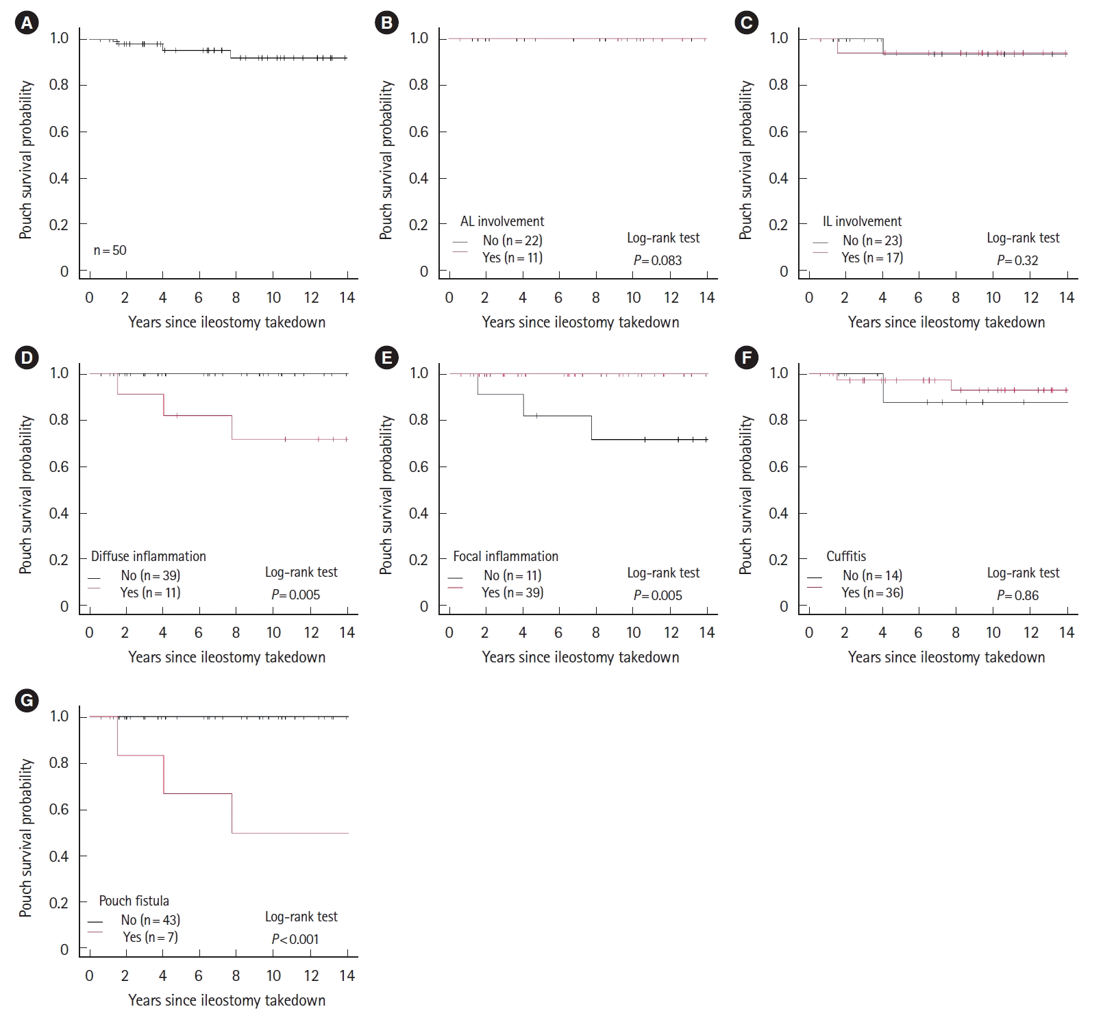
Pouch failure occurred in 5 patients (10%). While no patients required pouch excision, DLI was performed for all patients with pouch failure (Table 1). The 10-year pouch survival rate was 92% (95% confidence interval [CI], 76%–97%) (Fig. 1). Patients with diffuse inflammation, pouch fistula were at significantly increased risk of pouch failure (P= 0.005 and P< 0.001, respectively) (Fig. 1). Notably, the 10-year pouch survival rates in patients with diffuse inflammation and pouch fistula were 72% (95% CI, 35%–90%) and 50% (95% CI, 11%–80%), respectively. Patients with focal inflammation had favorable pouch outcomes, with a 10-year pouch survival of 100% (Fig. 1). The rates of diffuse inflammation and pouch fistula were significantly higher in patients with pouch failure compared to patients without pouch failure (P= 0.006 and P< 0.001, respectively) (Supplementary Table 6). Twenty-four patients developed chronic pouchitis and the rates of diffuse inflammation and/or pouch fistulas were significantly higher in the patients with chronic pouchitis compared with those without chronic pouchitis (Supplementary Table 7).

3. Higher Proportion of Sulfomucin-Producing GCs in Diffuse Inflammation and Pouch Fistula

To evaluate colonic metaplasia of GCs in the J pouch, we performed histological assessments of pouch biopsies with HID staining. We identified a total of 113 available biopsy specimens from 64 endoscopic examinations in 28 patients. Twenty-three specimens obtained from the rectal cuff or polyps were excluded. Of the remaining 90 biopsy specimens, 87 specimens were obtained from the pouch body (97%) and 3 were obtained from the AL (3.2%). Eight samples did not include GCs (7 from the pouch body and one from the AL).
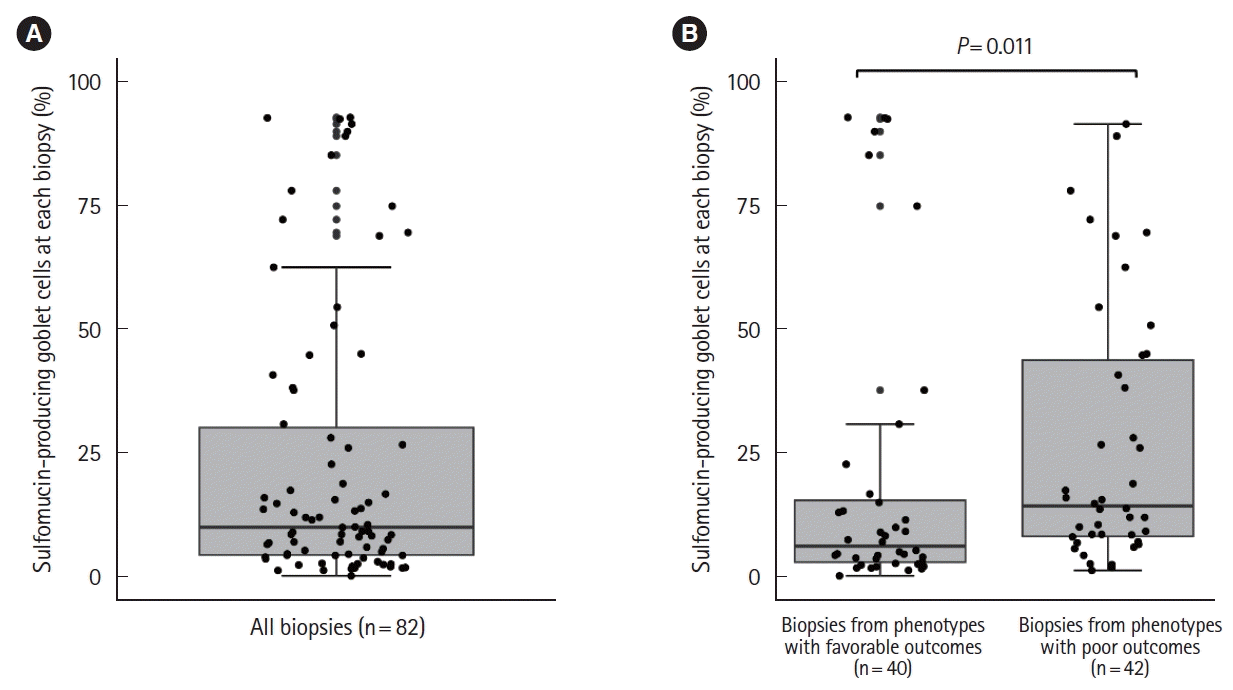
The final cohort included 82 samples obtained from 52 endoscopic examinations in 23 patients. In almost all endoscopic examinations (96%), biopsies were performed to assess the degree of mucosal inflammation. Samples of the colon and small intestine obtained from patients with UC were used as positive and negative controls, respectively. The proportions of HID+ GCs in the colon and small intestine were 99% and 1.9%, respectively (Supplementary Fig. 4). The median proportion of HID+ GCs in biopsies obtained from the J pouch was 9.9% (range, 0%–93%) (Fig. 2A).
We divided the cohort into patients with greater than 10% HID+ GCs in at least one biopsy (high HID group, n = 13) and patients with no biopsies with greater than 10% HID+ GCs (low HID group, n = 10). We then compared demographic data and endoscopic phenotypes according to the Chicago classification between the high and low HID groups. Age at diagnosis and at colectomy were lower in the high HID group (21 [9.0–47] years and 30 [16–51] years, respectively) compared to the low HID group (29.5 [13–43] years and 38 [14–46] years, respectively). Interestingly, the high HID group had significant longer disease duration until colectomy (7.1 [0.25–14] years) compared with the low HID group (1.1 [0.08–15] years). No significant difference in the follow-up time from the date of ileostomy takedown to the last clinical visit was observed between the 2 groups (high HID group, 12 [2.4–16] years; low HID group, 12 [8.5–16] years). The rate of 2-stage IPAA in the low HID group was higher than that in the high HID group. The high HID group had numerically higher risk of chronic pouchitis compared with the low HID group (69% vs. 50%), although not statistically significant. Otherwise, no significant differences in preoperative disease extent, infliximab use, anastomosis type, postoperative antibiotics, use of mesalamine, use of topical mesalamine or steroids, or DLI were observed between the 2 groups (Tables 3, 4).
Regarding endoscopic phenotypes in this patient cohort, the most frequent overall phenotype was cuffitis (74%), followed by focal inflammation (70%), IL involvement (43%), AL involvement (35%), diffuse inflammation (30%), and pouch fistula (13%). The number of endoscopic examinations per patient was similar between the 2 groups (high HID group, 11 [1–18] examinations; low HID group, 11.5 [9–14] examinations). The proportions of patients with diffuse inflammation or pouch fistula in the high HID group (46%) was 2-fold higher than in the low HID group (20%). The frequencies of focal inflammation or AL involvement were lower in the high HID group (62% and 30%, respectively) than in the low HID group (80% and 50%, respectively) (Table 4).
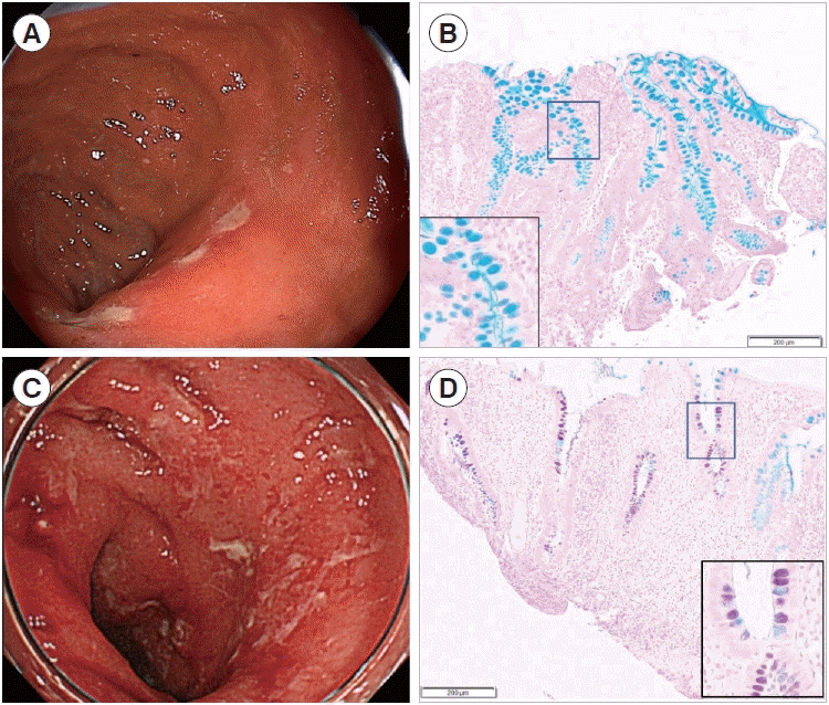
To evaluate the association between a higher proportion of sulfomucin-producing GCs on pouch biopsy and endoscopic pouch phenotypes with poor outcomes, we compared HID positivity in GCs between biopsies obtained from patients with diffuse inflammation or fistula (42 specimens from 26 endoscopic examinations in 8 patients) and patients with other endoscopic phenotypes (40 specimens from 26 endoscopic examinations in 15 patients). The median proportions of HID+GCs were 25.9% (range, 2.3%–91.5%, 25 samples in 5 patients) in diffuse inflammation without fistula, 10.3% (range, 1.1%–40.7%, 17 samples in 3 patients) (Supplementary Fig. 5) in pouch fistula ( ± diffuse inflammation), and 6.0% (range, 0%–93%) in the other endoscopic phenotypes. The median proportion of HID+ GCs was significantly higher in diffuse inflammation or pouch fistula compared to other endoscopic phenotypes (14% [range 1.1%–92%] vs. 6.0% [range 0%–93%]; P= 0.011) (Figs. 2B, 3).

DISCUSSION

The present study assessed clinical characteristics, endoscopic pouch phenotypes according to the Chicago classification, and pouch outcomes in UC patients with IPAA treated at a single study center. The findings of the present study demonstrate that diffuse inflammation of the pouch body and pouch fistula were significantly associated with pouch failure as well as chronic pouchitis, corroborating the results of the original study describing the Chicago classification [11]. We further investigated colonic phenotypic changes in ileal mucosa obtained from the J pouch using HID staining. While the median proportions of sulfomucin-producing GCs in the colon and small intestine were > 98% and < 3%, respectively, the proportion in the J pouch was approximately 10%. We further compared patient demographics and endoscopic phenotypes between low and high HID groups. We observed that the high HID group had a significantly longer duration of UC until colectomy and numerically higher risk of chronic pouchitis compared with the low HID group. Furthermore, the proportion of sulfomucin-producing GCs was greater in biopsies obtained from patients with diffuse inflammation or pouch fistula compared to biopsies from the other endoscopic phenotypes, indicating that the proportion of HID-positive GCs is higher in pouch biopsies from UC patients with endoscopic phenotypes associated with poor clinical outcomes.
According to a previous study assessing a prospectively maintained ileal pouch database at the Cleveland Clinic, the risk of pouchitis increases with every 5-year increase in the preoperative duration of UC, indicating the long-term effects of a chronic inflammatory state in patients with UC [30]. A separate study investigated the role of HID positivity on the pathogenesis of pouchitis and observed a significant association between HID positivity and increased colonization of sulfate-reducing bacteria [22]. Bacteria producing hydrogen sulfide [31] have been shown to induce epithelial apoptosis and mucosal depletion [32]. As these processes have been posited to contribute to the pathogenesis of pouchitis in UC, increased duration of UC prior to surgery may increase the risk of pouchitis by increasing colonization of sulfate-reducing bacteria as a result of changes in HID positivity among ileal GCs. While biologics and small molecules have recently demonstrated efficacy in reducing inflammation and delaying the timing of colectomy even in patients with medically-refractory UC, further studies are required to determine the most effective timing of colectomy to reduce the risk of pouchitis and poor associated outcomes [30].
The results of the present study demonstrate a strong association between HID positivity and endoscopic phenotypes associated with poor outcomes, indicating that colonization of sulfate-reducing bacteria and hydrogen sulfide production may increase the risk of diffuse pouchitis or pouch fistula. Accordingly, the Chicago classification of endoscopic phenotypes may have utility in identifying pathological differences between pouch phenotypes in UC in addition to predicting clinical outcomes. As the use of antibiotics can provide sufficient time for mucosal regeneration [33] and improve the clinical symptoms of pouchitis [2], early detection and antibiotic therapy may have greater benefit in cases of pouchitis with greater HID positivity, thereby improving pouch outcomes.
We found that 15% of patients with focal inflammation on initial endoscopy subsequently developed diffuse inflammation, suggesting that pouch monitoring for early detection of the endoscopic phenotype is an important component of strategies for maintaining healthy pouch condition. Patients with pouch fistula are known to have poor outcomes [6]. We identified 7 patients with pouch fistula and the proportion of diffuse inflammation of the pouch body was 71% in this group, suggesting fistulas developed due to chronic inflammation such as pouch with Crohn’s disease-like features [5]. While 5 out of 7 patients with fistula (71%) required DLI in the present study, the University of Chicago study reported that 21 out of 78 patients with pouch fistula (27%) required DLI [11]. This difference may be attributable to differences in the use of anti-tumor necrosis factor therapies. Indeed, 60% of patients with pouch fistula in the University of Chicago study were receiving anti-tumor necrosis agents [11], whereas only 14% of patients were receiving infliximab in the present study. We believe endoscopic pouch monitoring with assessment of biopsy specimens using HID staining represents a reasonable approach to targeting early therapeutic intervention in patients with adverse endoscopic phenotypes.
The present study had several strengths and limitations. The major strength of the present study is the first demonstration of an association between sulfomucin-producing GCs and endoscopic pouch phenotypes with poor outcomes using the Chicago classification. Additionally, each patient included in the present study had substantial endoscopic and histological data available, with a median number of 7 endoscopic examinations per patient which is 2-fold higher than the original study describing the Chicago classification [11].
A limitation was that this was a single-center retrospective study. Therefore, the number of included patients was relatively small and only 5 patients experienced pouch failure requiring DLI, implying the difficulty to adjust the confounding factors such as postoperative treatments using multivariate analysis. For instance, each patient had heterogenous endoscopic phenotypes and multivariate analysis including sufficient data on phenotypes may be needed. However, due to the small number of outcomes, it was difficult to conduct meaningful multivariate analysis for any outcome. Furthermore, endoscopic data, particularly findings in the AL and IL of the pouch, were not available for all patients included in the present study. Regarding histological assessments, we retrospectively evaluated biopsy samples which were obtained by endoscopic examinations performed in routine clinical settings. Therefore, biopsies typically demonstrated inflamed mucosa and the timing and number of biopsies varied among patients. The difference in the number of biopsies among phenotypes could skew the results. Although we performed an analysis showing a proportion of HID+ GC cells per biopsy due to the limited number of biopsy samples, per patient analysis may be ideal to better understand the association between HID positivity and endoscopic pouch phenotypes. Thus, multi-center prospective studies with larger sample sizes are required to confirm the findings of the present study and validate the clinical utility of HID staining for pouch monitoring.
In conclusion, the results of the present study demonstrate that patients with greater disease duration until colectomy are more likely to have a higher proportion of sulfomucin-producing GCs in pouch biopsy specimens. The proportion of HID+ GCs was significantly higher in patients with high-risk endoscopic phenotypes, such as diffuse pouchitis or pouch fistula, compared to other endoscopic phenotypes associated with favorable outcomes, implying each phenotype of the Chicago classification may have distinct pathophysiology. Our findings indicate patients with endoscopic phenotypes showing high HID positivity (colonic metaplasia of GCs) may benefit from early detection and intervention to improve pouch outcomes. Further studies are required to further elucidate the pathogenesis of specific endoscopic phenotypes described by the Chicago classification and determine the clinical utility of endoscopic pouch monitoring with assessment of pouch biopsy specimens using HID staining.

Notes

Funding Source

The work was supported by the Japan Foundation for Applied Enzymology, JSIBD Grants-in-Aid for IBD Research and JSPS Grant-in-Aid for Scientific Research C (22K08026).

Conflict of Interest

Tsuchiya K has received honoraria (lecture fees) from Takeda Pharmaceutical Co., Ltd. No other potential conflicts of interest relevant to this article were reported.

Data Availability Statement

All data generated or analyzed during this study are included in this published article and its supplementary information files.

Author Contributions

Conceptualization: Akiyama S, Tsuchiya K. Data curation; Formal analysis: Akiyama S, Onoda T, Moue S. Data interpretation: Akiyama S, Onoda T, Moue S, Enomoto T, Tsuchiya K. Funding acquisition: Akiyama S, Onoda T, Moue S, Sakamoto N, Sakamoto T, Suzuki H, Enomoto T, Matsubara D, Oda T, Tsuchiya K. Writing – original draft: Akiyama S, Onoda T, Moue S. Writing – review & editing: Akiyama S, Onoda T, Moue S, Enomoto T, Tsuchiya K. Approval of final manuscript: all authors.

Additional Contributions

The authors would like to thank Tsukuba Pathological Analysis Support Service (http://www.hosp.tsukuba.ac.jp/outpatient/facility/kisokenkyu) for the histological staining and Enago (www.enago.jp) for the English language revie

Supplementary Material

Supplementary materials are available at the Intestinal Research website (https://www.irjournal.org).
Supplementary Table 1.
Surgery-Related Parameters and Pouch Outcomes (n=50)
ir-2023-00105-Supplementary-Table-1.pdf
Supplementary Table 2.
Preoperative Therapies (n=50)
ir-2023-00105-Supplementary-Table-2.pdf
Supplementary Table 3.
Frequency of Endoscopic Pouch Phenotype (per Scope) (n=390)
ir-2023-00105-Supplementary-Table-3.pdf
Supplementary Table 4.
Postoperative therapies (n=50)
ir-2023-00105-Supplementary-Table-4.pdf
Supplementary Table 5.
Therapies and Type of Pouch Fistula (n=7)
ir-2023-00105-Supplementary-Table-5.pdf
Supplementary Table 6.
Comparative Analysis of Endoscopic Phenotype between Patients with and without Pouch Failure (n=50)
ir-2023-00105-Supplementary-Table-6.pdf
Supplementary Table 7.
Comparative Analysis of Endoscopic Phenotype between Patients with and without Chronic Pouchitis (n=50)
ir-2023-00105-Supplementary-Table-7.pdf
Supplementary Fig. 1.
Graphical representation of the J pouch.
ir-2023-00105-Supplementary-Fig-1.pdf
Supplementary Fig. 2.
Endoscopic pouch phenotype according to the Chicago classification. (A) Normal pouch, (B) afferent limb involvement (an arrowhead shows an erosion), (C) inlet involvement (arrowheads show erosions), (D) diffuse inflammation of the pouch body, (E) focal inflammation of the pouch body, (F) cuffitis, and (G) pouch fistula (an arrowhead shows a fistula).
ir-2023-00105-Supplementary-Fig-2.pdf
Supplementary Fig. 3.
Bar graph showing the frequency of endoscopic pouch phenotype (%) in data from the University of Tsukuba (white bar) and University of Chicago (black bar) studies. AL inv, afferent limb involvement; IL inv, inlet involvement.
ir-2023-00105-Supplementary-Fig-3.pdf
Supplementary Fig. 4.
Alcian blue (pH, 2.5) and high-iron diamine (HID) staining. (A-C) Three representative images of resected ileum from a patient with ulcerative colitis. Almost all goblet cells were negative for HID (blue). (D-F) Three representative images of resected colon from a patient with ulcerative colitis. Almost all goblet cells were positive for HID (black). Scale bar=100 μm.
ir-2023-00105-Supplementary-Fig-4.pdf
Supplementary Fig. 5.
(A, B) Representative endoscopic images of pouch fistula. A fistula developed in the distal pouch body (arrowheads show a fistula). (C, D) The proximal pouch body showed many focal ulcerations. (E) Corresponding biopsy demonstrates 15.8% of goblet cells were positive for high-iron diamine (scale bar=200 μm).
ir-2023-00105-Supplementary-Fig-5.pdf

REFERENCES

1. Fumery M, Singh S, Dulai PS, Gower-Rousseau C, Peyrin-Biroulet L, Sandborn WJ. Natural history of adult ulcerative colitis in population-based cohorts: a systematic review. Clin Gastroenterol Hepatol. 2018; 16:343–356.
crossref
2. Akiyama S, Rai V, Rubin DT. Pouchitis in inflammatory bowel disease: a review of diagnosis, prognosis, and treatment. Intest Res. 2021; 19:1–11.
crossref
3. Kayal M, Plietz M, Rizvi A, et al. Inflammatory pouch conditions are common after ileal pouch anal anastomosis in ulcerative colitis patients. Inflamm Bowel Dis. 2020; 26:1079–1086.
crossref
4. Barnes EL, Herfarth HH, Kappelman MD, et al. Incidence, risk factors, and outcomes of pouchitis and pouch-related complications in patients with ulcerative colitis. Clin Gastroenterol Hepatol. 2021; 19:1583–1591.
crossref
5. Akiyama S, Dyer EC, Rubin DT. Diagnostic and management considerations for the IPAA with Crohn’s disease-like features. Dis Colon Rectum. 2022; 65(S1):S77–S84.
crossref
6. Ricardo AP, Kayal M, Plietz MC, et al. Predictors of pouch failure: a tertiary care inflammatory bowel disease centre experience. Colorectal Dis. 2023; 25:1469–1478.
crossref
7. Shen B, Kochhar GS, Kariv R, et al. Diagnosis and classification of ileal pouch disorders: consensus guidelines from the International Ileal Pouch Consortium. Lancet Gastroenterol Hepatol. 2021; 6:826–849.
crossref
8. Shen B, Kochhar GS, Rubin DT, et al. Treatment of pouchitis, Crohn’s disease, cuffitis, and other inflammatory disorders of the pouch: consensus guidelines from the International Ileal Pouch Consortium. Lancet Gastroenterol Hepatol. 2022; 7:69–95.
9. Zhu H, Wu XR, Queener E, Kiran RP, Remzi FH, Shen B. Clinical value of surveillance pouchoscopy in asymptomatic ileal pouch patients with underlying inflammatory bowel disease. Surg Endosc. 2013; 27:4325–4332.
crossref
10. Kayal M, Plietz M, Radcliffe M, et al. Endoscopic activity in asymptomatic patients with an ileal pouch is associated with an increased risk of pouchitis. Aliment Pharmacol Ther. 2019; 50:1189–1194.
crossref
11. Akiyama S, Ollech JE, Rai V, et al. Endoscopic phenotype of the J pouch in patients with inflammatory bowel disease: a new classification for pouch outcomes. Clin Gastroenterol Hepatol. 2022; 20:293–302.
crossref
12. Xu W, Wang Y, Hua Z, et al. Risk factors and quality of life in patients with diffuse pouchitis after ileal pouch anal anastomosis according to the Chicago classification for J pouch: a retrospective multicenter cohort study in China. J Gastrointest Surg. 2023; 27:766–776.
crossref
13. Akiyama S, Rodriguez TG, Rubin DT. Stratifying risks of ileal pouch outcomes. Clin Gastroenterol Hepatol. 2022; 20:1620–1621.
14. Kayal M, Ungaro RC, Colombel JF. The Chicago classification of pouchitis: an important step toward a needed consensus. Clin Gastroenterol Hepatol. 2022; 20:281–282.
15. Deplancke B, Gaskins HR. Microbial modulation of innate defense: goblet cells and the intestinal mucus layer. Am J Clin Nutr. 2001; 73:1131S–1141S.
16. Akiyama S, Mochizuki W, Nibe Y, et al. CCN3 expression marks a sulfomucin-nonproducing unique subset of colonic goblet cells in mice. Acta Histochem Cytochem. 2017; 50:159–168.
17. Croix JA, Carbonero F, Nava GM, Russell M, Greenberg E, Gaskins HR. On the relationship between sialomucin and sulfomucin expression and hydrogenotrophic microbes in the human colonic mucosa. PLoS One. 2011; 6:e24447.
crossref
18. Filipe MI, Fenger C. Histochemical characteristics of mucins in the small intestine: a comparative study of normal mucosa, benign epithelial tumours and carcinoma. Histochem J. 1979; 11:277–287.
crossref
19. Apel R, Cohen Z, Andrews CW Jr, McLeod R, Steinhart H, Odze RD. Prospective evaluation of early morphological changes in pelvic ileal pouches. Gastroenterology. 1994; 107:435–443.
crossref
20. Garcia-Armengol J, Hinojosa J, Lledo S, Roig JV, Garcia-Granero E, Martinez B. Prospective study of morphologic and functional changes with time in the mucosa of the ileoanal pouch: functional appraisal using transmucosal potential differences. Dis Colon Rectum. 1998; 41:846–853.
crossref
21. Chadha K, Mukherjee B, Subramanya H, Singh KV. Restorative proctocolectomy with ileo-anal reservoir, a histopathological, histochemical, and electron microscopic study. Med J Armed Forces India. 2003; 59:306–309.
crossref
22. Bambury N, Coffey JC, Burke J, Redmond HP, Kirwan WO. Sulphomucin expression in ileal pouches: emerging differences between ulcerative colitis and familial adenomatous polyposis pouches. Dis Colon Rectum. 2008; 51:561–567.
crossref
23. Sandborn WJ, Tremaine WJ, Batts KP, Pemberton JH, Phillips SF. Pouchitis after ileal pouch-anal anastomosis: a Pouchitis Disease Activity Index. Mayo Clin Proc. 1994; 69:409–415.
crossref
24. Shen B, Achkar JP, Connor JT, et al. Modified Pouchitis Disease Activity Index: a simplified approach to the diagnosis of pouchitis. Dis Colon Rectum. 2003; 46:748–753.
25. Shen B, Remzi FH, Lavery IC, Lashner BA, Fazio VW. A proposed classification of ileal pouch disorders and associated complications after restorative proctocolectomy. Clin Gastroenterol Hepatol. 2008; 6:145–158.
crossref
26. Satsangi J, Silverberg MS, Vermeire S, Colombel JF. The Montreal classification of inflammatory bowel disease: controversies, consensus, and implications. Gut. 2006; 55:749–753.
crossref
27. Spicer SS. Diamine methods for differentialing mucosubstances histochemically. J Histochem Cytochem. 1965; 13:211–234.
28. Kanda Y. Investigation of the freely available easy-to-use software ‘EZR’ for medical statistics. Bone Marrow Transplant. 2013; 48:452–458.
crossref
29. Akiyama S, Ollech JE, Traboulsi C, et al. Histopathology of colectomy specimens predicts endoscopic pouch phenotype in patients with ulcerative colitis. Dig Dis Sci. 2022; 67:4020–4031.
crossref
30. Lavryk OA, Stocchi L, Hull TL, et al. Impact of preoperative duration of ulcerative colitis on long-term outcomes of restorative proctocolectomy. Int J Colorectal Dis. 2020; 35:41–49.
crossref
31. Ohge H, Furne JK, Springfield J, Rothenberger DA, Madoff RD, Levitt MD. Association between fecal hydrogen sulfide production and pouchitis. Dis Colon Rectum. 2005; 48:469–475.
crossref
32. Coffey JC, Bennett MW, Wang JH, et al. Upregulation of FasFas-L (CD95/CD95L)-mediated epithelial apoptosis: a putative role in pouchitis? J Surg Res. 2001; 98:27–32.
crossref
33. Kmiot WA, Youngs D, Tudor R, Thompson H, Keighley MR. Mucosal morphology, cell proliferation and faecal bacteriology in acute pouchitis. Br J Surg. 1993; 80:1445–1449.
crossref

Fig. 1.
Kaplan-Meier curves for pouch survival. (A) All participants and patients with (B) afferent limb (AL) involvement, (C) inlet (IL) involvement, (D) diffuse inflammation of the pouch body, (E) focal inflammation of the pouch body, (F) cuffitis, and (G) pouch fistula.
ir-2023-00105f1.tif
Fig. 2.
Comparison of high-iron diamine positive (HID+) goblet cells between phenotypes with favorable and poor outcomes. (A) Boxand- whisker plot showing the proportion of sulfomucin-producing (HID+) goblet cells in J pouch biopsies from patients with ulcerative colitis. (B) Box-and-whisker plots comparing the proportion of sulfomucin-producing (HID+) goblet cells between samples obtained from patients with endoscopic phenotypes associated with poor outcomes (diffuse inflammation of the pouch body or pouch fistula) and from patients with other endoscopic phenotypes.
ir-2023-00105f2.tif
Fig. 3.
Representative endoscopic image and high-iron diamine (HID) staining. (A) Representative endoscopic image of focal inflammation of the pouch body and (B) corresponding biopsy demonstrating almost all goblet cells were negative for HID staining (scale bar=200 μm). (C) Representative endoscopic image of diffuse inflammation of the pouch body and (D) corresponding biopsy demonstrating more than 60% of goblet cells were positive for HID (scale bar=200 μm).
ir-2023-00105f3.tif
Table 1.
Patient Demographics
Variable Category Value (n = 50)
Age at diagnosis (yr) 31.5 (9–57)
Age at colectomy (yr) 42.0 (14–66)
Sex Male 25 (50)
Female 25 (50)
Body mass index (kg/m2) 20.3 (15.9–26.9)
Race Asian 50 (100)
Primary sclerosing cholangitis Yes 0
No 50 (100)
Current smoker Yes 1 (2)
No 47 (94)
NA 2 (4)
Ever-smoker Yes 12 (24)
No 36 (72)
NA 2 (4)
Family history of IBD Yes 3 (6)
No 44 (88)
NA 3 (6)
Disease duration until surgery (yr) 4.92 (0.08–34.80)
Disease extent E1 0
E2 6 (12)
E3 44 (88)
Dysplasia/colorectal cancer Yes 9 (18)
No 41 (82)
Preoperative Clostridioides difficile infection Yes 1 (2)
No 47 (94)
NA 2 (4)
Pouch outcomes
 Pouch excision Yes 0
No 50 (100)
 Diverting loop ileostomy Yes 5 (10)
No 45 (90)
 Chronic pouchitis Yes 24 (48)
No 26 (52)

Values are presented as median (range) or number (%).

IBD, inflammatory bowel disease; NA, not available (missing data).

Table 2.
Endoscopic Parameters
Parameter Category Initial endoscopy (n = 50) Overall (n = 50)
Duration between ileostomy takedown and initial endoscopy (mo) 8.10 (0.43–180)
PDAI score at the initial endoscopy 1.0 (0.0–4.0)
No. of endoscopic examinations per patient 7.0 (1.0–18.0)
Follow-up time (yr) 8.9 (0.6–16.9)
No. of endoscopic examinations per yr 0.94 (0.07–3.90)
Endoscopic phenotype of the Chicago classification (per patient)
 Normal Yes 2 (4) 0
No 48 (96) 50 (100)
NA 0 0
 Afferent limb involvement Yes 2 (4) 11 (22)
No 15 (30) 22 (44)
NA 33 (66) 17 (34)
 Inlet involvement Yes 6 (12) 17 (34)
No 21 (42) 23 (46)
NA 23 (46) 10 (20)
 Diffuse inflammation Yes 4 (8) 11 (22)
No 44 (88) 39 (78)
NA 2 (4) 0
 Focal inflammation Yes 41 (82) 39 (78)
No 7 (14) 11 (22)
NA 2 (4) 0
 Cuffitis Yes 27 (54) 36 (72)
No 18 (36) 14 (28)
NA 5 (10) 0
 Pouch fistula Yes 2 (4) 7 (14)
No 48 (96) 43 (86)
NA 0 0

Values are presented as median (range) or number (%).

PDAI, Pouchitis Disease Activity Index; NA, not available (missing data).

Table 3.
Comparative Analysis between the High HID Group and Low HID Group (Patient Background)
Variable Category Low HID group (n = 10) High HID group (n = 13) P-valuea
Age at diagnosis (yr) 29.5 (13–43) 21.0 (9–47) 0.054
Age at colectomy (yr) 38 (14–46) 30.0 (16–51) 0.402
Sex Male 4 (40) 6 (46.2) 1.000
Female 6 (60) 7 (53.8)
Body mass index (kg/m2) 21.7 (19.1–25.8) 20.3 (17.0–24.2) 0.063
Ever-smoker Yes 2 (20) 2 (15.4) 1.000
No 8 (80) 11 (84.6)
Family history of IBD Yes 0 2 (15.4) 0.486
No 10 (100) 11 (84.6)
Disease duration until surgery (yr) 1.1 (0.08–14.8) 7.1 (0.25–14.3) 0.032
Disease extent E1 0 0 1.000
E2 1 (10) 1 (7.7)
E3 9 (90) 12 (92.3)
Dysplasia/colorectal cancer Yes 0 2 (15.4) 0.486
No 10 (100) 11 (84.6)
Stage of IPAA 1-stage 0 5 (38.5) 0.014
2-stage 10 (100) 6 (46.2)
3-stage 0 2 (15.4)
Anastomosis type Staple 10 (100) 11 (84.6) 0.486
Hand-sewn 0 2 (15.4)
Use of preoperative anti-TNF inhibitors Yes 1 (10) 1 (7.7) 1.000
No 9 (90) 12 (92.3)

Values are presented as median (range) or number (%).

a Categorical data were compared using Fisher exact test while continuous data were compared using the Mann-Whitney U test.

HID, high-iron diamine; IBD, inflammatory bowel disease; IPAA, ileal pouch-anal anastomosis; TNF, tumor necrosis factor.

Table 4.
Comparative Analysis between the High HID Group and Low HID Group (Postoperative Data)
Variable Category Low HID group (n=10) High HID group (n=13) P-valuea
Duration between ileostomy takedown and initial endoscopy (mo) 7.0 (0.50–22.9) 8.4 (3.1–180) 0.556
PDAI score at initial endoscopy 0.50 (0–2.0) 1.0 (0–2.0) 0.426
No. of endoscopic examinations per patient 11.5 (9.0–14) 11 (1.0–18) 0.574
Follow-up time (yr) 12.4 (8.5–15.7) 12.4 (2.0–16.3) 0.877
Postoperative ciprofloxacin Yes 1 (10) 3 (23.1) 0.604
No 9 (90) 10 (76.9)
Postoperative metronidazole Yes 5 (50) 11 (84.6) 0.169
No 5 (50) 2 (15.4)
Postoperative oral mesalamine Yes 5 (50) 7 (53.8) 1.000
No 5 (50) 6 (46.2)
Postoperative mesalamine enema or suppository Yes 1 (10) 6 (46.2) 0.089
No 9 (90) 7 (53.8)
Postoperative steroid enema or suppository Yes 2 (20) 2 (15.4) 1.000
No 8 (80) 11 (84.6)
Diverting loop ileostomy Yes 1 (10) 1 (7.7) 1.000
No 9 (90) 12 (92.3)
Chronic pouchitis Yes 5 (50) 9 (69.2) 0.417
No 5 (50) 4 (30.8)
Chicago classification Normal 0 0 -
Afferent limb involvement 5 (50) 3 (30.0) 0.650
Inlet involvement 5 (50) 5 (45.5) 1.000
Diffuse inflammation 2 (20) 5 (38.5) 0.405
Focal inflammation 8 (80) 8 (61.5) 0.405
Cuffitis 7 (70) 10 (76.9) 1.000
Fistulas 1 (10) 2 (15.4) 1.000
Diffuse inflammation and/or fistulas 2 (20) 6 (46.2) 0.379

Values are presented as median (range) or number (%).

a Categorical data were compared using Fisher exact test while continuous data were compared using the Mann-Whitney U test.

HID, high-iron diamine; PDAI, Pouchitis Disease Activity Index.

TOOLS
Similar articles