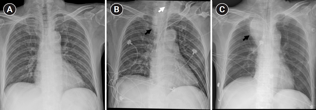
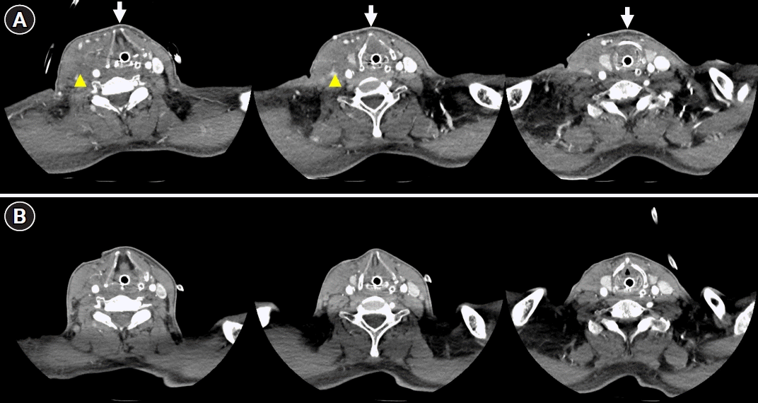
A 61-year-old man presented to the emergency room with left hemiparesis and dysarthria. Brain magnetic resonance imaging showed right middle cerebral artery territory infarction with proximal internal carotid artery stenosis. Carotid endarterectomy (CEA) was performed on the 8th day of admission. After 3 hours of CEA, the patient complained of dyspnea, and stridor was developed. Chest X-ray was performed immediately (Fig. 1), and emergent endotracheal intubation was performed under suspicion of airway obstruction due to hematoma. Neck computed tomography (CT) confirmed hematoma formation around the carotid vessels (Fig. 2). Blood pressure was strictly controlled without discontinuation of clopidogrel, and hematoma was noted to be reduced on a CT scan 10 days after intubation. The patient was extubated without airway problems.
CEA is the standard treatment for symptomatic carotid stenosis [1,2]. A previous study reported that perioperative hematoma occurred in 7.1% of patients after CEA and was associated with increased perioperative stroke and mortality [3]. Neck hematoma is potentially life-threatening because it can cause respiratory failure and often requires airway management [2]. Suspicion of neck hematoma is crucial if the patient complains of respiratory discomfort after CEA, and airway management should be performed immediately [2]. Checking the chest X-ray can provide guidance in the differential diagnosis of respiratory discomfort after CEA.
Notes
Ethics statement
This study was reviewed and approved by the Institutional Review Board of Dong-A University Hospital (No. DAUHIRB-23-159). The need for informed consent from the patient was waived by the board.
REFERENCES
1. Brott TG, Hobson RW 2nd, Howard G, Roubin GS, Clark WM, Brooks W, et al. Stenting versus endarterectomy for treatment of carotid-artery stenosis. N Engl J Med. 2010; 363:11–23.
2. Shakespeare WA, Lanier WL, Perkins WJ, Pasternak JJ. Airway management in patients who develop neck hematomas after carotid endarterectomy. Anesth Analg. 2010; 110:588–93.
3. Ferguson GG, Eliasziw M, Barr HW, Clagett GP, Barnes RW, Wallace MC, et al. The North American symptomatic carotid endarterectomy trial: surgical results in 1415 patients. Stroke. 1999; 30:1751–8.
Fig. 1.
(A) Chest X-ray taken before carotid endarterectomy (CEA) shows normal findings. (B) Chest X-ray taken during dyspnea after CEA shows soft tissue swelling (black arrow) with tracheal deviation (white arrow). (C) Chest X-ray taken the day after intubation shows aggravation of soft tissue swelling (black arrow).


 PDF
PDF Citation
 Print
Print


 XML Download