Introduction
Recently, we encountered a case of challenging chronic obstructive iliofemoral venous disease (COVD) presenting as postthrombotic syndrome (PTS) with a venous ulcer. The lesion was treated with a combination of a dedicated venous stent and a braided-type Wall stent. This case report was written after obtaining informed consent from the patient for the publication of this article.
Go to : 
Case report
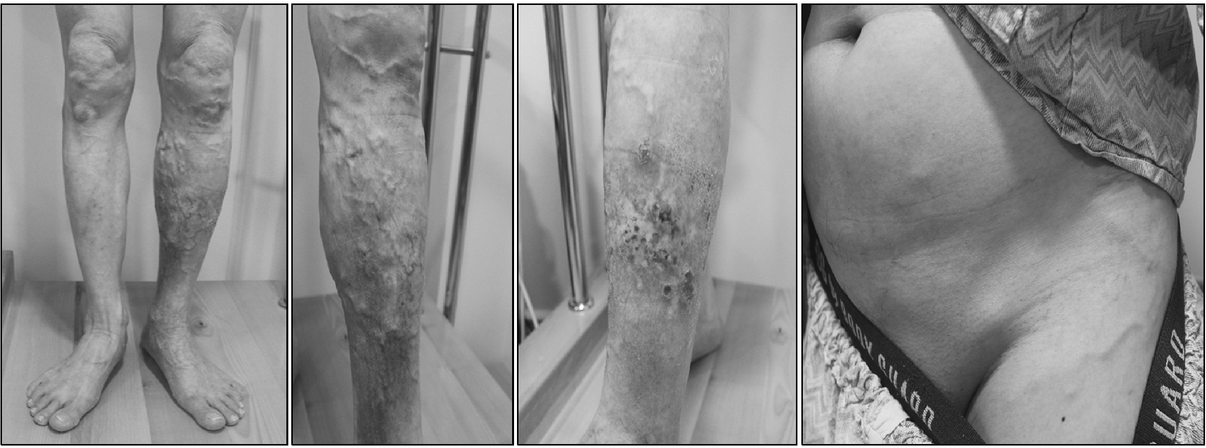
A 59-year-old man visited our hospital due to recurrent leg venous ulcers with a history of left-sided deep vein thrombosis (DVT) following pin insertion into a femur fracture caused by a motorcycle accident 35 years ago. The patient’s medical history includes high blood pressure, a smoking habit of 40 packs per year, and medication intake following coronary stent insertion due to angina. Physical examination revealed swelling in the left calf area, skin discoloration, ulcers, and varicosity in the calf and lower abdomen (Fig. 1). Venous duplex ultrasound showed evidence of venous reflux in the left great saphenous vein and small saphenous vein.
A CT venogram revealed occlusion in the proximal femoral vein, common femoral vein, and external iliac vein, and collateral veins in the abdomen. New thrombi were found in the proximal femoral vein.
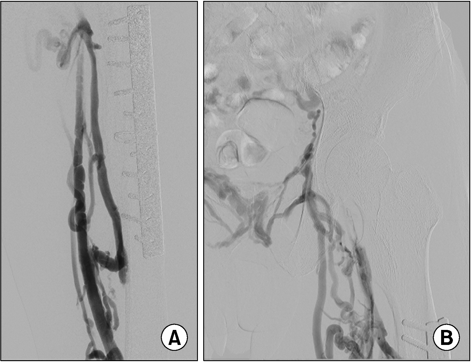
A venogram was performed by inserting a 5Fr sheath into the left small saphenous vein under intravenous anesthesia. It revealed multiple stenoses in the left superficial femoral vein, with the common femoral vein and iliac vein not visible (Fig. 2).
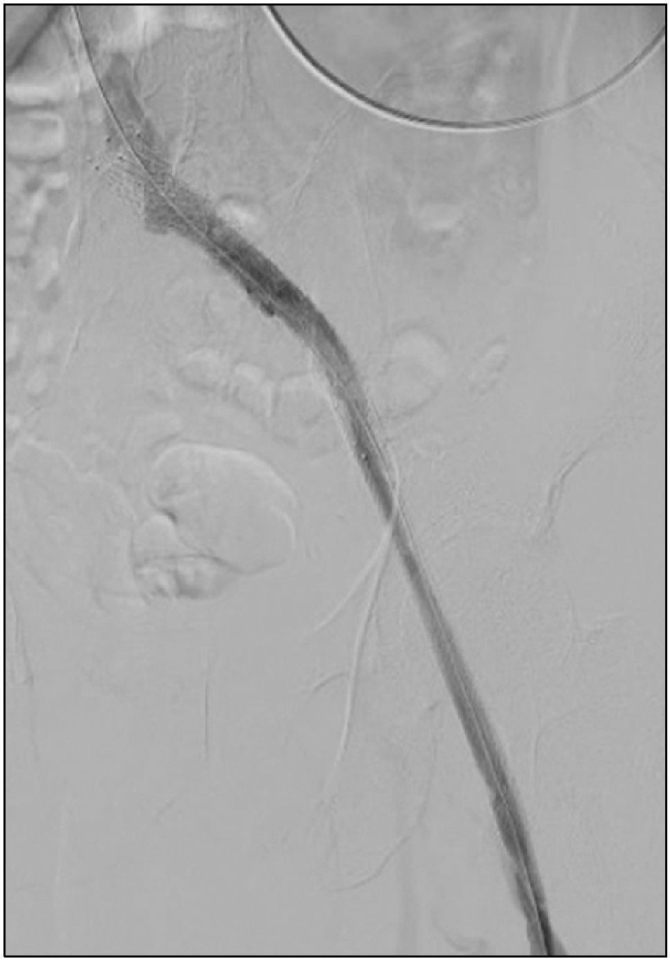
After the passage of a guide wire through occluded veins using various catheters and guide wires, a 14 mm×100 cm dedicated venous Venovo stent (Bard, Becton, Dickinson and Company, NJ) was implanted in the left iliac vein, and a 14 mm×90 cm braided-type Wall stent (Boston Scientific, Marlborough, MA) was implanted in the common to proximal superficial femoral veins (Fig. 3).
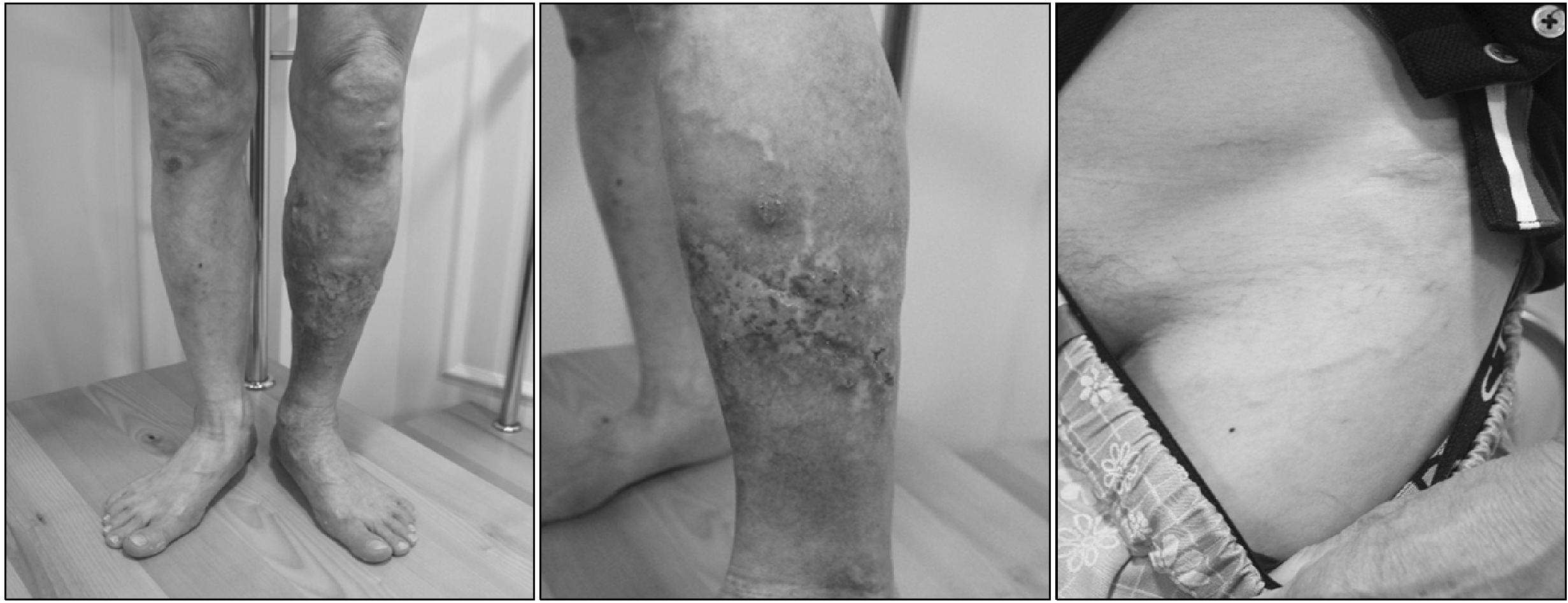
After one month, the edema and ulcer of the patient are improving (Fig. 4). He was planned to continue oral anticoagulants as part of the treatment for deep vein thrombosis. Accordingly, anticoagulant therapy was initiated. Following that, the patient is being observed periodically.
Go to : 
Discussion
Chronic venous ulcers are a debilitating condition that often significantly impacts the quality of life due to their tendency to recur. Lawrence et al. (1) recommended various treatments for patients with chronic venous ulcers caused by iliac vein obstruction when conventional treatments fail. Particularly, when stents are inserted into occluded iliac and femoral veins, the venous ulcer is often quickly cured, as observed in our patient.
1) Safety
In recent data, for PTS patients with stents placed above the inguinal ligament, the primary patency rates were 77% (95% CI: 69%∼83%) at 1 year. Similarly, in those with a stent placed across the ligament, the 1-year primary patency was 78% (95% CI: 73%∼82%), making it known as a safe procedure (2). When using a stent for the iliac vein, the dedicated venous stent has recently been reported to exhibit good performance. If a stent needs to be inserted into the infra-inguinal area, the braided stent is recommended if possible, as fractures typically occur less frequently. Braided stents available in Korea include the Supera stent or Wall stent, and among them, the appropriate size is the Wall stent. Additionally, in recent publications by Powell et al. (3), although there was no statistical difference, the results appeared slightly favorable. In this case, the Venovo stent was used for the common iliac and external iliac veins, while the Wall stent was used for the common femoral vein and proximal superficial femoral vein.
2) Medication
According to Raju’s opinion (4), for perioperative thromboprophylaxis, low molecular weight heparin 60 mg is given subcutaneously preoperatively, in addition to heparin 5000 units given intravenously in the operating room prior to the start of the procedure. Following iliofemoral stenting in patients with postthrombotic syndrome, therapeutic anticoagulation is continued.
3) Follow-up
CT venography is performed 30 days post-operatively. Venous duplex ultrasound is conducted every 3 months to obtain post-procedure metrics, including stent patency, and to assess stent compression and/or in-stent restenosis.
4) Outcome of the case
Iliofemoral vein stenting resulted in the rapid healing of this patient’s ulcer, as well as a significant improvement in limb pain and swelling. These outcomes have persisted without further reports of recurrent open ulcers. His venous stasis hyperpigmentation improved partially, and his leg swelling was significantly reduced. Stent patency was confirmed by duplex ultrasonography every 3 months thereafter.
Go to : 


 PDF
PDF Citation
 Print
Print


 XML Download