See the reply "READER’S FORUM" in Volume 54 on page 137.
Abstract
Since its inception in Europe in the 1950s, alveolar molding treatment for neonates with complete cleft lip and palate has undergone significant evolution in both design and application methodology, demonstrating effectiveness in normalizing the alveolar cleft and nasal shape. However, excessively wide alveolar clefts accompanied by disproportionately wide total maxillary arch pose significant challenges when utilizing conventional alveolar molding methods involving cyclical adding and grinding of acrylic on molding plates. The current report introduces a novel alveolar molding method named Biocreative Alveolar Molding Plate Treatment (BioAMP), which can normalize the maxillary alveolar cleft and arch shape without laborious conventional acrylic procedures. BioAMP sets the target arch form and provides unrestricted space for natural growth of the maxillary alveolar bones while systematically reducing the total maxillary arch width in precise increments. Two exemplary cases are presented as proof-of-concept, showcasing the clinical innovation of BioAMP.
Controlling the orofacial environment using an alveolar molding plate that redirects a neonate’s natural growth of the maxillary alveolar bones and facilitates closure of the interalveolar gap prior to primary cleft lip repair surgery is recognized as a reasonable treatment in modern neonatal cleft care.1-5 This approach has also been proven effective in enhancing oral feeding experiences.6 Since its inception in Scotland in 1950,1 alveolar molding treatment for neonates with unilateral cleft lip and palate (UCLP) has evolved in design and application methodology with different levels of efficacy and efficiency in normalizing maxillary alveolar clefts. Notably, the interalveolar gap at the alveolar cleft can result from a combination of discrepancies in the anteroposterior, transverse, and/or vertical dimensions. For example, an excessively wide alveolar cleft over 15 mm of the interalveolar gap predominantly occurs in the transverse dimension, accompanied by a disproportionately wide total maxillary arch width relative to the mandibular arch, resulting in a scissor bite. Alveolar clefts with this complexity become challenging to treat using conventional alveolar molding methods of repeated adding and grinding of acrylic to the molding plates. Additionally, any large residual interalveolar gap prior to lip repair surgery can also increase the risk of forming a palatal fistula, which may require subsequent repair surgeries later in the child’s life.7 To improve the treatment experience for neonates, families, and providers, this article presents a novel alveolar molding plate treatment called Biocreative Alveolar Molding Plate Treatment (BioAMP) that can efficiently reduce the interalveolar gap and normalize the maxillary arch shape in harmony with the mandibular arch without laborious conventional acrylic manipulation.
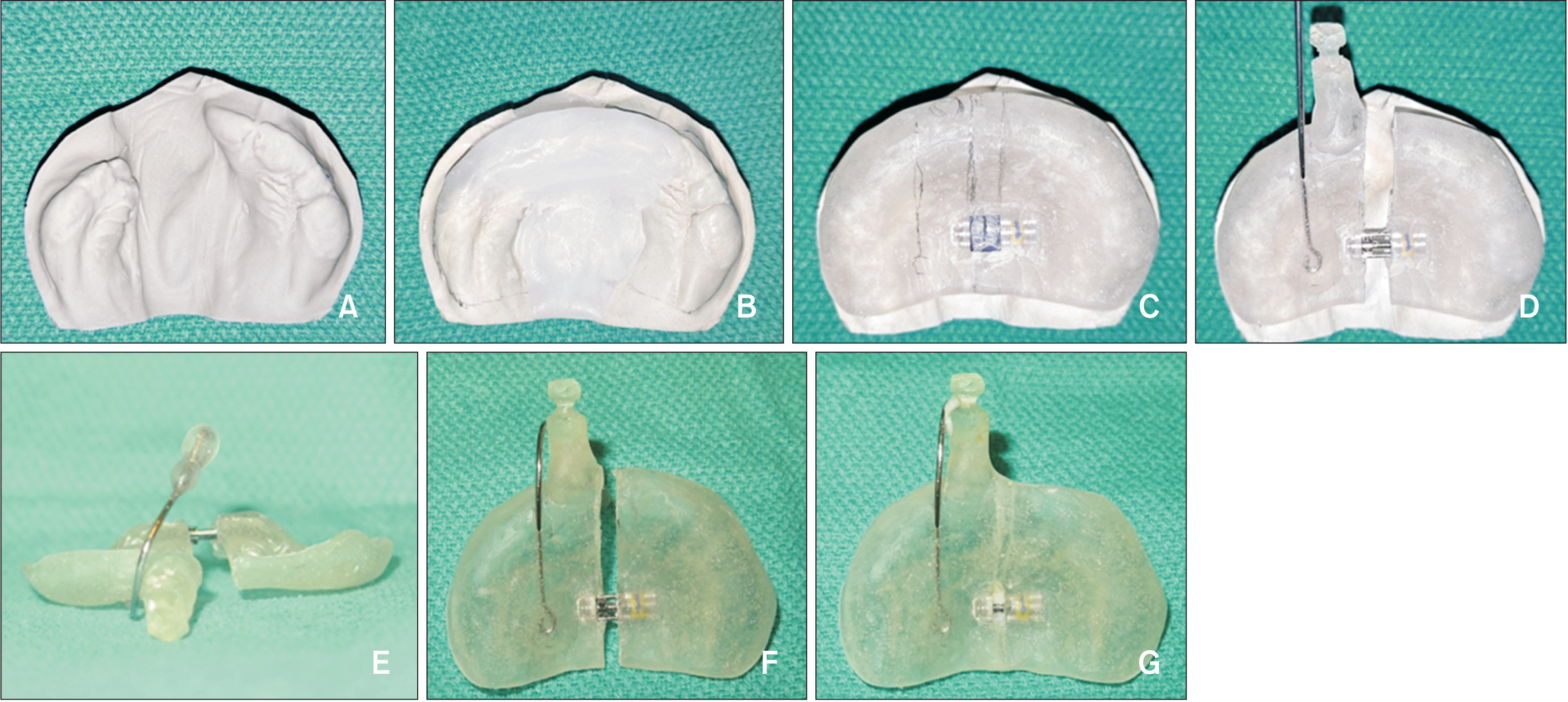
A target maxillary arch form is built on a maxillary stone model using utility wax (Figure 1A and 1B).8 The wax rim simulates the missing alveolar ridge at the cleft. The resultant arch shape is often a flattened ¾ oval rather than a ¾ circle in non-cleft neonates (Figure 1B). Next, the undercuts are completely blocked out to promote the medial growth of the palatal shelves and the anterior growth of the smaller alveolar bone. A constriction screw (maximum constriction capacity: 3 mm; Dentaurum, Ispringen, Germany) is positioned at the center of the maxillary arch. Following resin work for the molding plate, a button and nasal stent wire are added to the BioAMP as the last step in an orthodontic laboratory (Figure 1C and 1D).
On the day of delivery, the nasal wire establishes passive contact with the nasal dome while avoiding contact with the alar rim. This helps establish a superior boundary and minimizes the yaw movement of the BioAMP. An elastic face tape is then applied across the labial cleft with tension to simulate the orbicularis oris muscle. Conversely, a pair of tapes connecting the button of the BioAMP to the cheeks (one on each side of the face) must remain passive at all times to provide a stable position of the BioAMP anteriorly and vertically during treatment. The posterior border of the BioAMP may drop away from the maxillary arch when the patient opens their mouth; however, it can be comfortably re-seated on the maxillary arch with the assistance of the anterior and superior boundaries defined by the face tapes and nasal stent wire.
When a systematic constriction of the total maxillary arch width is needed, the BioAMP is split in half and the constriction screw is activated at a rate of anywhere from 1 to 7 turns at a time (approximately 0.1 mm/turn), followed by re-unification of the split halves using fresh hard acrylic (Figure 1D-1G). This procedure is repeated weekly until systematic constriction is no longer required. If > 3 mm of constriction is required, a new constriction screw can be installed in place of the old one.
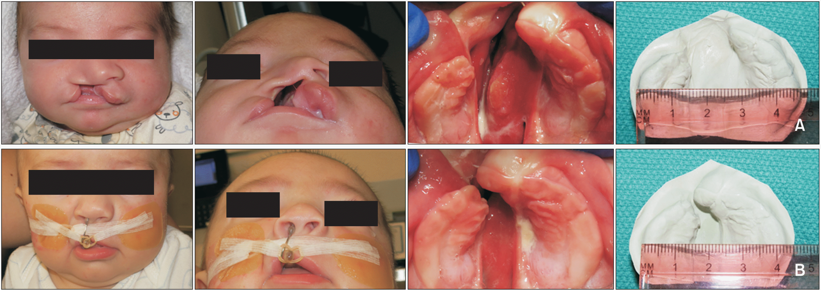
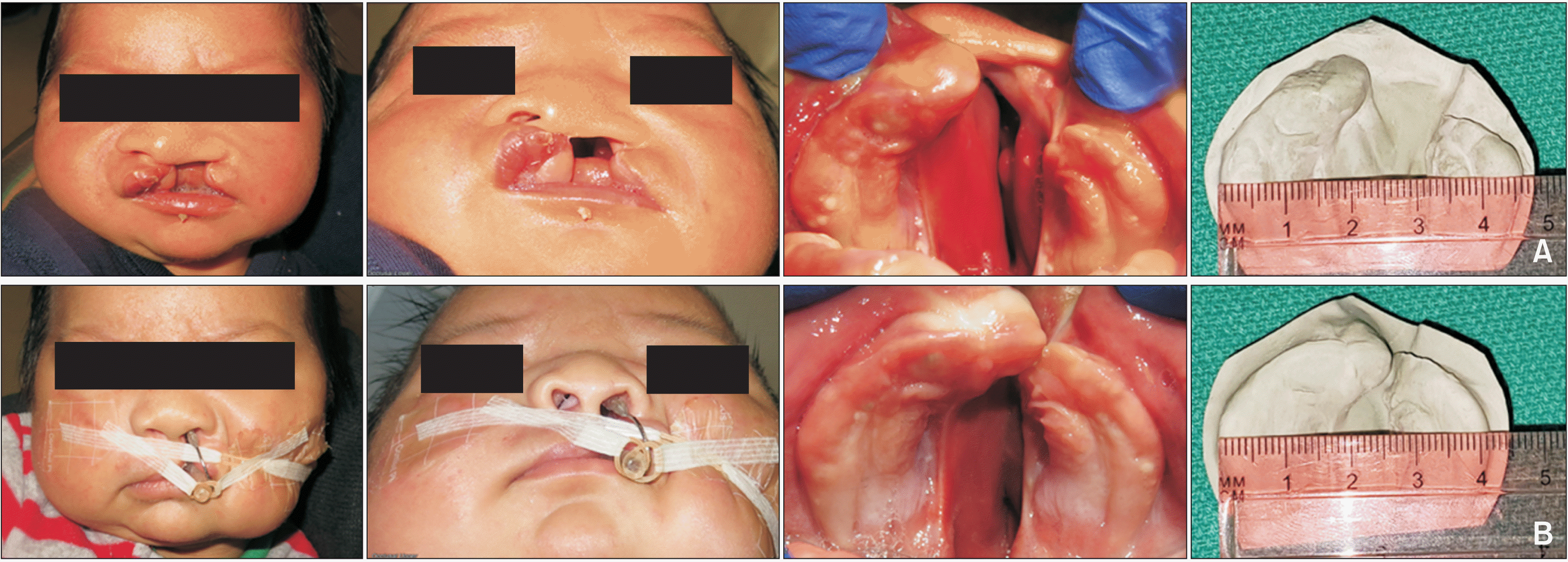
We present 2 exemplary treatments for neonates with an excessively wide interalveolar gap accompanied by an excessively wide total maxillary arch width. Written informed consent was obtained from the parents of all infants. The pre-treatment distance between the lateral sulci was 38.5 mm in patient 1 (Figure 2) and 40 mm in patient 2 (Figure 3). The mean distance was 25.44 mm in the non-cleft neonates at this age.9 The interalveolar gap was greater than 15 mm in both patients. The total maxillary arch width of patient 1 decreased from 47 to 42.5 mm, while the interalveolar gap decreased from 16 to 1 mm (Figure 2). Similarly, the total maxillary arch width of patient 2 decreased from 46 to 41 mm, while the interalveolar gap decreased from 15 to 0 mm (Figure 3). There was noticeable anterior and medial growth of the smaller alveolar bone, which favored the natural closure of the interalveolar gap. Concurrently, the larger alveolar bone grew medially and anteriorly with rotational movement in response to lip taping. The nasal dome on the cleft side rose naturally as the interalveolar gap decreased. Ultimately, nasal symmetry was achieved without actively pushing or shaping the nose (Figures 2 and 3). The nasal stent wire was adjusted to remain passive to the nasal dome at all times to secure a pyramid-shaped spatial retention. Duration of the treatment was 2.5 months for patient 1 and 3 months for patient 2. Both patients underwent primary cleft lip repair surgery before the age of 3.5 months. Both patients completed treatment with only one BioAMP, without requiring new impressions of the maxillary arch or new alveolar molding plates.
Neonates facing the anatomic challenge of an excessively wide alveolar cleft often suffer from discordant oral feeds, suboptimal primary cleft lip repair, or increased risk of palatal fistula formation later in life.6,7 Conventional alveolar molding methods utilizing repeated adding-and-grinding of acrylic to the molding plate are effective but are known to be labor-intensive and lack accuracy in quantifying adjusted amounts, especially when managing an excessively wide alveolar cleft. This often requires additional maxillary impressions and new molding plates for midcourse corrections.2,5
Successful applications of orthodontic constriction screws in retracting the pre-maxilla or expansion screws in expanding the two palatal segments in bilateral cleft lip and palate have been reported, with an emphasis on customized adjustment needs.10-12 However, none of these studies proposed a pre-treatment wax-up approach to pre-program a target maxillary arch shape. Another report presented a potential application of three-dimensional printing technology.13 However, the simulation did not incorporate the pre-programmed final arch shape from the onset of treatment. Instead, it utilized a conventional adding and grinding technique for the simulation.
To date, there has been no report suggesting the applicability of a constriction screw in the treatment of an excessively wide alveolar cleft while simultaneously providing sufficient room for natural growth of the maxillary alveolar bones at the onset of treatment. While the total maxillary arch width of patient 1 reduced by 4.5 mm by the construction screw mechanism, for example, the pre-treatment interalveolar gap of 16 mm reduced to 1 mm at post-treatment, not 11.5 mm. We speculate that this dramatic reduction during the 2.5 months of treatment was attributed not only to the constriction screw mechanism but also to the pre-treatment wax-up approach, which provided sufficient room for unrestricted growth of the maxillary alveolar bones from the onset of the treatment, without periodically requiring a small arbitrary increase in space.
In this report, we showcase the key characteristics of a novel alveolar molding plate treatment named BioAMP. First, its precise incremental constriction mechanism allows for predictable and accountable treatment outcomes by reducing the maxillary arch width to match that of the mandibular arch, thereby establishing occlusal harmony. Second, its ability to pre-program a target maxillary arch shape through wax-up on the pre-treatment maxillary model, enabled by a spatial retention mechanism incorporating the nasal stent wire from the first day of treatment, allows for unrestricted maxillary growth during treatment.
Through this method, spatial equilibrium is formed in the shape of a pyramid by four muscular or cartilaginous boundaries: the tongue as the inferior boundary, the bilateral pterygomandibular raphes as the posterior boundary, face tapes across the labial cleft as the anterior boundary, and the nasal dome as the superior boundary. Notably, the sole purpose of the nasal stent wire is to serve as the superior border of the spatial retention, playing a crucial role in preventing dislodgement and maintaining the BioAMP within a comfortable fitting range. Therefore, the nasal stent wire must remain in passive contact with the nasal dome throughout treatment.
The current report should be interpreted in light of brief report with two exemplary clinical presentations, which significantly limits generalizability of the data. Nevertheless, the BioAMP has potential applications in the management of excessively wide alveolar clefts accompanied by excessively wide arches in UCLP. We plan to validate these possibilities in future studies with larger sample sizes.
Notes
REFERENCES
1. McNEIL CK. 1950; Orthodontic procedures in the treatment of congenital cleft palate. Dent Rec (London). 70:126–32. https://pubmed.ncbi.nlm.nih.gov/24537837/.
2. Hotz M, Gnoinski W. 1976; Comprehensive care of cleft lip and palate children at Zürich university: a preliminary report. Am J Orthod. 70:481–504. https://doi.org/10.1016/0002-9416(76)90274-8. DOI: 10.1016/0002-9416(76)90274-8. PMID: 1068633.
3. Matsuo K, Hirose T. 1988; Nonsurgical correction of cleft lip nasal deformity in the early neonate. Ann Acad Med Singap. 17:358–65. https://pubmed.ncbi.nlm.nih.gov/3064700/.
4. Pool R, Farnworth TK. 1994; Preoperative lip taping in the cleft lip. Ann Plast Surg. 32:243–9. https://doi.org/10.1097/00000637-199403000-00003. DOI: 10.1097/00000637-199403000-00003. PMID: 8192382.
5. Grayson BH, Santiago PE, Brecht LE, Cutting CB. 1999; Presurgical nasoalveolar molding in infants with cleft lip and palate. Cleft Palate Craniofac J. 36:486–98. https://doi.org/10.1597/1545-1569_1999_036_0486_pnmiiw_2.3.co_2. DOI: 10.1597/1545-1569_1999_036_0486_pnmiiw_2.3.co_2. PMID: 10574667.
6. Clarren SK, Anderson B, Wolf LS. 1987; Feeding infants with cleft lip, cleft palate, or cleft lip and palate. Cleft Palate J. 24:244–9. https://pubmed.ncbi.nlm.nih.gov/3477346/.
7. Mak SY, Wong WH, Or CK, Poon AM. 2006; Incidence and cluster occurrence of palatal fistula after furlow palatoplasty by a single surgeon. Ann Plast Surg. 57:55–9. https://doi.org/10.1097/01.sap.0000205176.90736.e4. DOI: 10.1097/01.sap.0000205176.90736.e4. PMID: 16799309.
8. Choo H, Maguire M, Low DW. 2012; Modified technique of presurgical infant maxillary orthopedics for complete unilateral cleft lip and palate. Plast Reconstr Surg. 129:249–52. https://doi.org/10.1097/PRS.0b013e318230c8bb. DOI: 10.1097/PRS.0b013e318230c8bb. PMID: 22186514.
9. Bauer FX, Güll FD, Roth M, Ritschl LM, Rau A, Gau D, et al. 2017; A prospective longitudinal study of postnatal dentoalveolar and palatal growth: the anatomical basis for CAD/CAM-assisted production of cleft-lip-palate feeding plates. Clin Anat. 30:846–54. https://doi.org/10.1002/ca.22892. DOI: 10.1002/ca.22892. PMID: 28459132.
10. Tankittiwat P, Pisek A, Manosudprasit M, Punyavong P, Manosudprasit A, Phaoseree N, et al. 2021; Function of nasoalveolar molding devices in bilateral complete cleft lip and palate: a 3-dimensional maxillary arch analysis. Cleft Palate Craniofac J. 58:1389–97. https://doi.org/10.1177/1055665621990184. DOI: 10.1177/1055665621990184. PMID: 33657892.
11. Bhutiani N, Tripathi T, Rai P. 2022; Active nasoalveolar molding using 3-directional expansion screw for arch alignment in bilateral cleft lip and palate. AJO-DO Clin Companion. 2:20–5. https://doi.org/10.1016/j.xaor.2021.12.003. DOI: 10.1016/j.xaor.2021.12.003.
12. Suri S, Disthaporn S, Atenafu EG, Fisher DM. 2012; Presurgical presentation of columellar features, nostril anatomy, and alveolar alignment in bilateral cleft lip and palate after infant orthopedics with and without nasoalveolar molding. Cleft Palate Craniofac J. 49:314–24. https://doi.org/10.1597/10-204. DOI: 10.1597/10-204. PMID: 21981581.
13. Zheng J, He H, Kuang W, Yuan W. 2019; Presurgical nasoalveolar molding with 3D printing for a patient with unilateral cleft lip, alveolus, and palate. Am J Orthod Dentofacial Orthop. 156:412–9. https://doi.org/10.1016/j.ajodo.2018.04.031. DOI: 10.1016/j.ajodo.2018.04.031. PMID: 31474271.
Figure 1
Biocreative Alveolar Molding Plate (BioAMP) with a constriction screw: Fabrication and implementation protocol. A, Occlusal view of a pre-treatment maxillary arch shape of a neonate with an excessively wide alveolar cleft accompanied by excessive total maxillary arch width. B, Wax-up for a target maxillary arch shape. C, Hard acrylic plate with a constriction screw in place. D, A button and nasal stent wire were installed prior to the delivery of the plate. The palatal plate is split in half to activate the constriction screw. E, Frontal view of the final form of the BioAMP after the nasal stent wire was fitted to the nasal dome inside the nose. The palatal plate remained split prior to activating the constriction screw. F, Occlusal view of the BioAMP after activating the constriction. G, Occlusal view of the BioAMP after the gap between the split halves was re-united using fresh hard acrylic. The total maxillary arch width is now less than before.

Figure 2
Patient 1 was treated with the BioAMP with a constriction screw. The images present the frontal facial view, wormʼs eye view, occlusal view of the maxillary arch, and a stone model of the maxillary arch (arranged from left to right): A, Pre-treatment at the age of 1 week. B, Post-treatment at the age of 2.5 months. The total maxillary arch width decreased from 47 to 42.5 mm, and the interalveolar gap decreased from 16 to 1 mm. Permission to use the photos was granted by the patientʼs parents.

Figure 3
Patient 2 was treated with the BioAMP with a construction screw. The images present the frontal facial view, wormʼs eye view, occlusal view of the maxillary arch, and a stone model of the maxillary arch (arranged from left to right): A, Pre-treatment at the age of 2 weeks. B, Post-treatment at the age of 3 months. The total arch width decreased from 46 to 41 mm, and the interalveolar gap decreased from 15 to 0 mm. Permission to use the photos was granted by the patientʼs parents.


 PDF
PDF Citation
 Print
Print


 XML Download