

 PDF
PDF Citation
 Print
Print


Introduction
Surface roughness can lead to decreased gloss and discoloration or staining of materials, affecting the esthetic quality of restorations. Furthermore, it may promote dental plaque accumulation, leading to secondary caries and periodontitis. It is ideal to have composite restorations with smooth surfaces that do not deteriorate over the course of time.1 The smoothest surface of a composite resin is produced when the material is allowed to polymerize against the matrix. However, composites polymerized with a clear matrix on the surface will leave a resin-rich surface layer that is easily abraded in the oral environment exposing unpolished, rough, and inorganic filler material.2,3 Despite careful matrix placement, removing excess material and recontouring restorations is crucial in clinical practice. This step requires some degree of finishing and polishing, which may alter the smoothness obtained with the matrix.
Micro-defects on resin surfaces sequentially caused by polishing result in weaker matrix and filler deterioration, finally leading to loss, deformation, and marginal microleakage. Dickinson GL, et al. suggested the necessity of surface sealant which is used to fill up the microleakage and gaps.4,5 Reports show that surface sealant application functions effectively owing to its high fluidity and wettability in accordance with low molecular weight.6,7
Composite resins have variable polishing conditions according to the type of inorganic filler, the size of filler particles, and the quantity of mixed filler. Any discrepancies in microhardness between the resin matrix and annexed filler pose challenges in achieving flat polished surfaces after finishing and polishing, resulting in surface roughness and irregularities on composite resin surfaces. Research confirms that the differential surface characteristics of dental composite resins after polishing influence the discoloration or staining of restorative materials. Moreover, surface sealant application after finishing and polishing affects surface roughness and discoloration or staining of material surfaces.8
The present study aimed to evaluate the influence on surface roughness of the composite resin by applying variable types of surface sealant respectively to polished surface state.
Materials and Methods
Two composite resins, microfilled composite (Metafil CX) and hybrid composite (Aelite LS posterior) were tested. Three surface sealants (BisCover LV, Optiguard, and Seal-n-Shine) were used (Table 1).
Sixty samples per material (N = 120), 8 mm in diameter and 4 mm thick, were made in plastic molds placed on a glass plate. They were light-cured for 60 s using Optilux 501 (Demetron, Kerr, Orange, USA). Polishing was performed with 600, 1000, and 2000-grit sandpaper (Tamiya finishing abrasives, Tamiya Inc., Shizuoka, Japan). Considering the ground amount of the composite resin, polishing treatment with the sandpapers was performed 30 times for 600-grit, 40 times for 1000-grit, and 50 times for 2000-grit sandpapers.
Polished samples were placed in water in an ultrasonic cleaner (Biosonic UC100, Coltene Whaledent, Cabot, USA) for 10 min to remove polishing debris that may have collected on the surface. A 32% phosphoric acid solution (UNI-ETCH; Bisco, Schaumburg, USA) was applied with a brush for 15 s onto the surface of the composite resin. The specimens were rinsed with water and air-dried. A thin layer of BisCover LV (Bisco) was applied with a brush and light-cured for 30 s after waiting for 15 s (according to the manufacturer’s recommendations). A thin layer of Optiguard (Kerr, Orange, USA) was applied with a brush, lightly thinned with compressed air to obtain satisfactory spreading to the naked eye, and light-cured for 20 s. A thin layer of Seal-n-Shine (Pulpdent Co., Watertown, USA) was applied with a brush and light-cured for 20 s.
A surface profilometer (SJ-301, Mytutoyo, Tokyo, Japan) was used to measure the average roughness, characterized by the height parameter, Ra (μm). Four scans (8 mm long) were made on each specimen in a different direction. The average surface roughness (for each specimen) was taken as the average of the four Ra values.
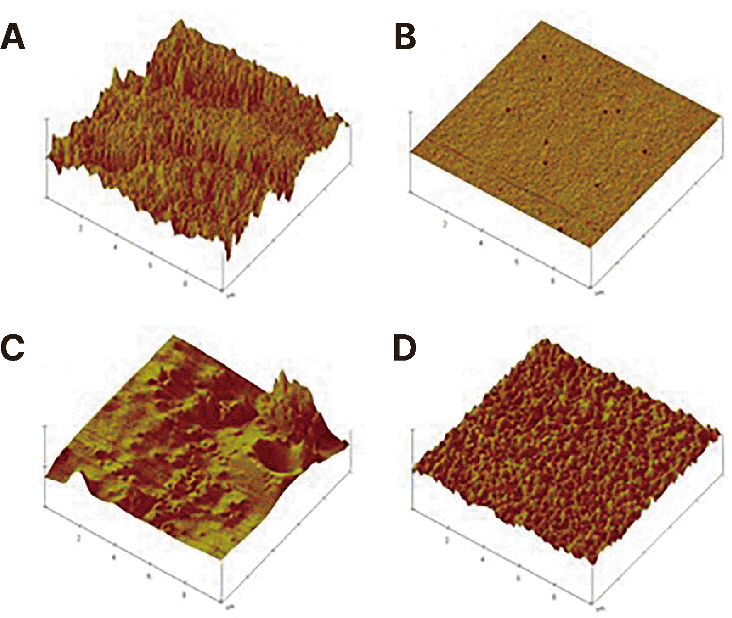
The samples were sputter-coated with gold and examined by scanning electron microscopy (SEM) (JSM-7500F, JEOL, Tokyo, Japan) before and after surface sealant application. Atomic force microscopy (AFM) was also performed before and after surface sealant application to assess surface morphology (10 µm × 10 µm). A MultiMode IV atomic force microscope (AFM) (Veeco Instruments Inc., Woodbury, USA) was used to obtain topographic images of the composite surface.
The data were analyzed by t-test followed by a Scheffe test (α = .05).
Results
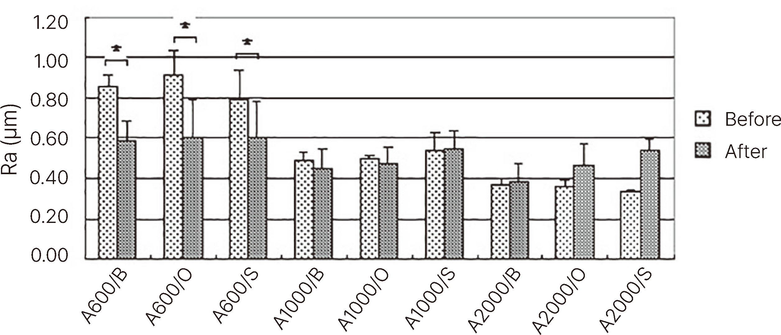
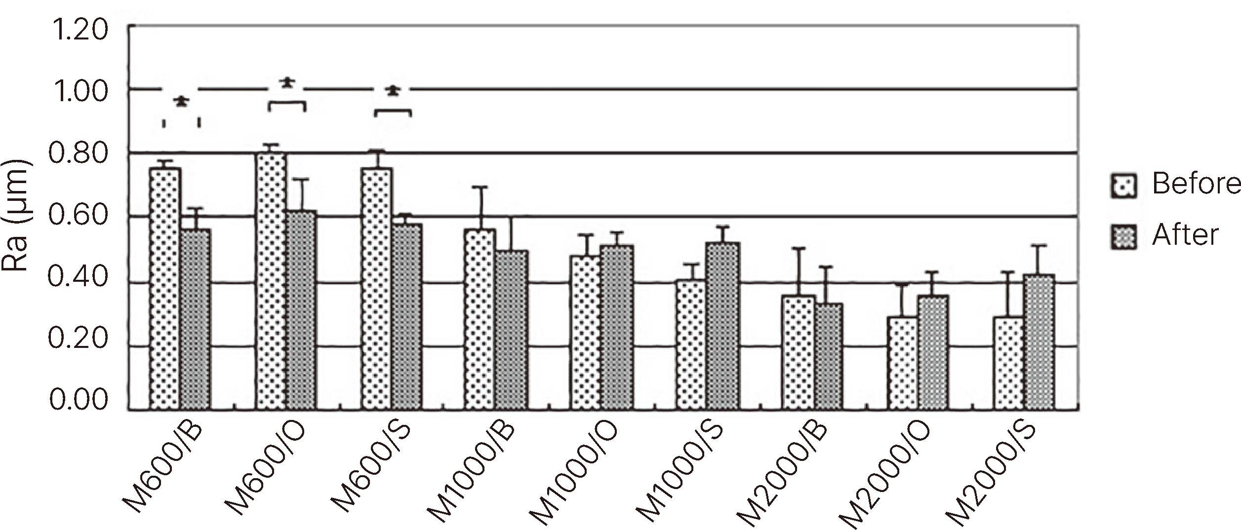
The lowest Ra values were obtained with specimens polished with 600-grit sandpaper and coated with surface sealant (P < 0.05). Specimens polished with 1000 and 2000-grit sandpaper showed no significant difference. On comparing types of surface sealants, there was no significant difference (Fig. 1, 2).
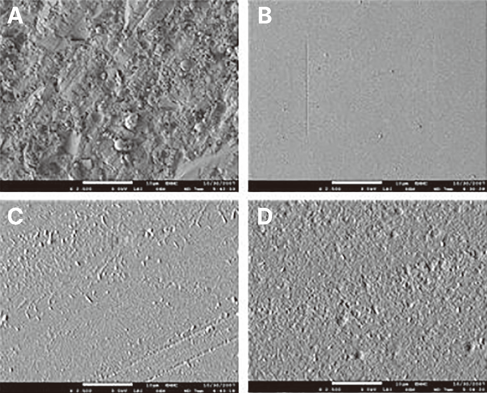
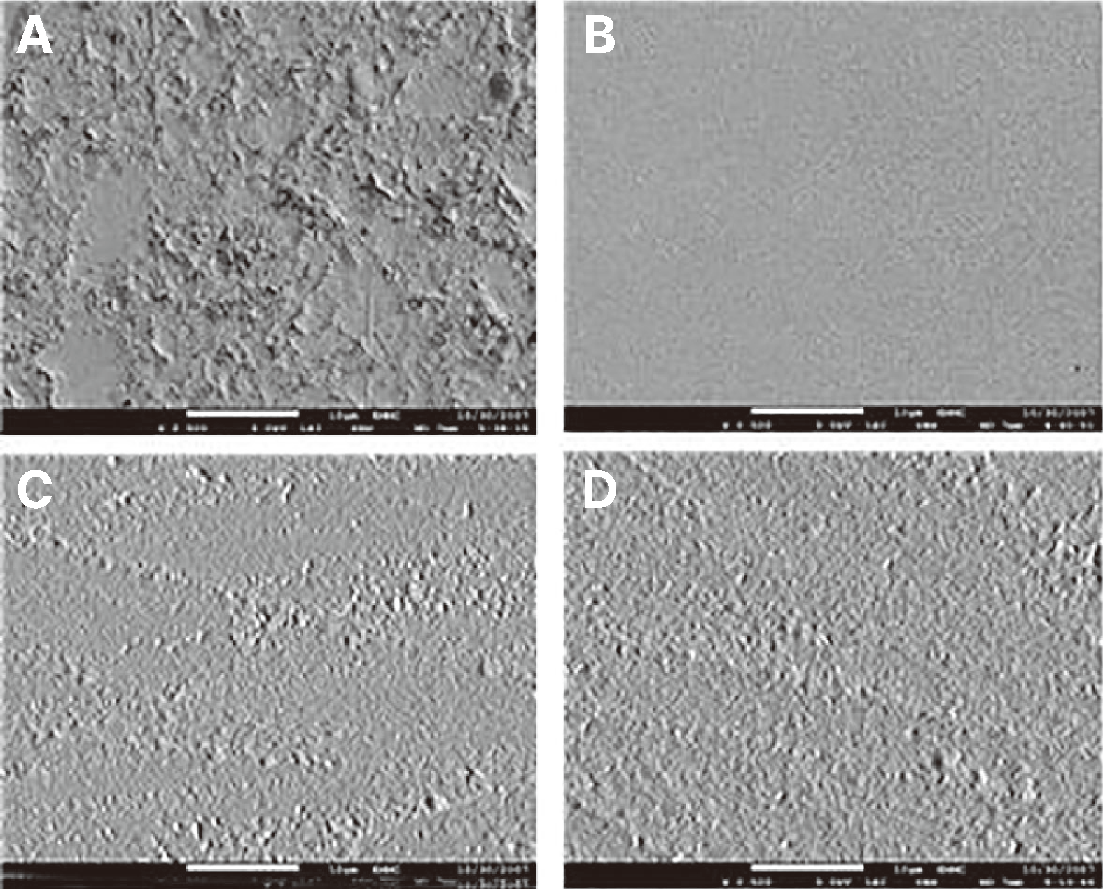
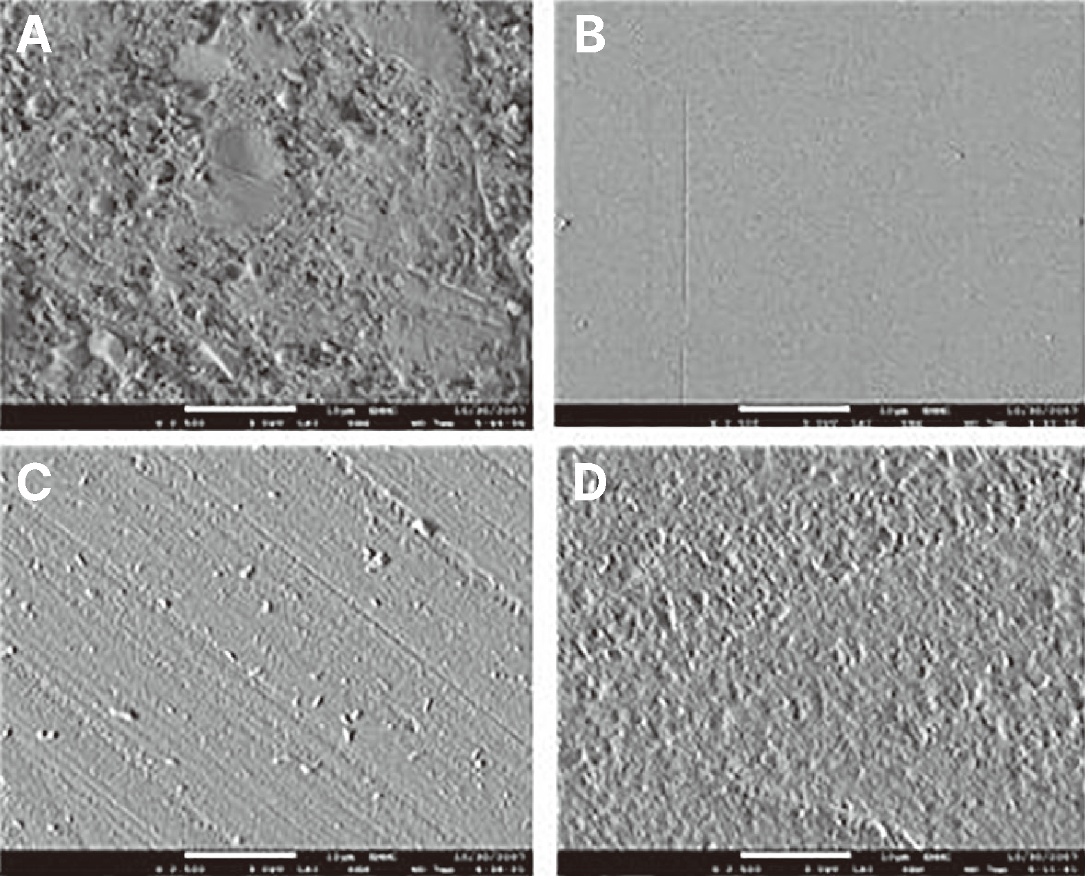
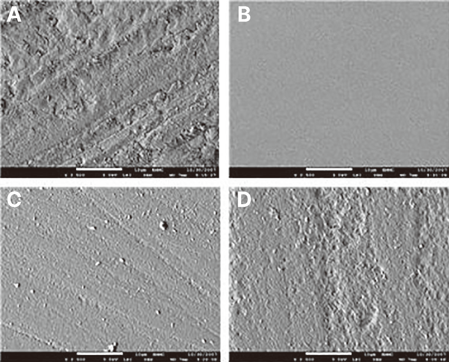
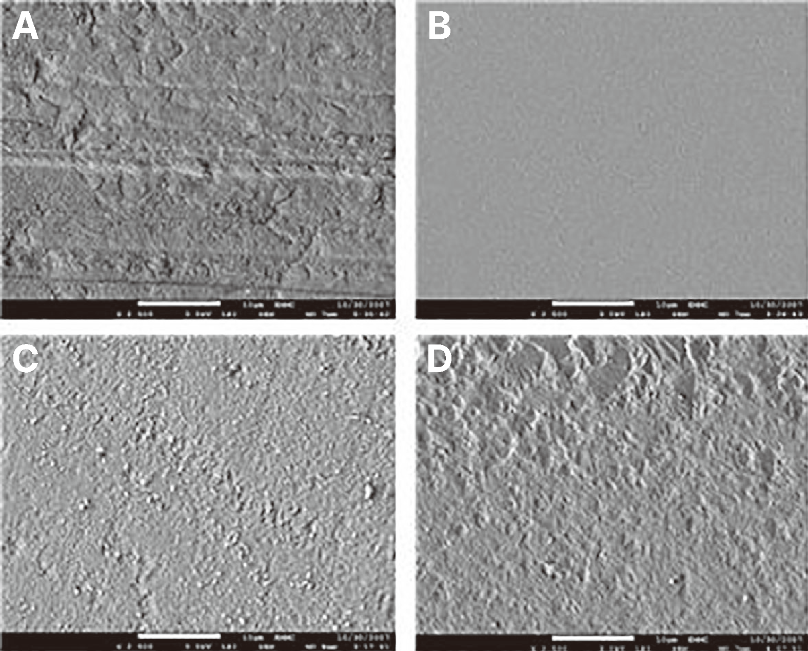
Scanning electron micrographs of the materials with surface sealant treatment are presented in Fig. 3 to 8. Composite resin surfaces were examined at 2500 × with a scanning electron microscope (JSM-7500F, JEOL) before surface sealant application (Fig. 3 - 8). The untreated surface had numerous voids and filler projections extruding from the surface. By comparison, the treated surface revealed substantially fewer voids, cracks, and other microstructural defects. Furthermore, the filler particles appeared less pronounced or extruded from the surface.
The SEM images for each material showed different surface characteristics. The surface of Aelite LS Posterior was less homogeneous than that of Metafil CX after polishing. The specimen without surface sealant application (after polishing) presented a rough surface texture induced by polishing procedures in the SEM images. Resins composed of more hybridized than microfilled fillers showed more irregular surface texture in SEM. The specimen coated with surface sealant showed a flat surface texture with more reduced microstructural defects on the subsurface than only polished specimens. BisCover LV-treated specimens showed numerous micro-voids regularly on the smooth surface. The specimen group coated with Optiguard or Seal-n-Shine showed a flat surface texture compared to others with only polishing treatment. However, micro-roughness came to exist among these specimens.
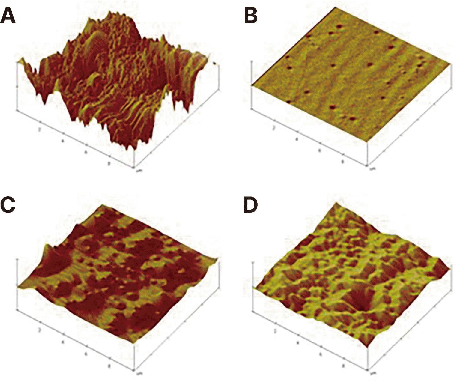
The specimens were divided into two groups, pre-treatment and post-treatment of surface sealant application, and monitored under 10 µm focusing conditions with AFM (MultiMode IV, Veeco Instruments, Plainview, USA) (Fig. 9, 10). The specimen with polishing treatment alone showed dense projections and pronouncement. The BisCover LV-treated specimens had voids on the flat and smooth surfaces. Optiguard and Seal-n-Shine-treated specimens had much flatter surfaces than the pre-treatment state but still showed micro-projections compared to BisCover LV-treated specimens.
In summary, the two different resins exhibited differential surface texture after polishing treatment alone; however, after surface sealant application, the surface texture of the two resins showed no definite difference.
Discussion
Surface roughness promotes initial bacterial adhesion and accumulation. Bollen et al. named threshold roughness value in the report that plaque came to have retention and develop to full grown phase at surface roughness 0.2 µm and above in vivo.9 Ra values observed after polishing treatment with various sandpapers were generally higher than the critical threshold surface roughness for bacterial adhesion, 0.2 µm. In sequence, surface smoothness is crucial for successful resin restorations.
Many researchers reported reduced surface roughness after surface sealant application, decreased marginal microleakage, and reduced amount of ground resin.10-12 During resin polishing, pronounced or extruded filler particles are generated because composite resin fillers have higher rigidity than the resin matrix. This phenomenon is apparently proportional to filler size.
This study also shows that a high filler particle size in hybrid resins is associated with irregular surfaces with dense projections rather than a small filler particle size in microfilled resins when monitoring surface microstructure by SEM and AFM. In evaluating the surface roughness of composite resins with a profilometer, the rougher surfaced group polished with 600-grit sandpaper tended to show a decrease in Ra values after surface sealant application. In contrast, the smoother surfaced group polished with 1000 or 2000-grit sandpaper showed no meaningful change in Ra values. However, SEM revealed that specimens with surface sealant application had a flatter texture and more reduced surface roughness than those that received polishing treatment only.
For advanced analysis, we worked with AFM, which shows the dimensional surface image of a specific specimen. On AFM images, specimens with surface sealant application exhibited notably flatter surface texture than those that received polishing treatment only but still showed voids and micro-projections on the surface. It is acceptable that the BisCover LV-treated specimens with highly smooth surfaces were influenced slightly by the contact of the profilometer exploring tip and voids on the surface when evaluating surface roughness. Optiguard® and Seal-n-Shine-coated specimens had flat surfaces but still showed structural limitations (micro-projections on the subsurface). In contrast to the fact that a thin layer is generated in BisCover LV-treated specimens, cracks and detachment are often present on the contact surface of Optiguard® and Seal-n-Shine. Optiguard treatment necessitates compressed air application on the surface before light curing. But in this procedure if there is a situation either excessive spraying or voids on resin surface, we found the defects of surface or complete absence of Optiguard of the surface.
It was impossible to achieve a consistent thickness in all products. Hence, there seems to be a limitation in attaining flat surfaces with existing methods such as brushing or air spraying. Marie-France et al. performed thickness measurements resulting in the same outcome and showed marked differences in not one but other specimens after surface sealant application. As a result, roughness on the whole surface increased after applying a surface sealant to specimens polished with 2000-grit sandpaper.
Research widely reports that composite resin curing is an imperfect procedure that leaves an uncured layer owing to the inhibitory effect of oxygen.13,14 Also, the other two surface sealants used in this study, except BisCover LV, created an uncured layer on the surfaces of the specimens. Although this layer readily disappeared by saliva, food debris, or tooth brushing, the surface was rinsed with running water for the time required to produce a satisfactory surface texture, any remaining uncured material was removed, and the surface was dried with compressed air.
To summarize, surface sealant application after finishing and polishing creates flat and smooth restoration surfaces, preventing plaque accumulation and attrition of resin restorations. In microscopic experiments, there is little effect on surface roughness with surface sealant application after highly leveled polishing. However, it is an affirmative effect to some extent on reduction of micro-projections extruding from the surface after polishing treatment only. Even though SEM or AFM shows the most determining aspect of the specimen surface, there need to be consideration about the unexpected voids, micro-defects or irregular structure which is not seen on the target screen of the imaging modalities.
Conclusion
Laboratory evidence after evaluating and observing the surface roughness and restoration surface after surface sealant application according to the surface polished level between two composite resins, microfilled and hybrid, showed the following results.
Surface sealant-coated specimens polished with 600-grit sandpaper showed lower Ra values than those without application (P < 0.05). There was no meaningful difference in specimen groups polished with 1000 or 2000-grit sandpaper.
SEM or AFM revealed that specimens with surface sealant application tended to have considerably decreased micro-defects on resin surfaces after finishing and polishing treatment.
The results show that surface sealant application influences the surface roughness of composite resins with coarse surfaces but not highly polished composite resins

 XML Download