Journal List > J Korean Med Sci > v.39(3) > 1516085826

Han, Kim, Shin, Kang, Choi, Lee, Kim, Joo, Chang, Park, Song, Ahn, Suh, Lee, Her, Jeong, Kim, Kim, Lim, Shin, Cho, and for the PTRG Investigator: Prognostic Implication of Platelet Reactivity According to Left Ventricular Systolic Dysfunction Status in Patients Treated With Drug-Eluting Stent Implantation: Analysis of the PTRG-DES Consortium

Abstract

Background

Coronary artery disease patients undergoing percutaneous coronary intervention (PCI) often exhibit reduced left ventricular ejection fraction (LVEF). However, the impact of LV dysfunction status in conjunction with platelet reactivity on clinical outcomes has not been previously investigated.

Methods

From the multicenter PTRG-DES (Platelet function and genoType-Related long-term prognosis in DES-treated patients) consortium, the patients were classified as preserved-EF (PEF: LVEF ≥ 50%) and reduced-EF (REF: LVEF< 5 0%) group by echocardiography. Platelet reactivity was measured using VerifyNow P2Y12 assay and high platelet reactivity (HPR) was defined as PRU ≥ 252. The major adverse cardiac and cerebrovascular events (MACCEs) were a composite of death, myocardial infarction, stent thrombosis and stroke at 5 years after PCI. Major bleeding was defined as Bleeding Academic Research Consortium bleeding types 3–5.

Results

A total of 13,160 patients from PTRG-DES, 9,319 (79.6%) patients with the results of both PRU and LVEF were analyzed. The incidence of MACCE and major bleeding was higher in REF group as compared with PEF group (MACCEs: hazard ratio [HR] 2.17, P < 0.001, 95% confidence interval [CI] 1.85–2.55; major bleeding: HR 1.78, P < 0.001, 95% CI 1.39–2.78). The highest rate of MACCEs was found in patients with REF and HPR, and the difference between the groups was statistically significant (HR 3.14 in REF(+)/HPR(+) vs. PEF(+)/HPR(-) group, P < 0.01, 95% CI 2.51–3.91). The frequency of major bleeding was not associated with the HPR in either group.

Conclusion

LV dysfunction was associated with an increased incidence of MACCEs and major bleeding in patients who underwent PCI. The HPR status further exhibited significant increase of MACCEs in patients with LV dysfunction in a large, real-world registry.

Trial Registration

ClinicalTrials.gov Identifier: NCT04734028

Graphical Abstract

jkms-39-e27-abf001.jpg

INTRODUCTION

Coronary artery disease (CAD) is one of the major causes of left ventricular (LV) systolic dysfunction, which subsequently leads to heart failure (HF) of ischemic origin (so-called “ischemic cardiomyopathy”). Patients who have experienced acute coronary syndrome or had CAD and were treated with percutaneous coronary intervention (PCI) often exhibit ischemic cardiomyopathy.123 In general, HF is also regarded as a prothrombotic condition with increased platelet reactivity and hypercoagulability due to various mechanisms.456 Antiplatelet agents, in particular P2Y12 inhibitors, have been reported to play an integral role in the secondary prevention of ischemic events after PCI in high-risk patients.78 The antiplatelet effect of P2Y12 inhibitors may be more closely related to clinical events in HF patients with a thrombogenic milieu.9 However, the responsiveness of P2Y12 in an individual patient is not uniform and is believed to be affected by clinical and genetic factors.
Based on platelet reactivity measured by platelet function assays, the concept of therapeutic range with either high platelet reactivity (HPR) or low platelet reactivity (LPR) has been suggested. HPR is associated with an increased incidence of ischemic events, such as myocardial infarction (MI) or death, in patients who previously underwent coronary interventions. On the other hand, LPR is associated with increased bleeding events.10111213141516
Currently, none of the studies have evaluated the association between platelet reactivity and LV systolic dysfunction status, and its prognostic implication in CAD patients treated with drug-eluting stent (DES) implantation. Therefore, we aimed to evaluate the prognostic implication of the association between platelet reactivity and LV systolic function in a large-scale cohort of CAD patients treated with DES implantation.

METHODS

Study design and participants

The PTRG-DES (Platelet function and genoType-Related long-term proGnosis in DES-treated patients with CAD) consortium was established to determine the association of platelet function test (PFT) and genotyping with long-term prognosis during clopidogrel treatment in a large-scale East Asian cohort treated with DES. This consortium was endorsed by the Korean Society of Interventional Cardiology (ClinicalTrials.gov Identifier: NCT04734028).17 A total of nine prospective registries from 32 Korean academic centers joined the PTRG-DES consortium and contributed data of 13,160 DES patients treated between July 2003 and August 2018. We obtained 11,714 PFT results measured by VerifyNow assay (PTRG-PFT cohort), and 8,163 genotyping results related to clopidogrel responsiveness (PTRG-Genotype cohort) from the consortium. Consecutive patients at each center were successfully treated with one or more DES approved by the US Food and Drug Administration or Conformité Européene mark. Patients adequately loaded with aspirin and clopidogrel were eligible for enrollment in the study, regardless of patient or lesion complexity. The exclusion criteria were occurrence of a major complication during the procedure, fibrinolytic therapy, and need for oral anticoagulant.

Procedures

All PCI procedures were performed in accordance with the standard technique.18 Following the procedure, patients were administered 100 mg of aspirin and 75 mg of clopidogrel per day. Patients were recommended aspirin indefinitely and clopidogrel for a duration of at least 1 year. All other treatments administered were according to the standard care. Clinical outcomes were evaluated until the last outpatient visit.17

PFT

Platelet reactivity was measured during the peri-procedural period using the VerifyNow assay (Accriva, San Diego, CA, USA). The measurements were performed after an adequate time duration to ensure full anti-platelet effect.19 Aspirin was administered as either (1) a coated 300-mg oral dose at least 6 hours or (2) a dose of 100 mg at least 5 days before PCI. Clopidogrel was administered in one of the following ways: 1) a dose of 600 mg at least 6 hours; 2) a dose of 300 mg at least 12 hours; or 3) a dose of 75 mg at least 5 days before PCI. If eptifibatide or tirofiban was used during PCI, a 24-hour washout period was required before VerifyNow testing. No patients receiving abciximab were enrolled as a long washout period would be needed.17
VerifyNow assay is a whole-blood, point-of-care, turbidimetric optical detection assay designed to measure agonist-induced platelet aggregation.19 Blood samples were collected in 3.2% citrate Vacuette tubes (Greiner Bio-One Vacuette North America, Monroe, NC, USA). Measurements were performed in accordance with the manufacturer’s protocol, the details of which have been described elsewhere.20 The cartridge comprised fibrinogen-coated polystyrene beads, 20 mmol/L adenosine diphosphate, and 22 nmol/L prostaglandin E1. The optical signal of this channel was reported as “P2Y12 reaction units (PRUs).” We assessed PRUs as continuous and categorical measures. Additionally, the cutoffs of HPR to ADP were defined according to the time-dependent receiver operating characteristic curve analysis from the PTRG-PFT cohort (≥ 252 PRUs).21

Status of LV systolic dysfunction

Echocardiography was performed using commercially available equipment during the peri-procedural period. Standard echocardiography and calculations were performed according to the recommendations of the American Society of Echocardiography.22 Echocardiography was performed by certified echocardiographers, and an echocardiography specialist supervised all the measurements independently. Left ventricular ejection fraction (LVEF) was calculated according to the modified Simpson rule using LV end-systolic and end-diastolic volumes.23
According to the current guidelines and status of LV systolic function, the study population was divided into two groups: preserved-ejection fraction (PEF: LVEF ≥ 50%) group and reduced-EF (REF: LVEF < 50%) group. Additionally, in a separate analysis, we classified LV systolic dysfunction based on LVEF: (1) preserved EF (PEF), LVEF ≥ 50%; (2) mildly reduced EF (mrEF), 40% < LVEF < 50%; and (3) reduced EF (rEF), LVEF ≤ 40%.2425

Clinical outcomes

The primary endpoint was occurrence of major adverse cardiac and cerebrovascular events (MACCEs) including all-cause death, MI, definite stent thrombosis (ST), or stroke. In addition, major bleeding was defined as Bleeding Academic Research Consortium bleeding types 3-5.26
All deaths were considered to have occurred due to cardiovascular (CV) causes unless a definite non-CV cause was established. Acute MI was defined as increased cardiac troponin values with ischemic symptoms or ischemic changes on electrocardiogram or imaging evidence of recent loss of viable myocardium or new regional wall motion abnormality, all of which were not related to the procedure.27 Stroke was defined as evidence of neurological deficit requiring hospitalization and presence of clinically documented lesions on brain computed tomography or magnetic resonance imaging. An independent clinical events committee masked to VerifyNow and LVEF results adjudicated all the clinical events using original source documents.

Statistical analysis

Kolmogorov–Smirnov test was performed to analyze normal distribution of continuous variables. Continuous variables were expressed as mean ± standard deviation or as medians (interquartile range [IQR]), while categorical variables were presented as absolute numbers and frequencies (%). Student’s unpaired t-test was used for parametric continuous variables and Mann–Whitney U test for non-parametric continuous variables. Analysis of variance was used for comparison amongst the three groups. Comparisons between categorical variables were performed using Pearson’s chi-square test or Fisher’s exact test when the Cochran rule was not met.
All demographic characteristics and laboratory measurements were evaluated in a univariate analysis. Variables with P < 0.1 in the univariate analysis were then entered into multivariate logistic backward elimination analysis to obtain odds ratios (ORs) and 95% confidence intervals (CIs). Univariate and multivariate Cox proportional hazard analyses were performed to identify proportional hazard risk on clinical events according to PRU levels and to adjust for known potential confounders (index MI presentation, age, sex, body mass index, hypertension, dyslipidemia, smoking, diabetes mellitus, chronic kidney disease, anemia, congestive HF, previous PCI, previous stroke, multivessel disease, PCI for left main or left anterior descending artery, use of 2nd DES, complex PCI, beta blocker, angiotensin blockade, statin, and proton pump inhibitor). A P value < 0.05 was considered statistically significant. All statistical analyses were performed using R (version 4.2.1, R Foundation for Statistical Computing, Vienna, Austria).

Ethics statement

The Institutional Review Board of each participating center reviewed and approved the registry and waived the requirement for written informed consent for access to an institutional registry (Kangnam Sacred Heart Hospital Institutional Review Board approval no. 2018-10-019). The study protocol conforms to the ethical guidelines of the 1975 Declaration of Helsinki as reflected in a priori approval by the institution’s human research committee.

RESULTS

Baseline characteristics

In the PTRG-PFT cohort (11,714 patients with VerifyNow result), the on-admission LVEF data was available for a total of 9,319 patients (79.6%) (81.8% [n = 7,620] in the PEF and 18.2% [n = 1,084] in the REF groups) (Fig. 1). According to the LVEF level, no statistically significant differences were observed between the PEF and REF groups in terms of PRU level (216 ± 77.3 vs. 220.5 ± 82.0; P = 0.070), but the prevalence of HPR was significantly different (33.2% vs. 35.4%; P = 0.008) (Table 1).
Fig. 1

Flow chart.

LVEF = left ventricular ejection fraction, PEF = preserved ejection fraction, REF = reduced ejection fraction, HPR = high platelet reactivity, mrEF = mildly reduced ejection fraction.
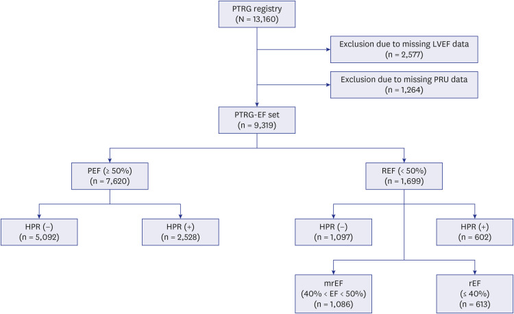
jkms-39-e27-g001
Table 1

Comparison of baseline characteristics between reduced and preserved ejection fraction groups

jkms-39-e27-i001
Variables Overall (N = 13,160) REF (n = 1,699) PEF (n = 7,620) P value
HPR (−) (n = 1,097) HPR (+) (n = 602) HPR (−) (n = 5,092) HPR (+) (n = 2,528)
Age, yr 64.2 ± 10.9 64.4 ± 12.0 68.0 ± 11.4 62.9 ± 10.8 67.1 ± 10.2 < 0.001
Male 8,848 (67.2) 790 (72.0) 336 (55.8) 3,731 (73.3) 1,397 (55.3) < 0.001
Body mass index, kg/m2 24.6 ± 3.1 24.1 ± 3.2 23.8 ± 3.2 24.7 ± 3.1 24.5 ± 3.2 < 0.001
Risk factors
Hypertension 7,933 (60.3) 610 (55.6) 361 (60.0) 2,993 (58.8) 1,660 (65.7) < 0.001
Dyslipidemia 8,303 (63.1) 711 (64.8) 386 (64.1) 3,411 (67.0) 1,636 (64.7) 0.128
Smoking 3,578 (27.2) 398 (36.3) 145 (24.1) 1,578 (31.0) 543 (21.5) < 0.01
Diabetes mellitus 4,619 (35.1) 393 (35.8) 145 (24.1) 1,597 (31.4) 939 (37.1) < 0.01
Chronic kidney disease 2,875 (21.8) 309 (28.2) 238 (39.5) 784 (15.4) 599 (23.7) < 0.01
History
Anemia 2,743 (20.8) 268 (24.4) 280 (46.5) 887 (17.4) 929 (36.7) < 0.001
PAD 1,550 (11.8) 207 (18.9) 127 (21.1) 529 (10.4) 326 (12.9) < 0.001
CHF 1,072 (8.2) 243 (22.2) 153 (25.4) 268 (5.3) 49 (1.9) < 0.001
MI 971 (7.4) 177 (16.1) 86 (14.3) 284 (5.6) 136 (5.4) < 0.001
PCI 1,737 (13.2) 173 (15.8) 101 (16.8) 645 (12.7) 330 (13.1) < 0.001
CABG 163 (1.2) 32 (2.9) 8 (1.3) 61 (1.2) 19 (0.8) < 0.001
Stroke 921 (7.0) 81 (7.4) 60 (10.0) 360 (7.1) 171 (6.8) 0.051
VerifyNow_PRU 217.8 ± 78.2 174.3 ± 58.1 304.7 ± 42.8 175.3 ± 55.3 299.6 ± 38.8 < 0.001
LVEF, % 58.8 ± 10.6 41.3 ± 7.2 41.2 ± 6.9 62.7 ± 6.5 62.6 ± 6.6 < 0.001
WBC, ×103/dL 7.8 ± 3.0 9.3 ± 3.9 9.0 ± 3.7 7.7 ± 2.8 7.5 ± 2.7 < 0.001
Hb, g/dL 13.6 ± 1.9 13.8 ± 2.0 12.5 ± 2.0 14.0 ± 1.7 12.8 ± 1.7 < 0.001
PLT count, ×103/dL 243.7 ± 82.1 237.3 ± 79.4 231.4 ± 73.3 234.0 ± 72.2 235.8 ± 76.5 0.323
GFR, mL/min/1.73 m3 77.3 ± 27.1 76.2 ± 31.4 68.5 ± 33.4 81.9 ± 25.6 76.8 ± 27.6 < 0.001
HbA1c, % 6.5 ± 1.4 6.6 ± 1.4 6.8 ± 1.4 6.5 ± 1.3 6.6 ± 1.3 < 0.001
TC, mg/dL 173.6 ± 44.4 174.6 ± 46.3 171.4 ± 42.6 175.8 ± 44.4 173.2 ± 45.9 0.037
LDL, mg/dL 106.5 ± 40.3 108.9 ± 52.6 106.0 ± 37.5 107.1 ± 38.0 105.8 ± 41.1 0.192
HDL, mg/dL 43.9 ± 12.7 42.6 ± 11.3 42.4 ± 11.3 44.2 ± 12.2 44.1 ± 15.2 < 0.001
TG, mg/dL 142.3 ± 97.1 132.2 ± 100.0 120.8 ± 77.0 148.5 ± 104.9 139.5 ± 90.0 < 0.001
Angiographic features
ACC/AHA lesion < 0.001
A/B1 5,626 (42.8) 481 (43.8) 241 (40.0) 2,494 (49.0) 1,197 (47.3)
B2/C 7,534 (57.2) 616 (56.2) 361 (60.0) 2,598 (51.0) 1,331 (52.7)
Number of diseased vessels < 0.001
One 7,755 (58.9) 631 (57.5) 328 (54.5) 3,194 (62.7) 1,481 (58.6)
Two 3,517 (26.7) 287 (26.2) 150 (24.9) 1,331 (26.1) 688 (27.2)
Three 1,888 (14.3) 179 (16.3) 124 (20.6) 567 (11.1) 359 (14.2)
Multivessel disease 5,967 (45.3) 466 (42.5) 274 (45.5) 1,898 (37.3) 1,047 (41.4) < 0.001
Bifurcation lesion 1,298 (9.9) 145 (13.2) 86 (14.3) 599 (11.8) 337 (13.3) 0.095
CTO lesion 789 (6.0) 147 (13.4) 66 (11.0) 357 (7.0) 151 (6.0) < 0.001
Procedural data
Multi-vessel PCI 3,234 (24.6) 311 (28.4) 184 (30.6) 1,284 (25.2) 675 (26.7) 0.010
Treated lesion
LM 659 (5.0) 52 (4.7) 38 (6.3) 264 (5.2) 116 (4.6) 0.316
LAD 7,757 (58.9) 698 (63.6) 382 (63.5) 3,039 (59.7) 1,465 (58.0) < 0.001
LCx 3,933 (29.9) 312 (28.4) 181 (30.1) 1,470 (28.9) 809 (32.0) 0.030
RCA 5,018 (38.1) 436 (39.7) 257 (42.7) 1,927 (37.8) 1,002 (39.6) 0.074
Stent type < 0.001
1st generation DES 1,402 (10.7) 41 (3.7) 43 (7.1) 271 (5.3) 187 (7.4)
2nd generation DES 9,181 (69.8) 1,056 (96.3) 559 (92.9) 4,821 (94.7) 2,341 (92.6)
Number of stents 1.6 ± 0.8 1.7 ± 0.8 1.7 ± 0.8 1.6 ± 0.8 1.6 ± 0.8 < 0.001
Stent length, mm 35.1 ± 22.1 39.5 ± 24.4 39.3 ± 24.1 36.0 ± 22.2 36.5 ± 22.7 < 0.001
Stent diameter, mm 3.0 ± 0.4 2.9 ± 0.4 2.9 ± 0.4 3.0 ± 0.5 3.0 ± 0.4 < 0.001
Concomitant medication
Aspirin 12,831 (97.5) 1,081 (98.5) 593 (98.5) 4,942 (97.1) 2,451 (97.0) 0.007
Clopidogrel 13,160 (100.0) 1,097 (100.0) 602 (100.0) 5,092 (100.0) 2,528 (100.0)
Cilostazol 1,292 (9.8) 90 (8.2) 47 (7.8) 439 (8.6) 223 (8.8)) 0.836
Beta-blocker 7,627 (58.0) 763 (69.6) 391 (65.0) 2,858 (56.1) 1,437 (56.8) < 0.001
RAS blockade 8,063 (61.3) 729 (66.5) 414 (68.8) 2,960 (58.1) 1,535 (60.7) < 0.001
Calcium channel blocker 3,118 (23.7) 177 (16.1) 99 (16.4) 1,309 (25.7) 667 (26.4) < 0.001
Statin 11,607 (88.2) 964 (87.9) 505 (83.9) 4,506 (88.5) 2,208 (87.3) 0.010
Proton-pump inhibitor 2,235 (17.0) 199 (18.1) 121 (20.1) 797 (15.7) 488 (19.3) < 0.001
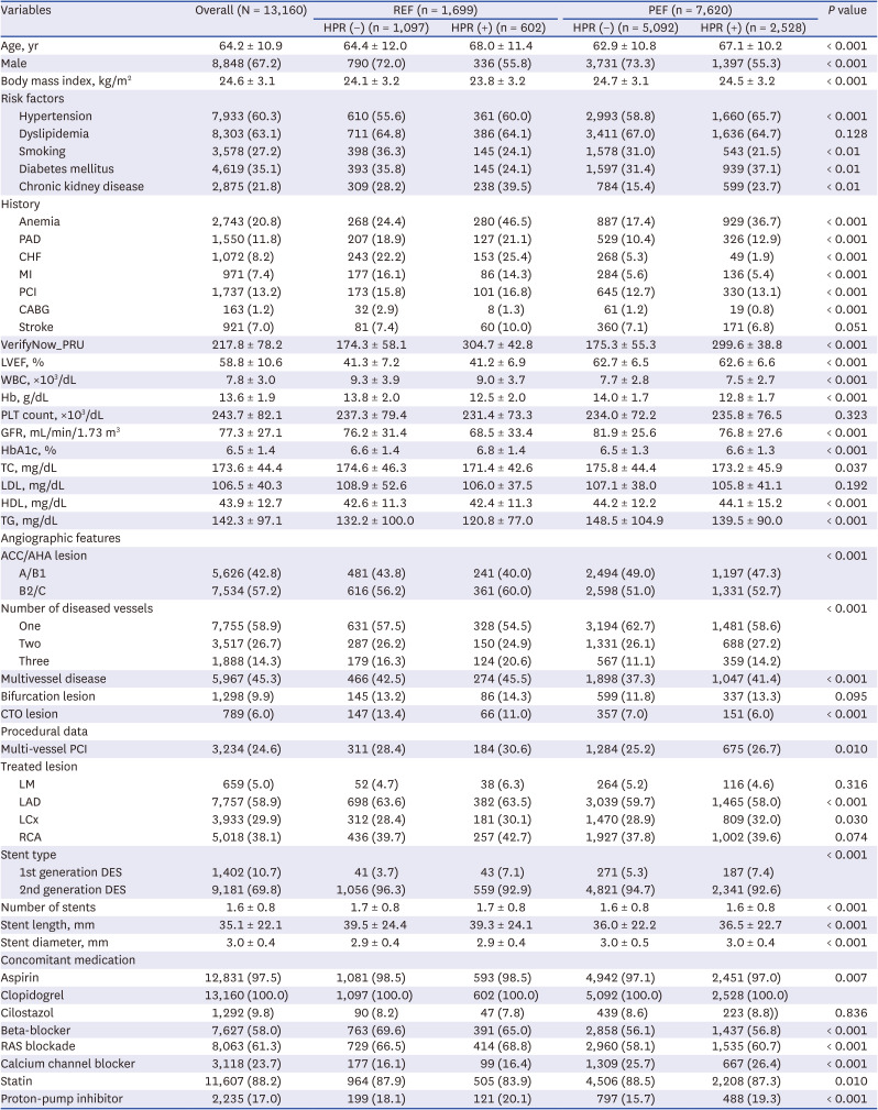
Values are presented as mean ± standard deviation or number (%).
REF = reduced ejection fraction, PEF = preserved ejection fraction, HPR = high platelet reactivity, PAD = peripheral artery disease, CHF = congestive heart failure, MI = myocardial infarction, PCI = percutaneous coronary intervention, CABG = coronary artery bypass graft, LVEF = left ventricular ejection fraction, WBC = white blood count, Hb = hemoglobin, PLT = platelet, GFR = glomerular filtration ratio, TC = total cholesterol, LDL = low-density lipoprotein, HDL = high-density lipoprotein, TG = triglyceride, CTO = chronic total occlusion, LM = left main, LAD = left anterior descending artery, LCx = left circumflex artery, RCA = right coronary artery, RAS = renin-angiotensin-aldosterone system, DES = drug-eluting stent.
In addition, no statistically significant differences in PRU level (216.5 ± 77.3 vs. 219.3 ± 82.5 vs. 222.7 ± 81.2 PRUs; P = 0.111) and HPR prevalence (33.2% vs. 35.0% vs. 36.2%; P = 0.179) were found amongst the three groups according to the LVEF level (Supplementary Table 1). We further investigated the relationship between PRUs and LVEF, which showed no association (Supplementary Fig. 1).

Clinical outcomes according to LV systolic function

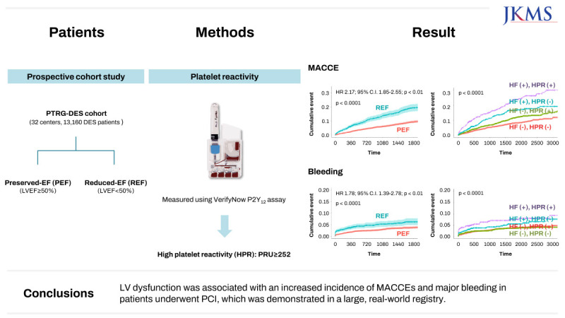
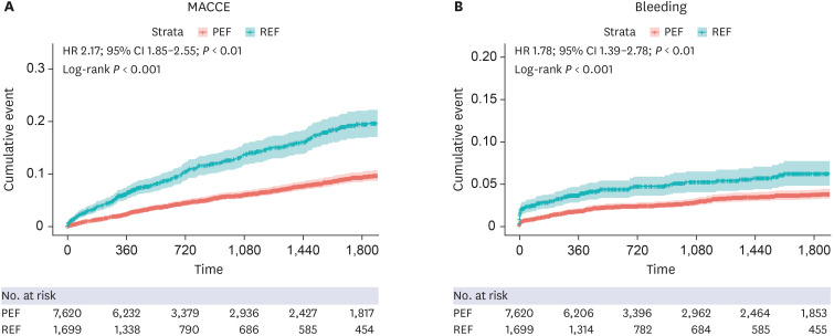
During the median follow-up period of 16.8 months (IQR, 12.0–59.8), a total of 455 MACCEs (6.0%) (224 deaths [2.9%], 118 non-fatal MI [1.5%], 37 ST [0.5%], and 132 non-fatal stroke [1.7%]) and 223 cases of major bleeding (2.9%) occurred in the PEF group. In the REF group, 227 cases of MACCEs (13.2%) (164 deaths [9.7%], 47 non-fatal MI [2.8%], 13 ST [0.8%], and 42 non-fatal stroke [2.5%]) and 88 cases of major bleeding (5.2%) was documented (Supplementary Table 2). The REF group had a significantly higher rate of occurrence of MACCEs compared with the PEF group (HR, 2.17; 95% CI, 1.85–2.55; P < 0.001). In addition, major bleeding occurred more frequently in patients with REF than those with PEF (HR, 1.78; 95% CI, 1.39–2.78; P < 0.001) (Fig. 2). In the multivariable analysis, REF was found to be an independent determinant of MACCE and major bleeding occurrence (Supplementary Table 3). Patients with mrEF (HR, 1.58; 95% CI, 1.29–1.95; P < 0.001) and rEF (HR, 3.38; 95% CI, 2.76–4.14; P < 0.001) showed an increased 5-year risk of MACCEs. Moreover, patients with mrEF (HR, 1.42; 95% CI, 1.03–1.95; P = 0.03) and rEF (HR, 2.46; 95% CI, 1.77–3.42; P < 0.001) also showed an increased risk of major bleeding (Supplementary Fig. 2).
Fig. 2

Kaplan-meier curves between PEF group and REF group.

PEF = preserved ejection fraction, REF = reduced ejection fraction, HR = hazard ratio, CI = confidence interval.
jkms-39-e27-g002

Prognostic implication of HPR status according to LV systolic dysfunction

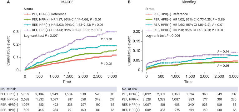
We analyzed the risks of primary outcome and major bleeding according to HPR status and LV systolic dysfunction. Irrespective of LV systolic dysfunction, the HPR phenotype significantly increased the risk of MACCEs (P interaction < 0.01), and this effect was more prominent in cases of all-cause death and ST (Tables 2 and 3). No association was found between the frequency of major bleeding and HPR phenotype in both PEF and REF groups (Fig. 3).
Table 2

Clinical outcomes in the reduced and preserved ejection fraction groups according to left ventricular ejection fraction and/or high platelet reactivity
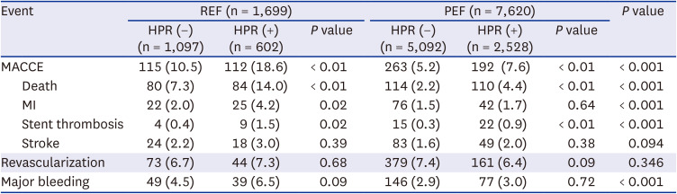
jkms-39-e27-i002
Event REF (n = 1,699) PEF (n = 7,620) P value
HPR (−) (n = 1,097) HPR (+) (n = 602) P value HPR (−) (n = 5,092) HPR (+) (n = 2,528) P value
MACCE 115 (10.5) 112 (18.6) < 0.01 263 (5.2) 192 (7.6) < 0.01 < 0.001
Death 80 (7.3) 84 (14.0) < 0.01 114 (2.2) 110 (4.4) < 0.01 < 0.001
MI 22 (2.0) 25 (4.2) 0.02 76 (1.5) 42 (1.7) 0.64 < 0.001
Stent thrombosis 4 (0.4) 9 (1.5) 0.02 15 (0.3) 22 (0.9) < 0.01 < 0.001
Stroke 24 (2.2) 18 (3.0) 0.39 83 (1.6) 49 (2.0) 0.38 0.094
Revascularization 73 (6.7) 44 (7.3) 0.68 379 (7.4) 161 (6.4) 0.09 0.346
Major bleeding 49 (4.5) 39 (6.5) 0.09 146 (2.9) 77 (3.0) 0.72 < 0.001
REF = reduced ejection fraction, PEF = preserved ejection fraction, HPR = high platelet reactivity, MACCE = major adverse cardiac and cerebrovascular events, MI = myocardial infarction
Table 3

Clinical outcomes according to left ventricular ejection fraction and/or high platelet reactivity
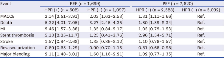
jkms-39-e27-i003
Event REF (n = 1,699) PEF (n = 7,620)
HPR (+) (n = 602) HPR (−) (n = 1,097) HPR (+) (n = 2,528) HPR (−) (n = 5,092)
MACCE 3.14 [2.51–3.91] 2.03 [1.63–2.53] 1.31 [1.11–1.66] Ref.
Death 5.32 [4.01–7.05] 3.27 [2.46–4.35] 1.80 [1.39–2.34] Ref.
MI 2.46 [1.57–3.88] 1.35 [0.84–2.17] 1.05 [0.72–1.53] Ref.
Stent thrombosis 5.13 [2.25–11.7] 1.25 [0.41–3.76] 2.96 [1.54–5.71] Ref.
Stroke 1.57 [0.94–2.62] 1.35 [0.86–2.12] 1.10 [0.78–1.57] Ref.
Revascularization 0.89 [0.65–1.22] 0.90 [0.70–1.15] 0.81 [0.68–0.98] Ref.
Major bleeding 2.11 [1.48–3.01] 1.60 [1.16–2.21] 1.02 [0.77–1.35] Ref.
Values are presented as hazard ratios and 95% confidence intervals.
REF = reduced ejection fraction, PEF = preserved ejection fraction, HPR = high platelet reactivity, MACCE = major adverse cardiac and cerebrovascular events, MI = myocardial infarction.
Fig. 3

Kaplan-meier curves related to left ventricular dysfunction and HPR status.

MACCE = major adverse cardiac and cerebrovascular event, REF = reduced ejection fraction, HPR = high platelet reactivity, HR = hazard ratio, CI = confidence interval, PEF = preserved ejection fraction.
jkms-39-e27-g003
We also investigated the association between PRUs and outcomes in each group using spline analysis. The curve showed a linear pattern in the PEF group. Although the spline analysis showed that the outcome decreased with reduction in PRUs in the REF group, the risk tended to rise below 100 PRUs. However, this was not significant (Supplementary Fig. 3).

DISCUSSION

To the best of our knowledge, this study is the first to evaluate the clinical impact of LV dysfunction and platelet reactivity on clinical outcomes (ischemic events and major bleeding) in a large-scale cohort of East Asian patients with CAD who underwent DES-based PCI. The principal findings of this study were as follows:
  • 1) Patients with < 50% LVEF (REF group) showed a two-fold increase in composite ischemic events (MACCEs) compared with the PEF group.

  • 2) The REF group showed a 65% increase in major bleeding events compared with the PEF group.

  • 3) HPR status and PRU levels ≥ 252 had an impact on MACCEs in both the REF and PEF groups; however, no such association was evident in terms of bleeding events.

  • 4) Further breakdown of the REF group into rEF (LVEF ≤ 40%) and mrEF (LVEF 41–50%) showed a difference in MACCE incidence.

Platelets play a key role in thrombotic occlusion during the rupture of a coronary atherosclerotic plaque, leading to myocardial ischemia and infarction.28 Moreover, thrombotic occlusion in epicardial arteries and platelet microembolization can induce tissue damage by microcirculatory arrest.29 Acute myocardial loss due to ischemia can induce myocardial remodeling.30 The mechanisms of remodeling are unclear; however, coronary microcirculation could mediate myocardial ischemic injury.3132 Endothelial cell swelling and sloughing along with platelet aggregates might lead to capillary obstruction. Moreover, advanced capillary impairment could result in intramural bleeding.33 Chronically ischemic myocardium may display structural remodeling of the coronary microvasculature, which appears as atrophy of larger microvessels and reduced vascular distensibility.3435 Moreover, myocardial reperfusion injury could occur paradoxically, resulting in fresh myocardial injury and cardiomyocyte death.36 This process leads to myocardial remodeling that results in reorganization of myocytes, intercellular matrix components, and vessels. Any of these can be reduced, normal, or increased, resulting in myocardial changes that are dependent on the loading conditions, neuroendocrine activation, and genetic factors. Failure to recover from these adverse remodeling events results in progressive dilatation, recruitment of border zone myocardium into the scar, and eventually deterioration in contractile function, known as LV dysfunction.3037
LV dysfunction is a criterion of HF. HF is associated with increased MACCE and thrombotic events, such as venous thromboembolism, due to platelet activation and hypercoagulability.383940414243 In line with these concepts, our data demonstrated that patients with LV dysfunction experienced increased ischemic events compared with those without LV dysfunction, and this difference was statistically significant.
Although the association between bleeding and HF is not well understood, some studies have reported that HF accompanied with atrial fibrillation and chronic kidney disease resulted in increased bleeding.444546 In this study, major bleeding was significantly higher in the REF group compared with the PEF group. The mechanisms of bleeding in HF have not been clearly identified. However, it might be associated with platelet dysfunction and microvascular damage with subsequent derangement of hemostasis, which might increase bleeding.47 Therefore, antiplatelet and anticoagulation therapy in HF patients should be carefully considered in the presence of concomitant vascular disease. Moreover, the balance between thromboembolic and bleeding risk should be carefully monitored.
HPR with the antiplatelet agent clopidogrel is a well-known risk factor for ischemic events in patients undergoing PCI.4849 We used 252 PRUs as the optimal cutoff value, which was higher than the cutoff values used in Europe and the United States.484950 Although the optimal PRU value was higher than that in the Western population, the incidence of ischemic events was lower, which was in concordance with the “East Asian concept.”51 Interestingly, the outcomes were consistent even when the Western cutoff (208 PRUs) was adopted. This demonstrates that the presence of HPR, regardless of whether the Western or East Asian cutoff is being applied, stands out as a significant predictor of future ischemic events (Supplementary Fig. 4). No consistent reports on the association between HPR and bleeding events exist in the literature.4852 The ADAPT-DES (Assessment of Dual AntiPlatelet Therapy With Drug Eluting Stents, N = 8,583) study reported a reduction in relevant bleeding in the HPR group compared with the no-HPR group. However, HPR status did not identify patients at risk for bleeding in a multicenter, prospective registry (j-CHIPS) in Japan, suggesting that ethnic differences might affect the results.4852 In our study, even though HPR status in patients with reduced EF resulted in increased MACCEs, it did not show any difference in terms of bleeding events. This was in accordance with a Japanese study.48 Interestingly, although the presence of HF demonstrated increased MACCE and bleeding risk in the study population, only MACCE, and not bleeding, risk was significantly increased in conjunction with HPR status. This suggests that HPR status may interfere with bleeding tendency in patients with HF, which needs to be validated in a prospective study.
This study has a few limitations. First, the PTRG-DES registry only included patients treated with clopidogrel (excluding potent P2Y12 inhibitors, such as prasugrel and ticagrelor). However, including only those treated with clopidogrel resulted in homogeneity in the study population. Second, owing to the non-randomized, observational cohort design, there may have been inherent selection bias and the possibility of residual confounding factors even after multivariable adjustment. Third, this analysis focused only on platelet reactivity and LV functional status. Further investigations incorporating genetic aspects, like CYP2C19 gene analysis, could be valuable. Finally, LVEF and PRU values were only assessed at the time of index PCI, which might have changed during the study period. Despite these limitations of restricting the generalizability of the results, this study has shown the impact of platelet reactivity on LV function status in a large-scale East Asian PCI cohort.
LV dysfunction was associated with an increased incidence of MACCEs and major bleeding in patients who underwent PCI. The HPR status further exhibited significant increase of MACCEs in patients with LV dysfunction in a large, real-world registry.

ACKNOWLEDGMENTS

The study was designed by the principal investigator and executive committee, and was supported by the Platelet Thrombosis Research Group under the Korean Society of Intervention Cardiology.
The corresponding author had full access to all pertinent data in the study and is responsible for making the final decision to submit for publication.

Notes

Disclosure: Dr. Jeong has received honoraria for lectures from AstraZeneca, Daiichi Sankyo, Sanofi-Aventis, Han-mi Pharmaceuticals, and Yuhan Pharmaceuticals, as well as research grants or support from Yuhan Pharmaceuticals and U&I Corporation. Dr. Song has received honoraria for lectures from AstraZeneca, Daiichi Sankyo, Sanofi-Aventis, Bayer Korea, and Samjin Pharmaceutical. Dr. Joo has received honoraria for lectures from AstraZeneca, Hanmi, Samjin, Dong-A, HK inno. N Pharmaceuticals, and DIO Medical Ltd. The other authors have no potential conflicts of interest to declare.

Data Sharing Statement: Data sharing statement is provided in Supplementary Data 1.

Author Contributions:

  • Conceptualization: Park Y, Jeong YH, Shin ES, Cho JR.

  • Data curation: Han D, Kim BK, Joo HJ, Chang K, Song YB, Ahn SG, Suh JW, Lee SY, Her AY, Jeong YH, Kim HS, Kim MH, Lim DS, Shin ES, Cho JR.

  • Formal analysis: Han D, Kim SH.

  • Investigation: Chang K.

  • Project administration: Kim BK, Joo HJ, Chang K, Park Y, Song YB, Ahn SG, Suh JW, Lee SY, Her AY, Jeong YH, Kim HS, Kim MH, Lim DS, Shin ES, Cho JR.

  • Resources: Kim BK, Joo HJ, Chang K, Park Y, Song YB, Ahn SG, Suh JW, Lee SY, Her AY, Jeong YH, Kim HS, Kim MH, Lim DS, Shin ES, Cho JR.

  • Supervision: Cho JR.

  • Visualization: Han D, Kim SH.

  • Writing - original draft: Han D, Kim BK, Cho JR.

  • Writing - review & editing: Shin DG, Kang MK, Choi S, Lee N, Joo HJ, Chang K, Park Y, Song YB, Ahn SG, Suh JW, Lee SY, Her AY, Jeong YH, Kim HS, Kim MH, Lim DS, Shin ES, Cho JR.

References

1. Lugo LM, Ferreiro JL. Dual antiplatelet therapy after coronary stent implantation: Individualizing the optimal duration. J Cardiol. 2018; 72(2):94–104. PMID: 29602648.
2. Valgimigli M, Bueno H, Byrne RA, Collet JP, Costa F, Jeppsson A, et al. 2017 ESC focused update on dual antiplatelet therapy in coronary artery disease developed in collaboration with EACTS: The Task Force for dual antiplatelet therapy in coronary artery disease of the European Society of Cardiology (ESC) and of the European Association for Cardio-Thoracic Surgery (EACTS). Eur Heart J. 2018; 39(3):213–260. PMID: 28886622.
3. Ammirati E, Guida V, Latib A, Moroni F, Arioli F, Scotti I, et al. Determinants of outcome in patients with chronic ischemic left ventricular dysfunction undergone percutaneous coronary interventions. BMC Cardiovasc Disord. 2015; 15(1):137. PMID: 26503520.
4. Shantsila E, Lip GY. Antiplatelet versus anticoagulation treatment for patients with heart failure in sinus rhythm. Cochrane Database Syst Rev. 2016; 9(9):CD003333. PMID: 27629776.
5. Marcano AL, Lugo LM, Besteiro A, Gomez-Lara J, Roura G, Fuentes L, et al. Association of fractalkine with functional severity of heart failure and impact on clopidogrel efficacy in patients with ischemic heart disease. Thromb Res. 2020; 196:215–221. PMID: 32916563.
6. Quyyumi AA, Cannon RO 3rd, Panza JA, Diodati JG, Epstein SE. Endothelial dysfunction in patients with chest pain and normal coronary arteries. Circulation. 1992; 86(6):1864–1871. PMID: 1451258.
7. Steinhubl SR, Berger PB, Mann JT 3rd, Fry ET, DeLago A, Wilmer C, et al. Early and sustained dual oral antiplatelet therapy following percutaneous coronary intervention: a randomized controlled trial. JAMA. 2002; 288(19):2411–2420. PMID: 12435254.
8. Wiviott SD, Braunwald E, McCabe CH, Montalescot G, Ruzyllo W, Gottlieb S, et al. Prasugrel versus clopidogrel in patients with acute coronary syndromes. N Engl J Med. 2007; 357(20):2001–2015. PMID: 17982182.
9. Park Y, Kim KH, Kang MG, Ahn JH, Jang JY, Park HW, et al. Antiplatelet therapy combinations and thrombogenicity in patients with non-valvular atrial fibrillation. Korean Circ J. 2017; 47(3):366–376. PMID: 28567087.
10. Matetzky S, Shenkman B, Guetta V, Shechter M, Beinart R, Goldenberg I, et al. Clopidogrel resistance is associated with increased risk of recurrent atherothrombotic events in patients with acute myocardial infarction. Circulation. 2004; 109(25):3171–3175. PMID: 15184279.
11. Gurbel PA, Bliden KP, Samara W, Yoho JA, Hayes K, Fissha MZ, et al. Clopidogrel effect on platelet reactivity in patients with stent thrombosis: results of the CREST Study. J Am Coll Cardiol. 2005; 46(10):1827–1832. PMID: 16286166.
12. Geisler T, Langer H, Wydymus M, Göhring K, Zürn C, Bigalke B, et al. Low response to clopidogrel is associated with cardiovascular outcome after coronary stent implantation. Eur Heart J. 2006; 27(20):2420–2425. PMID: 17005534.
13. Hochholzer W, Trenk D, Bestehorn HP, Fischer B, Valina CM, Ferenc M, et al. Impact of the degree of peri-interventional platelet inhibition after loading with clopidogrel on early clinical outcome of elective coronary stent placement. J Am Coll Cardiol. 2006; 48(9):1742–1750. PMID: 17084243.
14. Adamski P, Buszko K, Sikora J, Niezgoda P, Fabiszak T, Ostrowska M, et al. Determinants of high platelet reactivity in patients with acute coronary syndromes treated with ticagrelor. Sci Rep. 2019; 9(1):3924. PMID: 30850677.
15. Cornel JH, Ohman EM, Neely B, Jakubowski JA, Bhatt DL, White HD, et al. Relationship of platelet reactivity with bleeding outcomes during long-term treatment with dual antiplatelet therapy for medically managed patients with non-st-segment elevation acute coronary syndromes. J Am Heart Assoc. 2016; 5(11):e003977. PMID: 27815268.
16. Nishikawa M, Isshiki T, Kimura T, Ogawa H, Yokoi H, Miyazaki S, et al. No association between on-treatment platelet reactivity and bleeding events following percutaneous coronary intervention and antiplatelet therapy: a post hoc analysis. Thromb Res. 2015; 136(5):947–954. PMID: 26460279.
17. Her AY, Jeong YH, Kim BK, Joo HJ, Chang K, Park Y, et al. Platelet function and genotype after DES implantation in east asian patients: rationale and characteristics of the PTRG-DES consortium. Yonsei Med J. 2022; 63(5):413–421. PMID: 35512743.
18. Patel MR, Calhoon JH, Dehmer GJ, Grantham JA, Maddox TM, Maron DJ, et al. ACC/AATS/AHA/ASE/ASNC/SCAI/SCCT/STS 2017 appropriate use criteria for coronary revascularization in patients with stable ischemic heart disease: a report of the American College of Cardiology Appropriate Use Criteria Task Force, American Association for Thoracic Surgery, American Heart Association, American Society of Echocardiography, American Society of Nuclear Cardiology, Society for Cardiovascular Angiography and Interventions, Society of Cardiovascular Computed Tomography, and Society of Thoracic Surgeons. J Am Coll Cardiol. 2017; 69(17):2212–2241. PMID: 28291663.
19. Sibbing D, Aradi D, Alexopoulos D, Ten Berg J, Bhatt DL, Bonello L, et al. Updated expert consensus statement on platelet function and genetic testing for guiding P2Y12 receptor inhibitor treatment in percutaneous coronary intervention. JACC Cardiovasc Interv. 2019; 12(16):1521–1537. PMID: 31202949.
20. Jeong YH, Bliden KP, Antonino MJ, Park KS, Tantry US, Gurbel PA. Usefulness of the VerifyNow P2Y12 assay to evaluate the antiplatelet effects of ticagrelor and clopidogrel therapies. Am Heart J. 2012; 164(1):35–42. PMID: 22795280.
21. Lee SJ, Cha JJ, Jeong YH, Hong SJ, Ahn CM, Kim JS, et al. Platelet reactivity and clinical outcomes after drug-eluting stent implantation: results from the PTRG-DES consortium. JACC Cardiovasc Interv. 2022; 15(22):2253–2265. PMID: 36423968.
22. Porter TR, Shillcutt SK, Adams MS, Desjardins G, Glas KE, Olson JJ, et al. Guidelines for the use of echocardiography as a monitor for therapeutic intervention in adults: a report from the American Society of Echocardiography. J Am Soc Echocardiogr. 2015; 28(1):40–56. PMID: 25559474.
23. Folland ED, Parisi AF, Moynihan PF, Jones DR, Feldman CL, Tow DE. Assessment of left ventricular ejection fraction and volumes by real-time, two-dimensional echocardiography. A comparison of cineangiographic and radionuclide techniques. Circulation. 1979; 60(4):760–766. PMID: 476879.
24. Cannon CP, Brindis RG, Chaitman BR, Cohen DJ, Cross JT Jr, Drozda JP Jr, et al. 2013 ACCF/AHA key data elements and definitions for measuring the clinical management and outcomes of patients with acute coronary syndromes and coronary artery disease: a report of the American College of Cardiology Foundation/American Heart Association Task Force on clinical data standards (writing committee to develop acute coronary syndromes and coronary artery disease clinical data standards). J Am Coll Cardiol. 2013; 61(9):992–1025. PMID: 23369353.
25. McDonagh TA, Metra M, Adamo M, Gardner RS, Baumbach A, Böhm M, et al. 2021 ESC Guidelines for the diagnosis and treatment of acute and chronic heart failure. Eur Heart J. 2021; 42(36):3599–3726. PMID: 34447992.
26. Mehran R, Rao SV, Bhatt DL, Gibson CM, Caixeta A, Eikelboom J, et al. Standardized bleeding definitions for cardiovascular clinical trials: a consensus report from the Bleeding Academic Research Consortium. Circulation. 2011; 123(23):2736–2747. PMID: 21670242.
27. Thygesen K, Alpert JS, Jaffe AS, Chaitman BR, Bax JJ, Morrow DA, et al. Fourth universal definition of myocardial infarction (2018). J Am Coll Cardiol. 2018; 72(18):2231–2264. PMID: 30153967.
28. Gawaz M. Role of platelets in coronary thrombosis and reperfusion of ischemic myocardium. Cardiovasc Res. 2004; 61(3):498–511. PMID: 14962480.
29. Maxwell SR, Lip GY. Reperfusion injury: a review of the pathophysiology, clinical manifestations and therapeutic options. Int J Cardiol. 1997; 58(2):95–117. PMID: 9049675.
30. Sutton MG, Sharpe N. Left ventricular remodeling after myocardial infarction: pathophysiology and therapy. Circulation. 2000; 101(25):2981–2988. PMID: 10869273.
31. Lerman A, Holmes DR, Herrmann J, Gersh BJ. Microcirculatory dysfunction in ST-elevation myocardial infarction: cause, consequence, or both? Eur Heart J. 2007; 28(7):788–797. PMID: 17347176.
32. Prasad A, Stone GW, Holmes DR, Gersh B. Reperfusion injury, microvascular dysfunction, and cardioprotection: the “dark side” of reperfusion. Circulation. 2009; 120(21):2105–2112. PMID: 19933949.
33. Robbers LF, Eerenberg ES, Teunissen PF, Jansen MF, Hollander MR, Horrevoets AJ, et al. Magnetic resonance imaging-defined areas of microvascular obstruction after acute myocardial infarction represent microvascular destruction and haemorrhage. Eur Heart J. 2013; 34(30):2346–2353. PMID: 23594591.
34. Sorop O, Merkus D, de Beer VJ, Houweling B, Pistea A, McFalls EO, et al. Functional and structural adaptations of coronary microvessels distal to a chronic coronary artery stenosis. Circ Res. 2008; 102(7):795–803. PMID: 18292598.
35. Mills I, Fallon JT, Wrenn D, Sasken H, Gray W, Bier J, et al. Adaptive responses of coronary circulation and myocardium to chronic reduction in perfusion pressure and flow. Am J Physiol. 1994; 266(2 Pt 2):H447–H457. PMID: 8141345.
36. Heusch G, Libby P, Gersh B, Yellon D, Böhm M, Lopaschuk G, et al. Cardiovascular remodelling in coronary artery disease and heart failure. Lancet. 2014; 383(9932):1933–1943. PMID: 24831770.
37. Pfeffer MA, Braunwald E. Ventricular remodeling after myocardial infarction. Experimental observations and clinical implications. Circulation. 1990; 81(4):1161–1172. PMID: 2138525.
38. Lin FJ, Tseng WK, Yin WH, Yeh HI, Chen JW, Wu CC. Residual risk factors to predict major adverse cardiovascular events in atherosclerotic cardiovascular disease patients with and without diabetes mellitus. Sci Rep. 2017; 7(1):9179. PMID: 28835613.
39. Kim W, Kim EJ. Heart failure as a risk factor for stroke. J Stroke. 2018; 20(1):33–45. PMID: 29402070.
40. Mebazaa A, Spiro TE, Büller HR, Haskell L, Hu D, Hull R, et al. Predicting the risk of venous thromboembolism in patients hospitalized with heart failure. Circulation. 2014; 130(5):410–418. PMID: 24970782.
41. Chung I, Lip GY. Platelets and heart failure. Eur Heart J. 2006; 27(22):2623–2631. PMID: 17028108.
42. Gurbel PA, Tantry US. Antiplatelet and anticoagulant agents in heart failure: current status and future perspectives. JACC Heart Fail. 2014; 2(1):1–14. PMID: 24622113.
43. McMurray JJ, Ezekowitz JA, Lewis BS, Gersh BJ, van Diepen S, Amerena J, et al. Left ventricular systolic dysfunction, heart failure, and the risk of stroke and systemic embolism in patients with atrial fibrillation: insights from the ARISTOTLE trial. Circ Heart Fail. 2013; 6(3):451–460. PMID: 23575255.
44. Kirchhof P, Haas S, Amarenco P, Hess S, Lambelet M, van Eickels M, et al. Impact of modifiable bleeding risk factors on major bleeding in patients with atrial fibrillation anticoagulated with rivaroxaban. J Am Heart Assoc. 2020; 9(5):e009530. PMID: 32079476.
45. Mentias A, Briasoulis A, Shantha G, Alvarez P, Vaughan-Sarrazin M. Impact of heart failure type on thromboembolic and bleeding risk in patients with atrial fibrillation on oral anticoagulation. Am J Cardiol. 2019; 123(10):1649–1653. PMID: 30928033.
46. Melgaard L, Overvad TF, Skjøth F, Christensen JH, Larsen TB, Lip GY. Risk of stroke and bleeding in patients with heart failure and chronic kidney disease: a nationwide cohort study. ESC Heart Fail. 2018; 5(2):319–326. PMID: 29383860.
47. O’Rourke MF, Safar ME. Relationship between aortic stiffening and microvascular disease in brain and kidney: cause and logic of therapy. Hypertension. 2005; 46(1):200–204. PMID: 15911742.
48. Stone GW, Witzenbichler B, Weisz G, Rinaldi MJ, Neumann FJ, Metzger DC, et al. Platelet reactivity and clinical outcomes after coronary artery implantation of drug-eluting stents (ADAPT-DES): a prospective multicentre registry study. Lancet. 2013; 382(9892):614–623. PMID: 23890998.
49. Brar SS, ten Berg J, Marcucci R, Price MJ, Valgimigli M, Kim HS, et al. Impact of platelet reactivity on clinical outcomes after percutaneous coronary intervention. A collaborative meta-analysis of individual participant data. J Am Coll Cardiol. 2011; 58(19):1945–1954. PMID: 22032704.
50. Price MJ, Angiolillo DJ, Teirstein PS, Lillie E, Manoukian SV, Berger PB, et al. Platelet reactivity and cardiovascular outcomes after percutaneous coronary intervention: a time-dependent analysis of the Gauging Responsiveness with a VerifyNow P2Y12 assay: impact on thrombosis and safety (GRAVITAS) trial. Circulation. 2011; 124(10):1132–1137. PMID: 21875913.
51. Kang J, Park KW, Palmerini T, Stone GW, Lee MS, Colombo A, et al. Racial differences in ischaemia/bleeding risk trade-off during anti-platelet therapy: individual patient level landmark meta-analysis from seven RCTs. Thromb Haemost. 2019; 119(1):149–162. PMID: 30597509.
52. Nishikawa M, Takeda Y, Isomura N, Tanigawa T, Nanasato M, Tsukahara K, et al. Association between high platelet reactivity following dual antiplatelet therapy and ischemic events in japanese patients with coronary artery disease undergoing stent implantation. J Atheroscler Thromb. 2020; 27(1):13–24. PMID: 31092743.

SUPPLEMENTARY MATERIALS

Supplementary Table 1

Comparison of baseline characteristics among LV dysfunction states
jkms-39-e27-s001.doc

Supplementary Table 2

Event rate between PEF and REF groups
jkms-39-e27-s002.doc

Supplementary Table 3

Odds ratio of MACCE and Major bleeding
jkms-39-e27-s003.doc

Supplementary Table 4

Relative ratio of HPR and LV dysfunction
jkms-39-e27-s004.doc

Supplementary Fig. 1

Scattered plot about association between LVEF and PRU.
jkms-39-e27-s005.doc

Supplementary Fig. 2

Kaplan-meier curves related to 3 group of left ventricular dysfunction and HPR status.
jkms-39-e27-s006.doc

Supplementary Fig. 3

Spline curve according to LV dysfunction.
jkms-39-e27-s007.doc

Supplementary Fig. 4

Kaplan-meier curves related to left ventricular dysfunction and HPR status according to HPR cutoff 208.
jkms-39-e27-s008.doc

Supplementary Data 1

jkms-39-e27-s009.doc
TOOLS
Similar articles