Journal List > Pediatr Emerg Med J > v.11(1) > 1516085719

Cha, Kim, and Chung: Profile of pediatric ophthalmic referrals in a single emergency department in Korea

Abstract

Purpose

This study was performed to examine a comprehensive profile of ophthalmic (OPH) referrals based on diagnosis in a single emergency department (ED) in Korea.

Methods

We analyzed the clinical profiles of children (< 18 years) who underwent immediate OPH referrals in a tertiary hospital ED, from March 2013 through December 2022. Exclusion criteria were a visit related to procedural complication and a discharge before thorough evaluation. We focused on a diagnosis-based high OPH severity requiring emergency surgery or hospitalization. The profiles were compared according to the severity and age group (0-3, 4-6, 7-12, and 13-17 years).

Results

A total of 1,939 children consisted of 1,281 (66.1%) with injury and 658 (33.9%) with non-injury. Top 3 injuries were orbital fracture, hyphema, and corneal abrasion, whereas top 3 non-injuries were keratoconjunctivitis, cellulitis, and hordeolum. Children with high OPH severity (54.5%) had significantly lower proportions of girls (26.5% vs. 38.9%), visits on weekend/holiday (34.4% vs. 43.4%), and most non-injury chief complaints, and higher proportions of 13-17 years (41.1% vs. 23.6%), injury (87.7% vs. 40.1%), emergency surgery (4.9% vs. 0.1%), in-ED procedure (9.3% vs. 2.4%), hospitalization (4.0% vs. 0.8%), and most injury mechanisms. With increasing age, high OPH severity, orbital fracture, and hyphema increased in proportions, in contrast to a decreasing tendency in corneal abrasion, keratoconjunctivitis, cellulitis, and hordeolum (all Ps for trend ≤ 0.001). There were 610 (31.5%) children with low OPH severity or no OPH diagnosis.

Conclusion

True OPH emergencies may be more common in injured, older, or male children. This finding could be useful in focusing on emergencies while diverting less urgent cases to outpatient departments or outside ophthalmology clinics.

Introduction

In emergency departments (EDs), children or adolescents (henceforth children, unless otherwise specified) with eye-related symptoms account for approximately 30% of patients of all ages with similar symptoms (1). This aligns with the pediatric proportion of emergency patients of all ages, falling within the range of 25%-35% (2). Furthermore, these symptoms pose a threat to vision or provoke medical concerns for children or their guardians, prompting visits to EDs. However, this demand for emergency ophthalmic (OPH) care often goes unmet due to challenges in recruiting on-call ophthalmologists and frequent non-emergency visits reaching 44.3%-49.4% (1,3,4). Under these circumstances, emergency physicians and pediatricians should focus on true OPH emergencies while discerning non-emergencies. We aimed to provide a comprehensive profile of OPH referrals in a single ED in Korea based on diagnosis.

Methods

1. Study design, setting, and population

This retrospective study was based on the medical records of children (< 18 years) who underwent immediate OPH referrals in a tertiary hospital ED, from March 2013 through December 2022. The ED cares for approximately 21,000 children annually and provides on-call ophthalmologist services on a 24/7 basis. For similar symptom-related return visits within 1 week of the index visit, we analyzed data from the first visit to guarantee the independence of the visits of the same child. Exclusion criteria were a visit related to procedural complication and a discharge before a thorough evaluation. This study was approved by the institutional review board of Ajou University School of Medicine with a waiver for informed consent (IRB no. AJOUIRB-DB-2023-321).
In the ED, children presenting with eye-related symptoms are initially evaluated by attending emergency physicians or residents who perform essential examinations, such as pupil or slit lamp. If needed, the physicians refer the children to on-call ophthalmologists. Subsequently, the ophthalmologists usually check the children’s visual acuity, intraocular pressure, slit lamp, pupil, ocular movement, and fundus in the listed order, using age-appropriate methods (5,6). Imaging tests are performed at the discretion of the ophthalmologists or emergency physicians.

2. Data collection

Variables of interest included age (years) with age groups (0-3 [infants-toddlers], 4-6 [preschoolers], 7-12 [schoolers], and 13-17 years [adolescents]), sex, time of visit (night [18:00-07:59], weekend/holiday, and seasons [March-May, June-August, September-November, and December-February]), ED-assigned Korean Triage and Acuity Scale (KTAS) (7), mechanism and place of injury, chief complaint (for non-injury cases), OPH or imaging tests, OPH diagnosis, emergency surgery or in-ED procedure (performed < 48 hours of visits), and hospitalization to Department of Ophthalmology. Injury mechanisms were broken down as per a study performed in the ED of Wills Eye Hospital in Philadelphia, PA (Appendix 1, https://doi.org/10.22470/pemj.2023.00843) (8). A KTAS 1-2 was considered high ED-assigned acuity (9). The places of injury were categorized as home, playground, road, educational facility, and public.
For OPH diagnosis, we chose the diagnoses made on the same days by on-call ophthalmologists over the final diagnoses, because most children did not undergo follow-ups. Retrospectively, the diagnoses were assigned an OPH severity, ranging from high to low, as defined in Appendix 2 (https://doi.org/10.22470/pemj.2023.00843). Under this principle, specific diagnoses were classified by the OPH severity primarily based on relevant literature (1,11-13), and if remained uncertain, based on the opinion of a pediatric ophthalmologist (SA Chung). In cases where a child had multiple diagnoses, we chose a representative diagnosis, considering the severity and clinical context. Given the study objectives, diagnoses or procedures not requiring OPH referrals, such as an eyelid injury repair performed by an emergency physician, were not considered.

3. Statistical analysis

Data are presented as medians with interquartile ranges and as numbers and proportions for continuous and categorical variables, respectively. Variables were compared using Mann-Whitney U-tests, chi-square tests, chi-square tests for trend, or Fisher’s exact tests. Statistical significance was defined as P < 0.05. We used IBM SPSS Statistics for Windows, version 25.0 (IBM Corp) and MedCalc Statistical Software, version 18.6 (MedCalc Software bvba).

Results

1. Baseline characteristics of the study population

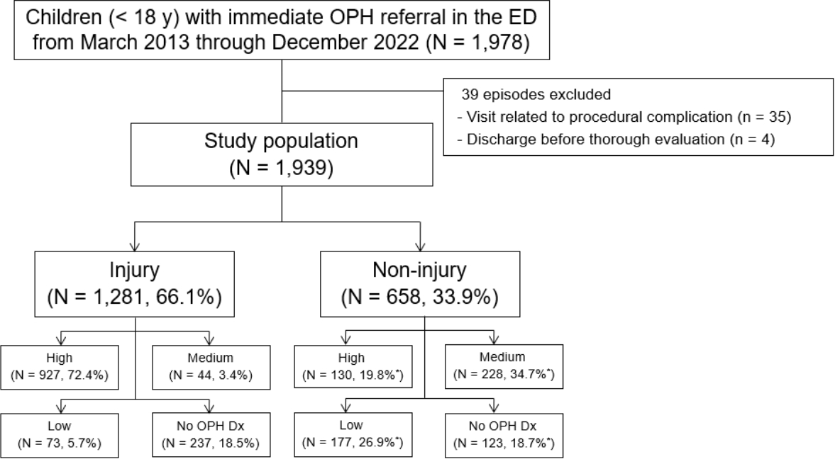
Of 1,978 eligible children, 1,939 were included in this study with a 66.1% proportion of injury (Fig. 1). The study population comprised 25.5% of the 7,595 children with eye-related symptoms who visited the ED during the study period. The median age of the population was 8.0 years (interquartile range, 3.0-14.0), and 623 children (32.1%) were girls. Visits at night, on weekend/holiday, and in fall accounted for 49.8%, 38.5%, and 32.1%, respectively. The most common injury mechanism and chief complaint were sports (14.4%) and swelling/erythema of the eye and adnexa (8.5%), respectively. The mechanisms are detailed in Appendix 3 (https://doi.org/10.22470/pemj.2023.00843). The most common place of injury was home (37.5%; Appendix 4, https://doi.org/10.22470/pemj.2023.00843). Most children underwent examinations for visual acuity, pupil, slit lamp, and fundoscopy, and 51.1% did computed tomography (Appendix 5, https://doi.org/10.22470/pemj.2023.00843). Emergency surgery, in-ED procedure, and hospitalization occurred in respectively 2.7%, 6.1%, and 2.5% (see details in Appendix 6, https://doi.org/10.22470/pemj.2023.00843).

2. OPH diagnosis and severity

Tables 1 and 2 list the representative diagnoses related to injury and non-injury, respectively. Top 3 injuries were orbital fracture, hyphema, and corneal abrasion whereas top 3 non-injuries were keratoconjunctivitis, cellulitis, and hordeolum. In cases of injuries, we additionally listed all coexisting diagnoses given the frequent multiple injuries (Appendix 7, https://doi.org/10.22470/pemj.2023.00843).
High OPH severity was associated with lower proportions of girls, visits on weekend/holiday, and most chief complaints, and with higher proportions of 13-17 years, injury, emergency surgery, in-ED procedure, hospitalization, and most injury mechanisms (Tables 3 and 4). There were 610 children (31.5%) with low OPH severity or no OPH diagnosis. No association was found between the high ED-assigned acuity (i.e., KTAS 1-2; 67 [3.5%]) and high OPH severity (1,057 [54.5%]) (Table 3). Most children (1,001 of 1,057 [94.7%]) with the high severity were assigned KTAS 3-4 (Appendix 8, https://doi.org/10.22470/pemj.2023.00843).

3. Comparisons of clinical features according to etiology and age groups

Table 5 shows that children with injury had a lower proportion of girls, visits on weekend/holiday, visits in summer, and a higher proportion of children aged 13-17 years, visits at night, visits in spring or fall, high OPH severity, emergency surgery, and in-ED procedure, compared to those with non-injury.
Table 6 shows a decrease in the proportion of girls and increases in injury and high OPH severity, with increasing age. Visits on weekend/holiday showed a decreasing trend with increasing age among children aged 4-17 years. Regarding the mechanism and chief complaint, sports, assault, eye pain, visual loss/blur, diplopia, and other visual symptoms tended to increase with age (Appendix 9, https://doi.org/10.22470/pemj.2023.00843). In contrast, the opposite trends were noted for poke, fall, cut/scratch/pierce, chemical/burn, swelling/erythema of the eye and its adnexa, red eye, itching sensation, and eye discharge. Older children tended to have injuries outside their homes (Appendix 4).
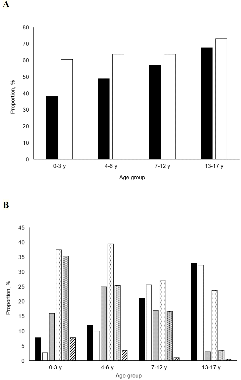
High OPH severity and the top 3 diagnoses are associated with the age groups (Fig. 2, Table 7, and Appendix 10 [https://doi.org/10.22470/pemj.2023.00843]). With increasing age, high OPH severity, orbital fracture, and hyphema increased in proportions whereas corneal abrasion, keratoconjunctivitis, cellulitis, and hordeolum tended to decrease (all Ps for trend ≤ 0.001).

Discussion

This study shows an association of high OPH severity with injury, older age, and boys. We discuss the dual implications for decision-making in children with eye-related symptoms who visit EDs. Children with high-to-medium severity probably have true OPH emergencies, which require emergency surgery, hospitalization, or expert opinion, compelling immediate referrals or transfers to ophthalmologists. Conversely, for children with low severity, emergency physicians or pediatricians may play some roles of on-call ophthalmologists in EDs, and divert the children to outpatient departments or outside clinics within a few days. The efficient use of emergency medicine resources is predicated on the non-ophthalmologists’ capability to make presumptive OPH diagnoses.
The study findings are consistent with those of a recent study performed in Wills Eye Hospital ED in terms of median age (9.8 years), the most common age group (15-18 years), injury mechanism (sports), and diagnosis (conjunctivitis) (8). As per a 2006-2014 United States Nationwide Emergency Department Sample (NEDS)-based study, “strike to the eye (22.5%)” was the most common mechanism, followed by “sports (14.2%),” which differs from the current study where sports were the predominant mechanism (13). This difference might stem from the higher proportion of injured children aged 10-17 years in our study (37.5% vs. 49.9% [639 of 1,281]) (13). Two Korean ED-based studies on ocular injury performed on patients of all ages listed “hyphema, eyelid laceration, and orbital fracture” and “corneal abrasion, orbital contusion, and hyphema” as the top 3 diagnoses, respectively (12,14). These diagnoses contain the top 3 injuries of the current study. Consistent with our study, the 2 Korean studies (12,13) reported football and baseball as the 2 most commonly causative sports activities (Appendix 3).
The association of injury with high OPH severity may be due to the following factors. First, trauma to the eye and its adnexa results in obvious wounds impacted by foreign bodies, or bleeding, which intuitively prompts emergency intervention. Second, OPH hospitalization is more frequent in injured children than in ill children (8). Third, by definition, an injury diagnosis is more likely to be regarded as highly severe than a non-injury diagnosis. As per a 2006-2011 United States NEDS-based study, a chief reference for the OPH severity, all diagnoses related to laceration or foreign bodies are categorized as “likely to be emergent (1).” In contrast, preseptal cellulitis, hordeolum, most conjunctivitis, and even optic neuritis are sorted as “nonemergent” or “could not be determined.” This categorization is in line with our rating of OPH severity (Tables 1 and 2).
The association of boys with high OPH severity may be related to the fact that 74.6% of the children with injuries are boys, which is inherently associated with the high severity of injuries (Table 5). Relevant studies show that boys account for 63%-68% of children with ocular injuries who visit EDs (13,15). The role of older age could be explained by the tendency for outside-the-home injuries in older children (Appendix 4) and age-related increases in sports, assault, eye pain, visual loss/blur, diplopia, and other visual symptoms (Appendix 9). For instance, if an adolescent with diplopia is diagnosed with benign oculomotor nerve palsy, he or she would be regarded as having a high OPH severity (1). The higher proportion of boys, older age, and higher OPH severity in our study are consistent with the findings of a Korean single ED-based study comparing children with general trauma and those with diseases (9).
In this study, 54.5%-68.5% of the children were considered candidates for OPH referrals. In EDs, in addition to high OPH severity, cases requiring only emergency OPH examinations (i.e., medium severity [14.0%]) can also be considered near emergencies. It is difficult to reduce such referrals. However, we found a minimal portion of potentially unnecessary referrals from children with low OPH severity or no OPH diagnosis (31.5%). The low-severity referrals can be decreased by enhancing emergency physicians’ or pediatricians’ knowledge and experience of OPH diseases and minor injuries. Depending on their clinical competence, the cutdown on unnecessary referrals may extend to some cases of medium OPH severity. The need for cutdown should be highlighted in infants or toddlers who require additional measures, such as procedural sedation and analgesia, for a detailed examination.
A discordance between KTAS 1-2 and OPH severity was observed, as exemplified by the fact that 3.6% of children with high severity were categorized as under KTAS 1-2 (Appendix 8). The discordance was also shown between KTAS 1-3 and the severity. This feature is likely due to the consideration of KTAS outside the primary diagnosis (e.g., vital signs) and the localized features of eye-related symptoms. Thus, emergency physicians or pediatricians may need to prioritize referrals or interventions for children with the symptoms, regardless of the initial KTAS.
Some miscellaneous findings are notable from the emergency medicine perspective. First, although most cases of conjunctivitis are classified as medium-to-low OPH severity, herpes simplex infection should be distinguished by the presence of vesicles or corneal dendrites owing to its potential for developing blindness and contraindications for frequently prescribed steroids. Second, we found 2 children with thelaziasis, infection by Thelazia callipaeda, of whom 1 underwent removal of the parasite (Appendix 6). This eye-specific parasite warrants attention in EDs, given its potential for causing keratitis and anxiety-provoking features in children or guardians. Third, because ball sports are commonly related to injuries, such as an orbital fracture or hyphema via ball-to-eye collision, protective eyewear should be used in a compulsory way (13,14).
This study has limitations. First, there might be inherent bias related to the single-center design. However, the study population is larger than that of the relevant Korean multicenter study (n = 446) (14). Second, given the lack of numerical data regarding visual acuity, we could not assess visual outcomes, irrespective of the diagnosis-based severity. Third, we could not determine the general conditions and diagnoses beyond the OPH scope because diagnoses made or procedures performed by ophthalmologists were the focus of data collection.
Briefly, true OPH emergencies may be more prevalent among injured, older, or male children. The study findings could help emergency physicians and pediatricians in directing their attention toward children exhibiting signs of true emergencies, thereby redirecting less urgent cases to outpatient departments or outside ophthalmology clinics.

Notes

Author contributions

Conceptualization, Data curation, Resources, and Software: Y Cha and JH Kim

Formal analysis, Investigation, and Methodology: all authors

Project administration and Validation: JH Kim and SA Chung

Supervision and Visualization: JH Kim

Writing-original draft: Y Cha and JH Kim

Writing-review and editing: all authors

All authors read and approved the final manuscript.

Conflicts of interest

JH Kim has served as an editor of Pediatric Emergency Medicine Journal since 2014, and was not involved in the review of this paper. Otherwise, no potential conflicts of interest relevant to this article were reported.

Funding sources

No funding source relevant to this article was reported.

References

1. Channa R, Zafar SN, Canner JK, Haring RS, Schneider EB, Friedman DS. Epidemiology of eye-related emergency department visits. JAMA Ophthalmol. 2016; 134:312–9.
2. Kwak YH. Current status and future direction of pediatric emergency medicine in Korea. Pediatr Emerg Med J. 2014; 1:1–10. Korean.
3. Hall LN, Jeng-Miller KW, Gardiner M, Kim EL. Utilization trends of an ophthalmology-specific emergency department: the Massachusetts Eye and Ear experience. Digit J Ophthalmol. 2021; 26:31–5.
4. Mott M. Who’s on call? Emergency care crisis looms [Internet]. American Academy of Ophthalmology; 2019 [cited 2023 Jul 15]. Available from: https://www.aao.org/eyenet/article/whos-on-call-emergency-care-crisis-looms.
5. Hutchinson AK, Morse CL, Hercinovic A, Cruz OA, Sprunger DT, Repka MX, et al. Pediatric eye evaluations peferred practice pattern. Ophthalmology. 2023; 130:P222–70.
6. Prentiss KA, Dorfman DH. Pediatric ophthalmology in the emergency department. Emerg Med Clin North Am. 2008; 26:181–98, vii.
7. Lim T, Park J, Je S. Pediatric Korean Triage and Acuity Scale. Pediatr Emerg Med J. 2015; 2:53–8. Korean.
8. Ramsay C, Murchison AP, Bilyk JR. Pediatric eye emergency department visits: retrospective review and evaluation. J Pediatr Ophthalmol Strabismus. 2021; 58:84–92.
9. Hwang Y, Jo HY, Yoo HW, Kim YM, Kim HY. Characteristics of children with trauma compared to those with disease in the emergency department: a Korean single regional emergency medical center study. Pediatr Emerg Med J. 2020; 7:108–13. Korean.
10. Kuhn F, Morris R, Witherspoon CD. Birmingham Eye Trauma Terminology (BETT): terminology and classification of mechanical eye injuries. Ophthalmol Clin North Am. 2002; 15:139–43, v.
11. Rai RS, Mehta N, Larochelle R, Rathi S, Schuman JS. A summary of eye-related visits to a tertiary emergency department. Sci Rep. 2021; 11:3823.
12. Oum BS, Lee JS, Han YS. Clinical features of ocular trauma in emergency department. Korean J Ophthalmol. 2004; 18:70–8.
13. Matsa E, Shi J, Wheeler KK, McCarthy T, McGregor ML, Leonard JC. Trends in US emergency department visits for pediatric acute ocular injury. JAMA Ophthalmol. 2018; 136:895–903.
14. Moon S, Ryoo HW, Ahn JY, Park JB, Seo KS, Shin SD, et al. Analysis on sports and recreation activity-related eye injuries presenting to the Emergency Department. Int J Ophthalmol. 2016; 9:1499–505.
15. Kinoshita M, Ihara T, Mori T. Characteristics of pediatric ocular trauma in a pediatric emergency department in Japan. Am J Emerg Med. 2023; 70:75–80.

Fig. 1.
Flowchart for selection of the study population. The “high,” “medium,” and “low” indicate the OPH severity. *The sum of proportions is not equal to 100% due to rounding. OPH: ophthalmic, ED: emergency department, Dx: diagnosis.
pemj-2023-00843f1.tif
Fig. 2.
Comparison of high ophthalmic severity and injury and non-injury top 3 diagnoses according to age groups. (A) With increasing age, high severity (shaded bars) and injury (open bars) increase in proportions. (B) Orbital fracture (black shaded bars) and hyphema (open bars) increase with age. This trend contrasts with decreases in corneal abrasion (gray shaded bars), keratoconjunctivitis (stippled bars), cellulitis (fine stippled bars), and hordeolum (hatched bars) (see numerical data in Table 7).
pemj-2023-00843f2.tif
Table 1.
List of representative OPH diagnoses related to injury (N = 1,281)
Diagnosis OPH severity Frequency*
Orbital fracture H 269 (21.0)
Hyphema H 261 (20.4)
Corneal abrasion H 165 (12.9)
Laceration H 91 (7.1)
 Corneal, penetrating-perforating H 15 (1.2)
 Corneal, lamellar H 10 (0.8)
 Canaliculi H 9 (0.7)
 Conjunctival H 39 (3.0)
 Eyelid H 18 (1.4)
Chemical burn H 61 (4.8)
Subconjunctival hemorrhage L 46 (3.6)
Foreign body in the cornea/conjunctiva/globe H 40 (3.1)
 Corneal H 29 (2.3)
 Conjunctival H 9 (0.7)
 Intraocular H 2 (0.2)
Corneal erosion L 24 (1.9)
Contusion of the orbit/eye M 20 (1.6)
Thermal burn H 16 (1.2)
Commotio retinae M 15 (1.2)
Other vitreous/retinal hemorrhages H 12 (0.9)
Conjunctival abrasion M 6 (0.5)
Abusive head trauma-related retinal hemorrhage H 5 (0.4)
Others - 13 (1.0)
No OPH diagnosis - 237 (18.5)

Values are expressed as numbers (%).

* The sum of proportions is not equal to 100% due to rounding.

All underwent emergency operations. Of these children, only 1 had blunt injury (i.e., ruptured globe).

In the order of high acuity, retinal detachment (H), 3; traumatic cataract (H), 1; trochlear nerve palsy (H), 1; abducens nerve palsy (H), 1; retrobulbar hemorrhage (H), 1; retinal tear/hole (M), 2; traumatic optic neuropathy (M), 1; pseudopapilledema (L), 1; epidermoid cyst rupture (L), 1; and allergic conjunctivitis (L), 1.

OPH: ophthalmic, H: high, M: medium, L: low.

Table 2.
List of representative OPH diagnoses related to non-injury (N = 658)
Diagnosis OPH severity Frequency*
Keratoconjunctivitis - 207 (31.5)
 Allergic L 56 (8.5)
 Viral or idiopathic M 54 (8.2)
 Epidemic keratoconjunctivitis L 48 (7.3)
 Herpes zoster ophthalmicus H 15 (2.3)
 Bacterial M 11 (1.7)
 Herpes simplex keratoconjunctivitis H 8 (1.2)
 Herpes simplex blepharitis M 8 (1.2)
 Others§ - 7 (1.1)
Cellulitis - 133 (20.2)
 Preseptal (periorbital) M 113 (17.2)
 Postseptal (orbital) H 11 (1.7)
 Dacryocystitis H 7 (1.1)
 Dacryoadenitis H 2 (0.3)
Hordeolum L 24 (3.6)
Corneal abrasion H 23 (3.5)
Corneal erosion L 18 (2.7)
Retinal detachment H 16 (2.4)
Optic neuritis M 14 (2.1)
Palsy of the cranial nerves|| H 14 (2.1)
Corneal ulcer H 12 (1.8)
Uveitis M 12 (1.8)
Other diseases of the conjunctiva - 9 (1.4)
Tumor or mass** H 5 (0.8)
Vitreous hemorrhage H 5 (0.8)
Others†† - 43 (6.5)
No OPH diagnosis - 123 (18.7)

Values are expressed as numbers (%).

* The sum of proportions is not equal to 100% due to rounding.

Including 8 keratitis.

Including 6 keratitis.

§ Glaucomatocyclitic crisis (H), 2; phlyctenular keratoconjunctivitis (L), 2; keratoconjunctivitis sicca (L), 2; and vernal keratoconjunctivitis (L), 1.

|| Abducens nerve, 11; trochlear nerve, 2; and oculomotor nerve, 1. All but 2 cases (1 midbrain infarction with oculomotor nerve palsy and 1 encephalitis with abducens nerve palsy) were benign.

Granuloma (L), 5; abrasion (M), 1; concretion (L), 1; epithelial inclusion cyst (L), 1; and hemangioma (L), 1.

** Lymphoma, 2; retinoblastoma, 1; lymphatic malformation, 1; and Rathke's cleft cyst, 1.

†† In the order of high acuity, Miller Fisher syndrome (H), 2; foreign body in the cornea/conjunctiva/globe (H), 2; amaurosis fugax (H), 2; branch retinal artery occlusion (H), 1; hyphema (H), 1; orbital myositis (H), 1; multiple sclerosis with internuclear ophthalmoplegia (H), 1; glaucoma (M), 4; ocular hypertension (M), 2; optic neuropathy (M), 2; central retinal vein occlusion (M), 2; papilledema, unspecified (M), 2; retinal tag (M), 1; acute posterior multifocal placoid pigment epitheliopathy (M), 1; myasthenia gravis (M), 1; mydriatic effect (L), 3; epiblepharon (L), 3; strabismus, unspecified (L), 3; cataract (L), 2; dacryostenosis (L), 2; thelaziasis (L), 2; dermatocele (L), 1; impetigo (L), 1; and astigmatism (L), 1.

OPH: ophthalmic, H: high, M: medium, L: low.

Table 3.
Comparison of clinical features according to the ophthalmic severity
Variable Total (N = 1,939) High (N = 1,057) Not high (N = 882)* P value
Age, y 8.0 (3.0-14.0) 11.0 (5.0-15.0) 6.0 (2.0-12.0) < 0.001
Age group, y < 0.001
 0-3 486 (25.1) 185 (17.5)* 301 (34.1)*
 4-6 314 (16.2) 154 (14.6)* 160 (18.1)*
 7-12 497 (25.6) 284 (26.9)* 213 (24.1)*
 13-17 642 (33.1) 434 (41.1)* 208 (23.6)*
Girls 623 (32.1) 280 (26.5) 343 (38.9) < 0.001
Injury 1,281 (66.1) 927 (87.7) 354 (40.1) < 0.001
Time of visit
 Night 965 (49.8) 521 (49.3) 444 (50.3) 0.645
 Weekend/holiday 747 (38.5) 364 (34.4) 383 (43.4) < 0.001
 Season 0.051
  Spring 479 (24.7)* 275 (26.0) 204 (23.1)
  Summer 497 (25.6)* 245 (23.2) 252 (28.6)
  Fall 623 (32.1)* 345 (32.6) 278 (31.5)
  Winter 340 (17.5)* 192 (18.2) 148 (16.8)
KTAS
 1-2 67 (3.5) 38 (3.6) 29 (3.3) 0.712
 1-3 937 (48.3) 444 (42.0) 493 (55.9) < 0.001
Emergency surgery 53 (2.7) 52 (4.9) 1 (0.1) < 0.001
In-ED procedure 119 (6.1) 98 (9.3) 21 (2.4) < 0.001
Hospitalization 49 (2.5) 42 (4.0) 7 (0.8) < 0.001

Values are expressed as medians (interquartile ranges) or numbers (%).

* The sums of proportions are not equal to 100% due to rounding.

To Department of Ophthalmology.

KTAS: Korean Triage and Acuity Scale, ED: emergency department.

Table 4.
Comparison of mechanism (for injury) or chief complaint (for non-injury) according to the ophthalmic severity
Variable Total (N = 1,939)* High (N = 1,057)* Not high (N = 882) P value
Mechanism
 Sports 280 (14.4) 227 (21.5) 53 (6.0) < 0.001
 Impact/compression 218 (11.2) 143 (13.5) 75 (8.5) < 0.001
 Poke 154 (7.9) 105 (9.9) 49 (5.6) < 0.001
 Fall 142 (7.3) 78 (7.4) 64 (7.3) 0.917
 Assault 141 (7.3) 90 (8.5) 51 (5.8) 0.021
 Cut/scratch/pierce 140 (7.2) 114 (10.8) 26 (2.9) < 0.001
 Chemical/burn 92 (4.7) 79 (7.5) 13 (1.5) < 0.001
 Motor vehicle accident 63 (3.2) 49 (4.6) 14 (1.6) < 0.001
 Foreign body 51 (2.6) 42 (4.0) 9 (1.0) < 0.001
Chief complaint
 Swelling/erythema 165 (8.5) 21 (2.0) 144 (16.3) < 0.001
 Red eye 83 (4.3) 6 (0.6) 77 (8.7) < 0.001
 Eye pain 63 (3.2) 20 (1.9) 43 (4.9) < 0.001
 Itching sensation 51 (2.6) 5 (0.5) 46 (5.2) < 0.001
 Visual loss/blur
  Painless 49 (2.5) 11 (1.0) 38 (4.3) < 0.001
  Painful 41 (2.1) 9 (0.9) 32 (3.6) < 0.001
 Other visual 45 (2.3) 13 (1.2) 32 (3.6) < 0.001
 Foreign body sense/mass 42 (2.2) 6 (0.6) 36 (4.1) < 0.001
 Rash/vesicle 40 (2.1) 17 (1.6) 23 (2.6) 0.123
 Eye discharge 24 (1.2) 3 (0.3) 21 (2.4) < 0.001
 Diplopia 27 (1.4) 13 (1.2) 14 (1.6) 0.504
 Other symptoms 28 (1.4) 6 (0.6) 22 (2.5) < 0.001

Values are expressed as numbers (%).

* The sums of proportions are not equal to 100% due to rounding.

Table 5.
Comparison of clinical features according to etiology (N = 1,939)
Variable Injury (N = 1,281) Non-injury (N = 658) P value
Girls 326 (25.4) 297 (45.1) < 0.001
Age, y 9.0 (4.0-14.0) 7.0 (3.0-13.0) < 0.001
Age group, y < 0.001
 0-3 294 (23.0) 192 (29.2)
 4-6 200 (15.6) 114 (17.3)
 7-12 317 (24.7) 180 (27.4)
 13-17 470 (36.7) 172 (26.1)
Time of visit
 Night 663 (51.8) 302 (45.9) 0.015
 Weekend/holiday 445 (34.7) 302 (45.9) < 0.001
 Season < 0.001
  Spring 339 (26.5) 140 (21.3)*
  Summer 286 (22.3) 211 (32.1)*
  Fall 427 (33.3) 196 (29.8)*
  Winter 229 (17.9) 111 (16.9)*
KTAS
 1-2 44 (3.4) 23 (3.5) 0.945
 1-3 539 (42.1) 398 (60.5) < 0.001
High OPH severity 927 (72.4) 130 (19.8) < 0.001
Medium OPH severity 44 (3.4) 228 (34.7) < 0.001
Emergency surgery 45 (3.5) 8 (1.2) 0.003
In-ED procedure 96 (7.5) 23 (3.5) 0.001
Hospitalization 38 (3.0) 11 (1.7) 0.085

Values are expressed as medians (interquartile ranges) or numbers (%).

* The sum of proportions is not equal to 100% due to rounding.

To Department of Ophthalmology.

KTAS: Korean Triage and Acuity Scale, OPH: ophthalmic, ED: emergency department.

Table 6.
Comparison of clinical features according to age groups (N = 1,939)
Variable 0-3 y (N = 486) 4-6 y (N = 314) 7-12 y (N = 497) 13-17 y (N = 642) P value
Girls 209 (43.0) 122 (38.9) 150 (30.2) 142 (22.1) < 0.001
Injury 294 (60.5) 200 (63.7) 317 (63.8) 470 (73.2) < 0.001
Time of visit
 Night 251 (51.6) 165 (52.5) 232 (46.7) 317 (49.4) 0.309
 Weekend/holiday 223 (45.9) 146 (46.5) 187 (37.6) 191 (29.8) < 0.001
 Season 0.213
  Spring 110 (22.6)* 72 (22.9) 133 (26.8) 164 (25.5)*
  Summer 114 (23.5)* 96 (30.6) 116 (23.3) 171 (26.6)*
  Fall 165 (34.0)* 90 (28.7) 162 (32.6) 206 (32.1)*
  Winter 97 (20.0)* 56 (17.8) 86 (17.3) 101 (15.7)*
KTAS
 1-2 16 (3.3) 4 (1.3) 19 (3.8) 28 (4.4) 0.098
 1-3 263 (54.1) 155 (49.4) 237 (47.7) 282 (43.9) 0.008
High OPH severity 185 (38.1) 154 (49.0) 284 (57.1) 434 (67.6) < 0.001
Medium OPH severity 93 (19.1) 54 (17.2) 68 (13.7) 57 (8.9) < 0.001
Emergency surgery 6 (1.2) 7 (2.2) 18 (3.6) 22 (3.4) 0.071
In-ED procedure 33 (6.8) 25 (8.0) 24 (4.8) 37 (5.8) 0.283
Hospitalization 5 (1.0) 7 (2.2) 22 (4.4) 15 (2.3) 0.008

Values are expressed as numbers (%).

* The sums of proportions are not equal to 100% due to rounding.

To the Department of Ophthalmology.

KTAS: Korean Triage and Acuity Scale, OPH: ophthalmic, ED: emergency department.

Table 7.
Comparison of OPH severity and top 3 diagnoses according to age groups
Variable Total (N = 1,939) 0-3 y (N = 486) 4-6 y (N = 314) 7-12 y (N = 497) 13-17 y (N = 642) P value*
High OPH severity 1,057 (54.5) 185 (38.1) 154 (49.0) 284 (57.1) 434 (67.6) < 0.001
Injury 1281 294 200 317 470
 Orbital fracture 269 (21.0) 23 (7.8) 24 (12.0) 67 (21.1) 155 (33.0) < 0.001
 Hyphema 261 (20.4) 8 (2.7) 20 (10.0) 81 (25.6) 152 (32.3) < 0.001
 Corneal abrasion 165 (12.9) 47 (16.0) 50 (25.0) 54 (17.0) 14 (3.0) < 0.001
Non-injury 658 192 114 180 172
 Keratoconjunctivitis 207 (31.5) 72 (37.5) 45 (39.5) 49 (27.2) 41 (23.8) 0.001
 Cellulitis 133 (20.2) 68 (35.4) 29 (25.4) 30 (16.7) 6 (3.5) < 0.001
 Hordeolum 24 (3.6) 15 (7.8) 4 (3.5) 2 (1.1) 3 (1.7) 0.001

Values are expressed as numbers only or numbers (%).

* Chi-square tests for trend.

Proportions were calculated with respect to injury. Resultantly, the proportions may differ from those in Appendix 10, in which the diagnoses are listed regardless of injury.

OPH: ophthalmic.

Appendices

Appendix 1

Definitions of injury mechanisms

Mechanism Definition
Sports Any injury that occurred while participating in a sport/leisure activity and could be directly attributed to the equipment for that activity or another participation in the activity
Impact/compression Blunt trauma (e.g., being struck by an object/body part)
Poke Injuries resulting from a poke from a blunt instrument (e.g., finger)
Fall Narrowly defined and self-explanatory
Assault Narrowly defined and self-explanatory
Cut/scratch/pierce Injuries resulting from sharp objects, which typically cause abrasions/lacerations
Chemical/burn Injuries caused by chemicals in the eye or by thermal burn
Motor vehicle accident Narrowly defined and self-explanatory
Foreign body Any foreign matter striking and retained in the eye/orbit

Modified from Ramsay et al. (8) with permission of SLACK Incorporated.

Appendix 2

Ophthalmic severity

Severity Definition Example
High Requiring emergency ophthalmic surgery/hospitalization Rupture globe
Medium Requiring emergency ophthalmic examination, without surgery/hospitalization Uveitis
Low Requiring outpatient or outside hospital follow-up Chalazion
Appendix 3

Injury mechanisms (N = 1,281)

Mechanism No.
Sports 280
 Football 101
 Baseball/softball 46
 Badminton 40
 Basketball 18
 Unknown ball 11
 Horizontal bar 7
 Boxing 6
 Volleyball 5
 Tennis 5
 Fitness equipment 5
 Discus 10
 Others* 26
Impact/compression 218
 Body parts 71
 Furniture 23
 Unknown toys 23
 Toy bow/gun 9
 Unknown 8
 Bottle 7
 Streetlamp/tree/pillar 6
 Book 6
 Wall 6
 Others 59
Poke 154
 Tree branch/wooden stick 16
 Finger 15
 Toy sword/arrow 12
 Chopstick/fork/spoon 10
 Picture card 10
 Bullet (sponge/rubber) 8
 Other plastic materials (toy, signpost, tube, pole, and ring) 8
 Pencil/pen 7
 Umbrella 7
 Unknown 7
 Straw 7
 Rubber band 5
 Doorknob 5
 Others 37
Fall 142
 Ground fall 44
 Bicycle 32
 Bed 16
 Unknown 13
 High fall 8
 Hugging/carrying 7
 Stairs 5
 Sofa/chair 6
 Swing/slide/rides 6
 Others (desk, stroller, and car seat) 5
Assault 141
 Punch 71
 Unknown 46
 Abuse 13
 Kick 8
 Strangulation 2
 Flying object 1
Cut/scratch/pierce 140
 Finger/nail 20
 Paper 16
 Wire/iron rod/awl 16
 BB bullet 15
 Tree branch/wooden stick 13
 Hanger 11
 Fragments (glass/wood/porcelain/stone) 10
 Pencil/pen 10
 Cutter knife/scissors 9
 Cat/dog/snake 7
 Others§ 13
Chemical/burn 92
 Detergent/bleach 17
 Disinfectant 7
 Contact lens cleaner 7
 Broth/tea 6
 Electrical 1
 Other chemical|| 37
 Other thermal 17
Motor vehicle accident 63
Foreign body 51
 Unknown 9
 Lens 8
 Adhesive 5
 Others** 29

* Tumbling, dodgeball, taekwondo, skating, hockey, futsal, trampoline, ping-pong, jump rope, foot volleyball, kendo, squash, jogging, and swimming.

Snowball, golf club, model rocket, door, wooden stick, fan, cell phone, car part, balloon, plastic stick, fruit, stone, block toy, remote control, hairband, television, bag, ball, swing, ice, eraser, vacuum cleaner, rubber bat, instrument, necklace, fountain, yo-yo, frame, milk carton, earphone, desk, kickboard, and propeller.

Book, plastic card, desk, bracelet, ballpoint pen spring, toothbrush, paper, hanger, clothes, balloon air injection device, glasses, fan, broomstick, Styrofoam, xylophone stick, zipper, children's scissors, tongs, blanket, kickboard handle, ping-pong table, hairpin, and pinwheel.

§ Pincette, toothpick, pin, snack bag, chestnut thorn, guitar string, top string, key, and unknown.

|| Fluorescent material, pesticide, odorant, gasoline, heavy metal, acetone, adhesive, cosmetic, burn ointment, sodium acetate, perm neutralizer, ammonium nitrate, toothpaste, water repellent coating agent, lotion, oil, sodium hydroxide, glacial acetic acid, polyvinyl alcohol, hair bleach, resin, mouthwash, and bath salt.

Oil, water, candle, squib, lighter, cigarette, chopstick, mascara, egg shell, tree branch, and plastic.

** Wood, sand, metal, pencil lead, paper, manicure, glass, vinyl, thread, seed, hair, cookie crumbs, fire extinguisher powder, hair band, grass, and powdered milk.

Appendix 4

Places of injury according to age groups

Variable Total (N = 1,281)* 0-3 y (N = 294) 4-6 y (N = 200) 7-12 y (N = 317)* 13-17 y (N = 470) P value
Home 481 (37.5) 245 (83.3) 106 (53.0) 81 (25.6) 49 (10.4) < 0.001
Playground 336 (26.2) 12 (4.1) 28 (14.0) 109 (34.4) 187 (39.8) < 0.001
Road 249 (19.4) 15 (5.1) 27 (13.5) 56 (17.7) 151 (32.1) < 0.001
Educational facility 137 (10.7) 9 (3.1) 21 (10.5) 39 (12.3) 68 (14.5) < 0.001
Public 78 (6.1) 13 (4.4) 18 (9.0) 32 (10.1) 15 (3.2) < 0.001

Values are expressed as numbers (%).

* The sums of proportions are not equal to 100% due to rounding.

Appendix 5

Frequency of ophthalmic and imaging tests

Variable No. (%)
Visual acuity 1,939 (100)
Pupil 1,939 (100)
Slit lamp 1,938 (99.9)
Fundoscopy 1,937 (99.9)
Tonometry 1,219 (62.9)
Extraocular movement 1,459 (75.2)
Computed tomography* 990 (51.1)
B-scan ultrasonography* 127 (6.5)
Magnetic resonance imaging* 58 (3.0)

* Any 1 of the 3 imaging tests were performed in 1,061 children (54.7%).

Appendix 6

List of emergency surgeries and in-emergency department procedures

Name of surgery or procedure No.
Emergency surgery 53
 Reconstruction of orbital fracture 17
 Primary closure of perforated laceration 13
 Circumferential segmental scleral buckling with silicone sponge 8
 Reconstruction of canalicular laceration with silicone tube insertion 7
 Trans pars plana vitrectomy 2
 Anterior chamber irrigation 2
 Removal of foreign body 1
 Phacoemulsification with extracapsular cataract extraction 1
 Intravitreal injection of ganciclovir with anterior chamber paracentesis 1
 Amniotic membrane transplant 1
In-emergency department procedure 119
 Removal of foreign body 33
 Primary wound repair 33
 Therapeutic contact lens 27
 Removal of pseudomembrane 16
 Laser photocoagulation 8
 Removal of parasite 1
 Conjunctival mass excision 1
Appendix 7

List of all co-existing diagnoses related to injury (N = 1,281)

Diagnosis OPH severity Frequency*
Hyphema H 337 (26.3)
Orbital fracture H 280 (21.9)
Corneal abrasion H 182 (14.2)
Commotio retinae M 125 (9.8)
Subconjunctival hemorrhage L 105 (8.2)
Laceration H 101 (7.9)
 Corneal, penetrating-perforating H 15 (1.2)
 Corneal, lamellar H 14 (1.1)
 Canaliculi H 9 (0.7)
 Conjunctival H 43 (3.4)
 Eyelid H 20 (1.6)
Chemical burn H 61 (4.8)
Corneal erosion L 50 (3.9)
Foreign body in the cornea/conjunctiva/globe H 40 (3.1)
 Corneal H 29 (2.3)
 Conjunctival H 9 (0.7)
 Intraocular H 2 (0.2)
Other vitreous/retinal hemorrhages H 24 (1.9)
Contusion of the orbit/eye M 20 (1.6)
Thermal burn H 16 (1.2)
Traumatic mydriasis M 15 (1.2)
Retinal tear/hole M 7 (0.5)
Conjunctival abrasion M 6 (0.5)
Abusive head trauma-related retinal hemorrhage H 5 (0.4)
Others - 20 (1.6)
No OPH diagnosis - 237 (18.5)

Values are expressed as numbers (%).

* Mutually inclusive.

All underwent emergency operations. Of these children, only 1 had blunt injury (i.e., ruptured globe).

In the order of high acuity, retinal detachment (H), 3; traumatic cataract (H), 2; trochlear nerve palsy (H), 1; abducens nerve palsy (H), 1; glaucoma (H), 1; retrobulbar hemorrhage (H), 1; traumatic optic neuropathy (M), 4; papilledema, unspecified (M), 1; uveitis (M), 1; pseudopapilledema (L), 1; macular hole (L), 1; iridodialysis (L), 1; epidermoid cyst rupture (L), 1; and allergic conjunctivitis (L), 1.

OPH: ophthalmic, H: high, M: medium, L: low.

Appendix 8

Association of KTAS with the OPH severity

KTAS Total (N = 1,939)* High (N = 1,057) Medium-low or no OPH diagnosis (N = 882)*
1 5 (0.3) 2 (0.2) 3 (0.3)
2 62 (3.2) 36 (3.4) 26 (2.9)
3 870 (44.9) 406 (38.4) 464 (52.6)
4 958 (49.4) 595 (56.3) 363 (41.2)
5 44 (2.3) 18 (1.7) 26 (2.9)

Values are expressed as numbers (%).

* The sums of proportions are not equal to 100% due to rounding.

KTAS: Korean Triage and Acuity Scale, OPH: ophthalmic.

Appendix 9

Comparison of mechanism (for injury) or chief complaint (for non-injury) according to age groups

Variable Total (N = 1,939)* 0-3 y (N = 486) 4-6 y (N = 314) 7-12 y (N = 497)* 13-17 y (N = 642)* P value
Mechanism
 Sports 280 (14.4) 2 (0.4) 9 (2.9) 87 (17.5) 182 (28.3) < 0.001
 Impact/compression 218 (11.2) 53 (10.9) 42 (13.4) 66 (13.3) 57 (8.9) 0.067
 Poke 154 (7.9) 45 (9.3) 51 (16.2) 43 (8.7) 15 (2.3) < 0.001
 Fall 142 (7.3) 63 (13.0) 20 (6.4) 30 (6.0) 29 (4.5) < 0.001
 Assault 141 (7.3) 14 (2.9) 4 (1.3) 13 (2.6) 110 (17.1) < 0.001
 Cut/scratch/pierce 140 (7.2) 45 (9.3) 40 (12.7) 40 (8.0) 15 (2.3) < 0.001
 Chemical/burn 92 (4.7) 47 (9.7) 10 (3.2) 17 (3.4) 18 (2.8) < 0.001
 Motor vehicle accident 63 (3.2) 8 (1.6) 11 (3.5) 11 (2.2) 33 (5.1) 0.005
 Foreign body 51 (2.6) 17 (3.5) 13 (4.1) 10 (2.0) 11 (1.7) 0.07
Chief complaint
 Swelling/erythema 165 (8.5) 84 (17.3) 36 (11.5) 38 (7.6) 7 (1.1) < 0.001
 Red eye 83 (4.3) 34 (7.0) 18 (5.7) 20 (4.0) 11 (1.7) < 0.001
 Eye pain 63 (3.2) 8 (1.6) 10 (3.2) 17 (3.4) 28 (4.4) 0.088
 Itching sensation 51 (2.6) 16 (3.3) 13 (4.1) 19 (3.8) 3 (0.5) < 0.001
 Visual loss/blur
  Painless 49 (2.5) 3 (0.6) 7 (2.2) 15 (3.0) 24 (3.7) 0.009
  Painful 41 (2.1) 1 (0.2) 3 (1.0) 18 (3.6) 19 (3.0) < 0.001
 Other visual 45 (2.3) 3 (0.6) 2 (0.6) 11 (2.2) 29 (4.5) < 0.001
 Foreign body sense/mass 42 (2.2) 16 (3.3) 7 (2.2) 9 (1.8) 10 (1.6) 0.228
 Rash/vesicle 40 (2.1) 8 (1.6) 5 (1.6) 8 (1.6) 19 (3.0) 0.281
 Eye discharge 24 (1.2) 15 (3.1) 6 (1.9) 2 (0.4) 1 (0.2) < 0.001
 Diplopia 27 (1.4) 0 (0) 3 (1.0) 11 (2.2) 13 (2.0) 0.009
 Other symptoms 28 (1.4) 4 (0.8) 4 (1.3) 12 (2.4) 8 (1.2) 0.183

Values are expressed as numbers (%).

* The sums of proportions are not equal to 100% due to rounding.

Appendix 10

List of diagnoses according to the age groups*

No. 0-3 y (N = 486) 4-6 y (N = 314) 7-12 y (N = 497) 13-17 y (N = 642)
1 Keratoconjunctivitis 73 (15.0) Corneal abrasion 55 (17.5) Hyphema 82 (16.5) Orbital fracture 155 (24.1)
2 Cellulitis 68 (14.0) Keratoconjunctivitis 45 (14.3) Orbital fracture 67 (13.5) Hyphema 152 (23.7)
3 Corneal abrasion 54 (11.1) Cellulitis 29 (9.2) Corneal abrasion 59 (11.9) Keratoconjunctivitis 41 (6.4)
4 Chemical burn 30 (6.2) Orbital fracture 24 (7.6) Keratoconjunctivitis 49 (9.9) Laceration 22 (3.4)
5 Orbital fracture 23 (4.7) Laceration 23 (7.3) Cellulitis 30 (6.0) Corneal abrasion 20 (3.1)
6 Laceration 22 (4.5) Hyphema 20 (6.4) Laceration 24 (4.8) SCH 17 (2.6)
7 SCH 19 (3.9) Foreign body 11 (3.5) Optic neuritis 12 (2.4) Corneal erosion 15 (2.3)
8 Foreign body 15 (3.1) Chemical burn 10 (3.2) Chemical burn 11 (2.2) Retinal detachment 13 (2.0)
9 Hordeolum 15 (3.1) Corneal erosion 7 (2.2) Corneal erosion 11 (2.2) Vitreous/retinal hemorrhage 12 (1.9)
10 Corneal erosion 9 (1.9) SCH 6 (1.9) Foreign body 8 (1.6) Commotio retinae 12 (1.9)
11 Thermal burn 8 (1.6) Others 23 (7.3) Abducens nerve palsy 6 (1.4) Chemical burn 10 (1.6)
12 Hyphema 8 (1.6) No OPH diagnosis 61 (19.4) Retinal detachment 5 (1.0) Foreign body 8 (1.2)
13 Contusion 6 (1.2) - Others§ 45 (9.1) Corneal ulcer 7 (1.1)
14 AHT-related retinal hemorrhage 5 (1.0) - No OPH diagnosis 88 (17.7) Uveitis 7 (1.1)
15 Others 18 (3.7) - - Contusion 7 (1.1)
16 No OPH diagnosis 113 (23.3) - - Thermal burn 6 (0.9)
17 - - - Cellulitis 6 (0.9)
18 - - - Others|| 34 (5.3)
19 - - - No OPH diagnosis 98 (15.3)

Values are expressed as numbers (%).

* The sums of proportions are not equal to 100% due to rounding.

In the order of frequency, lymphoma, 2; Miller Fisher syndrome, 2; cataract, 2; dacryostenosis, 2; epiblepharon, 2; retinal disorder, unspecified, 1; retinoblastoma, 1; corneal ulcer, 1; abducens nerve palsy, 1; conjunctival epithelial inclusion cyst, 1; conjunctival hemangioma, 1; conjunctival abrasion, 1; and thelaziasis, 1.

In the order of frequency, contusion, 4; hordeolum, 4; conjunctival abrasion, 4; conjunctival granuloma, 3; abducens nerve palsy, 1; trochlear nerve palsy, 1; uveitis, 1; vitreous/retinal hemorrhage, 1; central retinal vein occlusion, 1; myasthenia gravis, 1; epidermoid cyst rupture, 1; and conjunctival concretion, 1.

§ In the order of frequency, corneal ulcer, 4; uveitis, 4; vitreous/retinal hemorrhage, 4; SCH, 4; commotio retinae, 3; contusion, 3; thermal burn, 2; conjunctival abrasion, 2; conjunctival granuloma, 2; strabismus, unspecified, 2; hordeolum, 2; lymphatic malformation, 1; amaurosis fugax 1; traumatic optic neuropathy, 1; traumatic cataract 1; glaucoma, 1; papilledema, unspecified, 1; trochlear nerve palsy, 1; retrobulbar hemorrhage, 1; mydriatic effect, 1; thelaziasis, 1; dermatocele, 1; impetigo, 1; and astigmatism, 1.

|| In the order of frequency, abducens nerve palsy, 4; glaucoma, 3; hordeolum, 3; ocular hypertension, 2; optic neuritis, 2; optic neuropathy, 2; retinal tear/hole, 2; mydriatic effect, 2; amaurosis fugax 1; Rathke's cleft cyst, 1; acute posterior multifocal placoid pigment epitheliopathy, 1; branch retinal artery occlusion, 1; central retinal vein occlusion, 1; trochlear nerve palsy, 1; oculomotor nerve palsy, 1; epiblepharon, 1; strabismus, unspecified, 1; multiple sclerosis with internuclear ophthalmoplegia, 1; orbital myositis, 1; papilledema, unspecified, 1; pseudopapilledema, 1; and retinal tag, 1.

SCH: subconjunctival hemorrhage, AHT: abusive head trauma, OPH: ophthalmic.

TOOLS
Similar articles