

 PDF
PDF Citation
 Print
Print


INTRODUCTION
Upper gastrointestinal (GI) and lower GI hemorrhage are the most common causes of GI bleeding; small bowel hemorrhage accounts for 5% of GI bleeding. Small bowel hemorrhage is defined as bleeding distal to the ligament of Treitz, and small bowel hemorrhage can be attributed to various conditions, including vascular, inflammatory, iatrogenic, tumorous, or diverticular disorders.1-3 On the other hand, among patients with liver cirrhosis (LC), portal hypertension (PHT) can also cause portal hypertensive enteropathy, and its manifestation ranges from asymptomatic incidental findings to life-threatening small bowel variceal bleeding.3
The diagnosis of small bowel hemorrhage has been a challenging task for gastroenterologists. With the recent advances in imaging techniques, there has been a significant shift in assessing patients presenting with small bowel hemorrhage. Newer techniques, such as capsule endoscopy, double-balloon enteroscopy, single-balloon enteroscopy, spiral enteroscopy, and computed enterography, play a key role in the diagnosis of small bowel hemorrhage.4
A standard treatment for small bowel varices has not been established owing to its rarity. Managing small bowel variceal bleeding can vary depending on the indications, expertise, and availability and may include conservative, radiological, pharmacological, endoscopic, and surgical methods. This paper reports cases of small bowel variceal hemorrhage in patients with LC, which was managed safely and effectively by trans-arterial embolization (TAE).
CASE REPORT
Table 2 lists the baseline characteristics and clinical outcomes of three patients with TAE.
1. Case 1
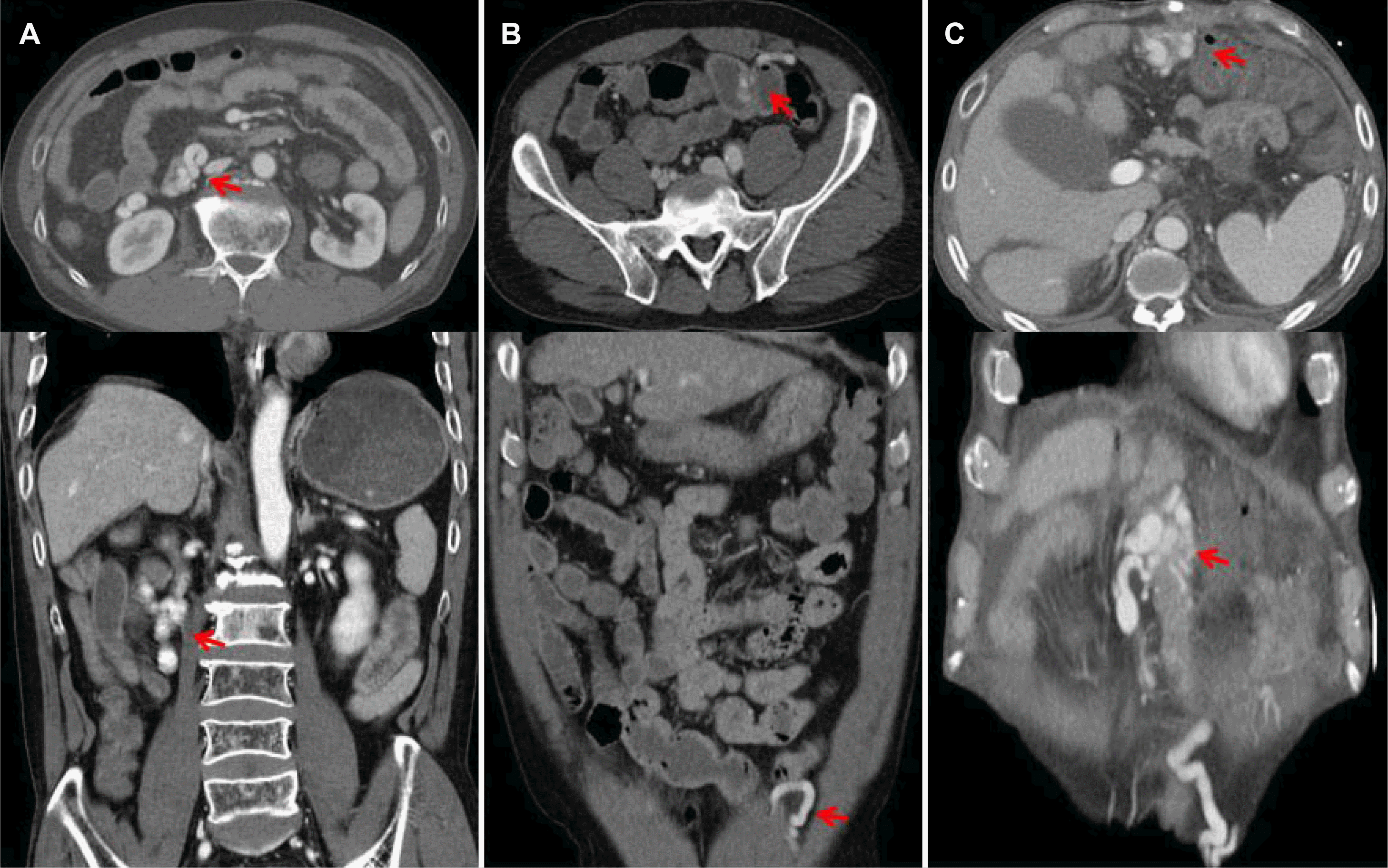
A 63-year-old male presented to the emergency department with hematemesis and hematochezia. The initial laboratory findings revealed anemia with a hemoglobin (Hb) level of 7.4 g/dL. Thus, abdominal-pelvic contrast tomography (AP-CT) was performed to identify the source of bleeding. The CT scan demonstrated a bulging contour and surface nodularity of the liver, suggesting LC with a suspicious mild esophageal varix (Fig. 1A).
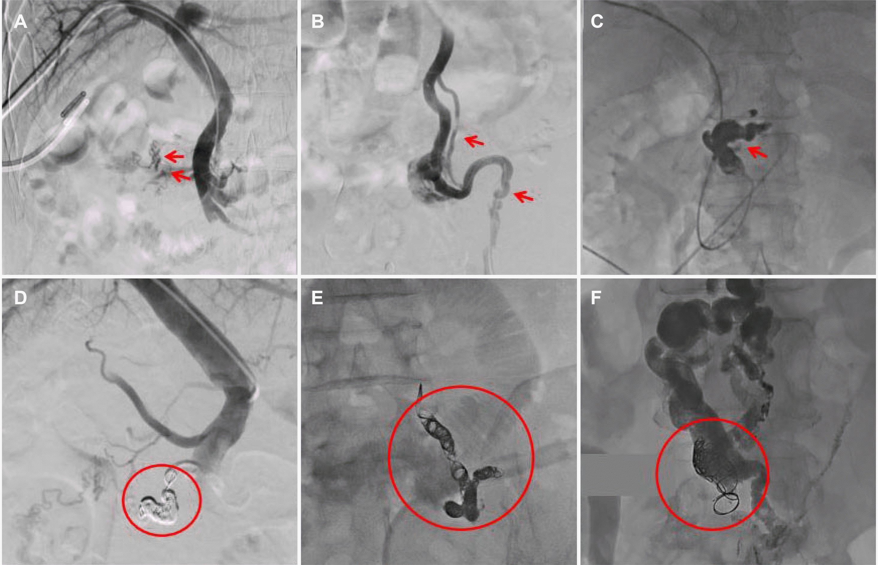
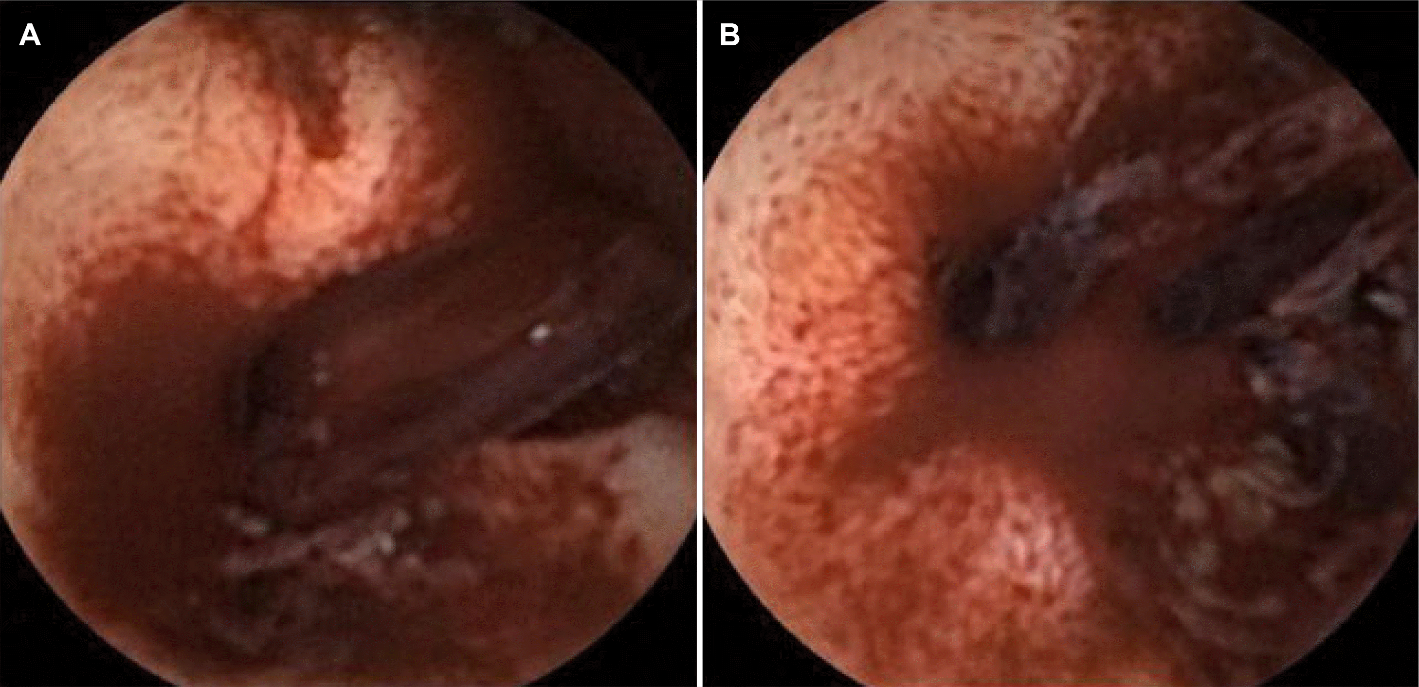
Esophagogastroduodenoscopy was performed considering the CT findings and the clinical image, which revealed a linear venous dilation with the stigmata of recent hemorrhage in the second portion of the duodenum. Endoscopic variceal ligation was initially performed as an immediate measure (Fig. 3). On the other hand, despite band ligation, the Hb level decreased to 6.5 g/dL, and serial hematemesis was observed. This prompted the need for angiography to identify any further sites of bleeding and to ensure hemostasis as a rescue therapy. Percutaneous access to a branch of the right portal vein was achieved under ultrasound guidance. The venogram revealed a tortuous, dilated varix in the second portion of the duodenum with partial thrombi. Embolization was performed successfully using a microcatheter and six Interlock microcoils (Fig. 2A, D). He was discharged from hospitalization a few days after treatment without complications.
2. Case 2
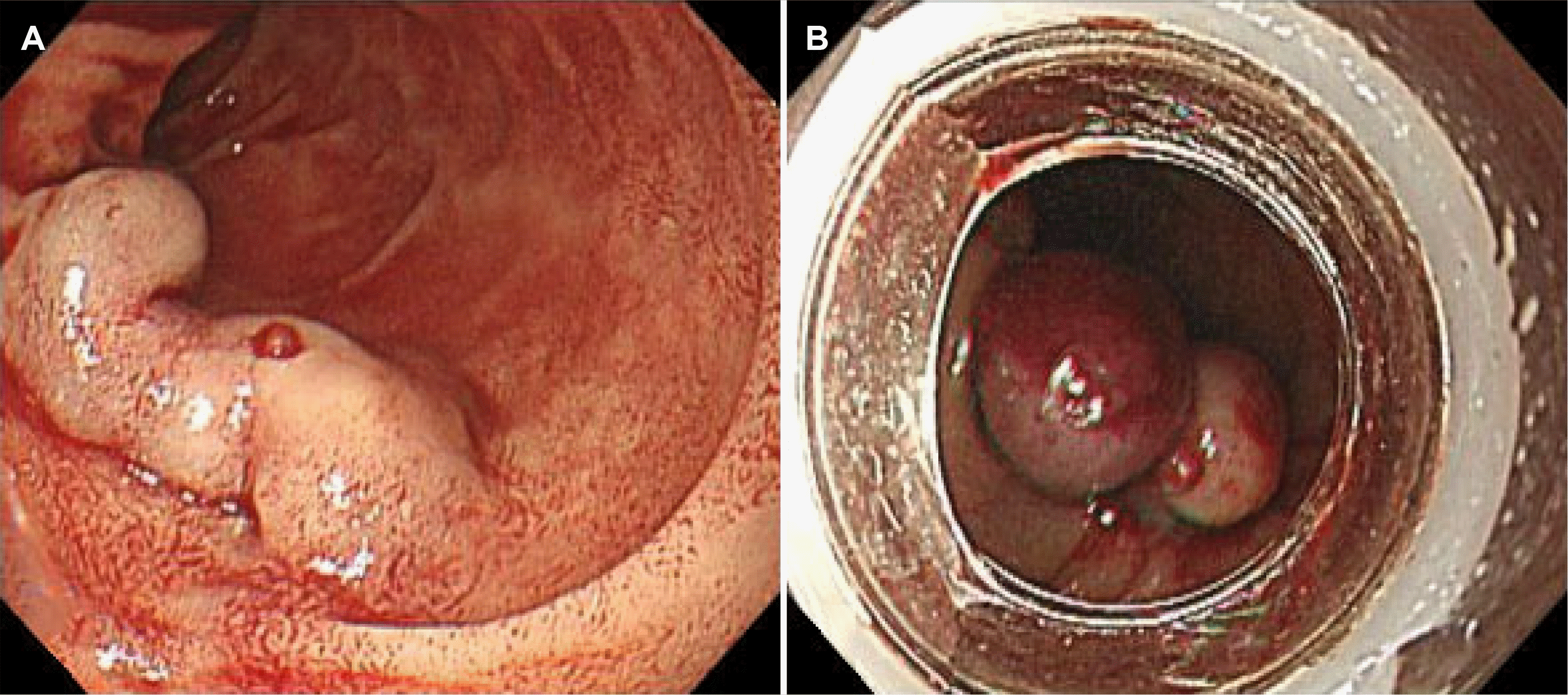
A 60-year-old male with a diagnosis of alcoholic LC, without regular outpatient follow-up, presented to the emergency department with a one-day history of hematochezia. The initial laboratory findings revealed anemia with a Hb level of 8.4 g/dL. AP-CT revealed active contrast extravasation between the inferior epigastric and superior mesenteric veins, suggesting angiodysplasia or a varix (Fig. 1B). A colonoscopy was performed to visualize the source directly, revealing no active bleeding. Capsule endoscopy revealed active bleeding in the small bowel caused by a ruptured ileal varix (Fig. 4). Angiography was urgently performed to identify the focus of the hemorrhage and revealed a varix located between the superior mesenteric vein (SMV) and the left inferior epigastric vein, with notable active contrast extravasation indicating ongoing bleeding (Fig. 2B). Angiography was performed with access from the left common femoral vein. A left inferior epigastric venogram revealed a varix between the SMV and the left inferior epigastric vein. Embolization was achieved using a microcatheter to deploy 17 Interlock microcoils, followed by the application of cyanoacrylate and gelfoam, ensuring a thorough occlusion of the bleeding varix (Fig. 2E). After the procedure, the patient's postoperative course was uneventful without complications. He steadily recovered over the following days with no further bleeding episodes. He was discharged in a stable condition on the fifth post-embolization day without complications.
3. Case 3
A 69-year-old male with a prior surgical history of resections for gastric cancer, hepatocellular carcinoma, and LC presented to the emergency department due to dizziness, which persisted for two days. The initial evaluation revealed anemia with a Hb level and blood pressure of 7.3 g/dL and 87/59 mmHg, respectively. AP-CT was performed to determine the cause of the anemia. The scan showed a prominent encircling varix at a jejunal loop with paraumbilical and left inferior epigastric varices (Fig. 1C). Angiography revealed a tortuous, dilated large varix in the small bowel, which appeared to drain into an abdominal wall varix (Fig. 2C). A decision was made to proceed with angiographic embolization, considering the risk of potential life-threatening hemorrhage from these varices. Under the transportal and transabdominal wall route, embolization was performed using a microcatheter and two Interlock microcoils. Cyanoacrylate was also applied to ensure complete occlusion (Fig. 2F). The procedure was carried out successfully without immediate complications. The patient's clinical condition improved steadily after the procedure. The patient experienced no further episodes of dizziness or other complications related to the embolization. Subsequent follow- up revealed stabilized Hb levels, and the patient was discharged.
DISCUSSION
Small bowel varices are a rare cause of GI hemorrhage, and there are various causes of small bowel varices. Commonly associated with PHT, small bowel varices represent potential channels to relieve the increased portal venous flow. Small bowel varices can also be observed in patients undergoing abdominal surgeries or at ostomy anastomoses.5 In these instances, the intestinal walls where the collateral circulation forms often adhere to previous suture sites. On rarer occasions, small bowel varices can be idiopathic. All the cases presented in this report were associated with PHT because of LC with Child-Pugh B classification.
Small bowel varices have been reported to occur with bleeding in approximately 25% of cases.6 A review of 169 patients with bleeding ectopic varices revealed 17%, 17%, and 26% of patients with varices in the duodenum, varices in the jejunum or ileum, and peristomal varices, respectively.7 The clinical manifestation could be characterized by three distinctive features: a history of abdominal surgery, PHT, and the presence of hematochezia without accompanying hematemesis.3 In the present case series, the bleeding locations were the duodenum, jejunum, and ileum, which is consistent with previous studies (Table 2). Future research with more cases will be needed to examine the locations of small bowel varices.
In some reports, significant bleeding was attributed to small bowel varices, with patients having hemodynamic instability and Hb concentrations of 6 g/dL or below.8 A recent study estimated a 5.3% to 18.4% mortality rate associated with this type of bleeding.6 In the present study, although all the patients had anemia with Hb levels in the range of 7.2–8.4 g/dL, only case three experienced shock with an initial blood pressure of 87/59 mmHg. None of them had serious complications, such as sepsis and death.
The management of small bowel varices remains challenging because of the localization of the lesion and the difficulty of the procedure. Therapeutic options include endoscopic treatment, surgical intervention, and interventional radiological approaches.3 Although the efficacy of medical management for ectopic varices or other manifestations of PHT has not been studied thoroughly, vasoactive agents, such as octreotide and non-selective beta-blockers, should be considered for both primary and secondary prophylaxis to reduce the splanchnic and portal pressure considering their proven effectiveness in managing esophageal and gastric varices.9,10
EVL and endoscopic variceal obliteration (EVO) with tissue glue monomers, such as cyanoacrylate, are recommended to manage duodenal variceal bleeding.11 Some research suggests EVO may have fewer complications than EVL, particularly highlighting the potential risk of re-bleeding from ulcers induced by EVL.12,13 In relation to this, in case 1, although EVL was initially used for hemostasis, signs of re-bleeding emerged, prompting the decision to proceed with embolization. Therefore, it might be prudent to consider EVO as a preferable option over EVL for duodenal varices. Coil embolization was performed in a previous report of jejunal varices, similar to case 3. The outcomes of coil embolization in this case were satisfactory in the short and long term. The results are consistent with an American study analyzing the outcomes of percutaneous treatment for patients with ectopic GI variceal bleeding.12
Coil embolization is a secure and straightforward method for treating patients with ectopic varices. On the other hand, high recurrent bleeding rates are a complication of this procedure (55% at six months and 92% at four years). The risk of recurrent bleeding is because PHT, the primary cause of varices, is not addressed. Other complications of embolization include hemoperitoneum, enterobacter sepsis, coil migration, acute renal and liver failure, and intrasplenic hematoma in patients undergoing percutaneous trans-splenic catheterization with a reported incidence of 3%.14 Fortunately, in the present case series, the patients did not experience any immediate complications after the procedure (Table 2). Further research with long-term follow-up is needed on the presence of other complications.
In conclusion, patients with small bowel variceal bleeding were treated successfully and safely with TAE. Studies involving a larger patient population are warranted in the future.
 XML Download