Abstract
The intercostobrachial nerve (ICBN) originates from the second intercostal nerve’s lateral cutaneous branch, while the median nerve (MN) typically arises from the brachial plexus’s lateral and medial roots. The medial cutaneous nerve of the arm, a branch of the medial cord of the brachial plexus, often connects with the ICBN. Variations were observed during the dissection of a 50-year-old male cadaver, including MN having two lateral roots (LR), LR1 and LR2, joining at different levels. Three ICBNs innervated the arm in this case, with the absence of the medial cutaneous nerve of the arm compensated by branches from the medial cutaneous nerve of the forearm. Understanding these anatomical variations is crucial for surgical procedures like brachioplasty, breast augmentation, axillary lymph node dissection, and orthopedic surgery. Surgeons and medical professionals must be aware of these variations to enhance preoperative planning, minimize complications, and improve patient outcomes in these procedures.
Go to : 
Nerves in the upper limb primarily originate from the brachial plexus, a significant nerve network that provides innervation to the upper limb. The brachial plexus is formed by the fusion of the anterior rami of the last four cervical (C5–C8) and the first thoracic (T1) nerves. It consists of roots, trunks, divisions, cords, and various branches. The cords are named posterior, lateral, and medial cords based on their positions relative to the axillary artery.
The formation of the cords from divisions of the trunks holds functional significance in the brachial plexus. The posterior cord, which originates from the posterior divisions of all trunks, supplies the extensor musculature. On the other hand, the lateral and medial cords, formed from the anterior divisions, exhibit variations and provide innervation to the flexor musculature of the upper limb [1].
The median nerve is formed within the axilla by a single root originating from both the medial and lateral cords of the brachial plexus. As it travels down the arm, the median nerve initially runs along the lateral side of the brachial artery until it reaches the middle of the arm. At this point, it crosses over to the medial side and closely follows the path of the brachialis muscle.
On the other hand, the medial cutaneous nerve of the forearm emerges from the medial region of the brachial plexus, receiving fibers from the C8 and T1 spinal nerves. It descends along the inner aspect of the forearm, coursing through the subcutaneous tissue until it reaches the wrist [2].
The intercostobrachial nerve (ICBN) is the lateral cutaneous branch originating from the second intercostal nerve. It exists as a prominent single branch that establishes connections with both the medial cutaneous nerve of the arm and the lateral cutaneous branch of the third intercostal nerve.
Furthermore, it is common for the lateral cutaneous branch of the third intercostal nerve to give rise to a secondary ICBN. However, the presence of a third ICBN is exceedingly rare in the human body [3].
The brachial plexus, responsible for innervating the upper limb, can also exhibit variations in its formation, branching patterns, or contributions from adjacent nerves. These variations can lead to different patterns of sensory and motor innervation and might be relevant during surgeries or diagnostic procedures.
In addition to nerve variations, surgeons should also be vigilant about the presence of diverse axillary artery and axillary vein configurations, as well as the occurrence of various patterns of axillary arch and extensions of breast tissue within the axilla.
One common variation is the branching pattern of the axillary artery. While the standard branching pattern involves the axillary artery giving rise to several branches, there can be variations in the number, origin, or course of these branches. Such variations can have implications for vascular access during medical procedures.
Muscular variations in the axilla can involve the presence, absence, or altered course of certain muscles, such as the pectoralis minor, subscapularis, or latissimus dorsi. These variations can affect shoulder function and may be important during surgical interventions.
These anatomical variations warrant careful attention during surgical procedures in the axillary region.
Go to : 
During a routine undergraduate medical dissection in 2022 at Govt. Medical College, Thrissur, Kerala, India, an examination of the cadaver of a 50-year-old male revealed unusual variations in the right axilla. Notably, the axilla on the left side exhibited no variations and appeared to be normal in comparison to the right side. Since its a cadaveric study, we have got clearance for publishing it for academic purpose.
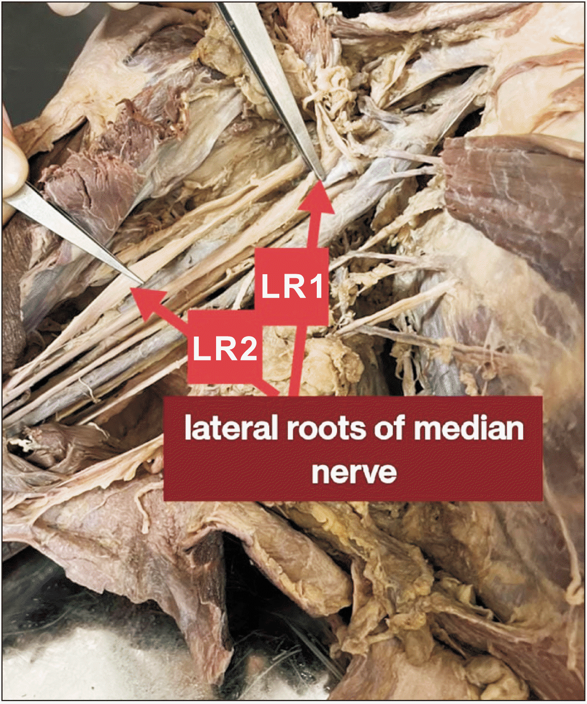
On the right side, the median nerve (MN) exhibited an uncommon presentation with the presence of two lateral roots (LR1 and LR2) instead of the usual single root, as depicted in Fig. 1. These two roots joined at different levels.
Notably, the proximal lateral root (LR1) of the median nerve was shorter in length compared to the distal lateral root (LR2), while both roots were approximately similar in thickness. The thickness of LR1 measured 1.4 cm, while LR2 measured 1.5 cm. The length of LR1, from the point of division of the lateral cord, to its attachment with the median nerve, was found to be 1.7 cm. In contrast, LR2 measured a length of 12.3 cm from the same point of division to its attachment with the median nerve.
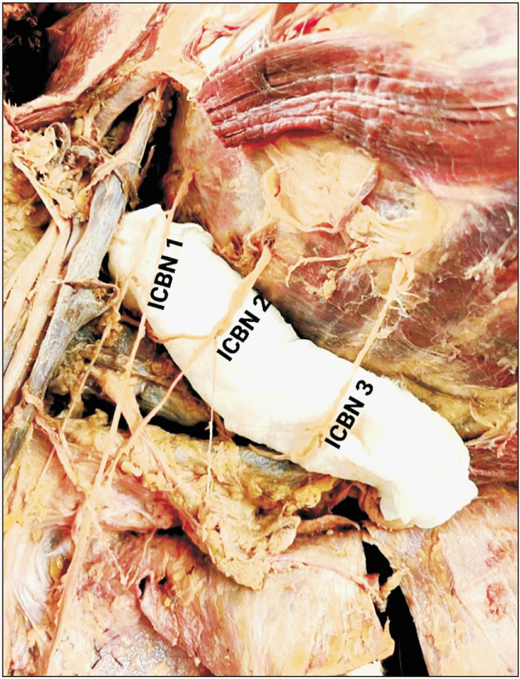
In the present cadaver, there were notable variations in the innervation of the arm. Typically, the second ICBN (ICBN 2) crosses the axilla and communicates with the medial cutaneous nerve of the arm, providing innervation to the upper medial portion of the arm. However, in this case, the first ICBN (ICBN 1) was observed to be innervating the arm directly instead of communicating with the medial cutaneous nerve of the arm.
ICBN 1 was long and thick, giving off branches at a distance of 16.5 cm from its origin in the intercostal space. Its total length from the lateral chest wall was 28.1 cm, with the main branch extending for 11.6 cm from the point of branching to the elbow.
Additionally, ICBN 2 was also involved in innervating the medial aspect of the arm. It emitted branches at a distance of 8 cm from the lateral thoracic wall. The total length of ICBN 2 from the lateral chest wall was 16.6 cm. Comparatively, ICBN 1 was thicker and longer than ICBN 2.
Furthermore, a third ICBN (ICBN 3) was identified supplying the arm, with a length of 9 cm. ICBN 3 was relatively shorter and slender when compared to ICBN 1 and ICBN 2. Thus, a total of three ICBNs were found to supply the arm, as depicted in Fig. 2. Notably, the medial cutaneous nerve of the arm was absent in this particular cadaver.
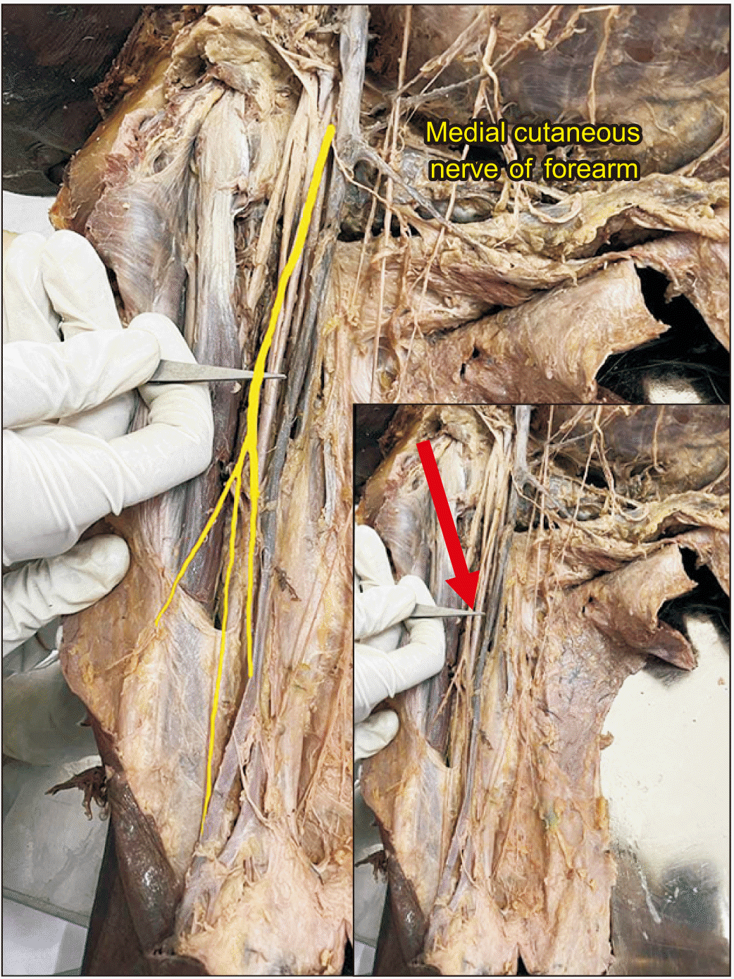
In the observed cadaver, the median cutaneous nerve of the forearm exhibited unusual variations in its course and branching pattern. Normally, this nerve originates from the medial cord of the brachial plexus, with its branches typically arising just prior to the elbow to innervate the forearm. However, in this case, the median cutaneous nerve of the forearm gave off branches not only in the forearm but also in the arm, as illustrated in Fig. 3. Approximately 12.8 cm from the point of branching of the medial cord, four branches were observed as if arising from the medial cutaneous nerve of the forearm.
Among these branches, the thicker and the most prominent branch measured 7.4 cm and continued towards the arm. The thinner branch, on the other hand, was longer, measuring 11.3 cm in length. It is worth noting that the left axilla showed normal characteristics in terms of nerve innervation. The lateral cord, medial cord, and posterior cord arose in the usual manner and formed their respective branches.
Go to : 
The present study provides valuable insights into the diverse variations that can occur in the innervation of the upper limb. Typically, the median nerve is formed by the contribution of a lateral root from the lateral cord and a medial root from the medial cord. However, in the examined specimen, an additional lateral root was observed, resulting in the presence of three roots for the median nerve instead of the usual two. This unique variation deviates from the normal anatomical configuration.
Interestingly, a similar variation, corroborating the findings of the present study. These observations highlight the complexity and potential for anatomical variations in the innervation of the upper limb. Understanding such variations is crucial for accurate clinical assessments and surgical procedures involving the upper limb. Further research in this area will contribute to the knowledge of these variations and their clinical implications [1].
Approximately 7% of cadavers have been reported to exhibit abnormal formation and course of the median nerve. The underlying cause of this variation can be better understood by tracing the development of the median nerve back to the embryonic stage. During limb development, the somites responsible for forming the limbs migrate along with their corresponding nerve supply, thereby maintaining their basic segmental innervation pattern.
In this context, it is important to note that the 7th cervical segmental artery gives rise to the axillary artery and is closely associated with the cords of the brachial plexus. However, it is also possible for the median nerve to derive its arterial supply from the 6th or 8th segmental arteries. These segmental arteries play a significant role in the development and variation of the median nerve’s course and distribution.
By considering the embryonic origins and the migration of nerve and arterial structures, we can better understand the variations observed in the formation and course of the median nerve. Further research and exploration of these developmental processes will enhance our knowledge of these variations and their clinical implications [4].
Variations of this nature have been extensively studied and documented worldwide, with a reported occurrence rate of approximately 15%. It is crucial to recognize that any iatrogenic injury to these extra or anomalous nerves can have significant clinical implications. Such injuries may lead to the manifestation of atypical symptoms that do not align with the typical symptoms associated with median nerve injuries.
Therefore, having knowledge of these variations is essential for clinicians as it enables them to accurately diagnose and differentiate these atypical symptoms. Additionally, surgeons can benefit from this knowledge by taking precautions to avoid iatrogenic damage to these nerves during procedures. Understanding these variations is of great relevance in the field of anesthesiology, as it expands the applications and considerations for anesthesiologists.
Overall, the awareness and understanding of these variations play a vital role in clinical practice, ensuring accurate diagnosis, preventing potential complications, and optimizing patient care [5].
Anomalous branches like the ones described can potentially manifest as compression syndromes, leading to ischemia and related symptoms. Understanding these variations in the brachial plexus is particularly valuable when assessing patients using magnetic resonance imaging techniques. Therefore, it is crucial for radiologists to possess knowledge and awareness of such variations to accurately interpret imaging findings and contribute to comprehensive patient care. By recognizing these variations, radiologists can provide vital insights and contribute to the accurate diagnosis and management of patients with brachial plexus abnormalities [6, 7].
According to Cunningham’s Manual of Practical Anatomy, the ICBN is a lateral cutaneous branch of the second intercostal nerve. It typically communicates with the medial cutaneous nerve of the arm and the lateral cutaneous branch of the intercostal nerves, collectively supplying the skin and medial side of the upper arm (brachium).
However, in the cadaver we dissected, we observed variations in the ICBN. Multiple ICBN nerves, including T1, T2, and T3, have been reported in a study of 40 cadavers, with 11 cadavers showing such variations. In this particular cadaver, the first ICBN was found to innervate the medial aspect of the arm along with the second and third ICBNs.
The brachial plexus serves as a pathway for nerve distribution, allowing for potential corrections in the pattern of branch distribution before reaching their target in the arm, forearm, or hand. However, in the present case, the absence of the medial cutaneous nerve of the arm was observed. Nevertheless, compensation may occur through collateral innervation from neighbouring nerves or branches, such as the ICBNs or branches originating from the medial cutaneous nerve of the forearm as is seen in the present case.
The medial cutaneous nerve of the forearm typically arises from the medial cord of the brachial plexus and branches just before the elbow, primarily innervating the forearm. However, in the present cadaver, we observed that the medial cutaneous nerve of the forearm began branching in the arm itself.
Apart from the above mentioned variations, bilateral absence of musculocutaneous nerve with unusual branching pattern of lateral cord and median nerve of brachial plexus was reported by Bhanu [9].
Another interesting variation noted by Ebenezer et al. [10] was bifurcated posterior cord with an axillary arch and a common arterial trunk from the axillary artery located between them, and struthers ligament giving rise to muscle fibers blending with the bicipital aponeurosis.
First, our study was limited to a single specimen, reducing the generalizability of our findings. Second, the scope of our observations was confined to the axilla and arm, restricting the broader understanding of the subject matter.
In conclusion, further investigation is warranted to explore potential associated anomalies in the cadaver. In addition to the variations in the medial cutaneous nerve of the arm, ICBN, and median nerve, several other anatomical considerations are crucial for various surgical interventions.
A comprehensive understanding of musculocutaneous nerve variations is essential for procedures involving the anterior compartment of the arm, such as biceps tendon repairs, coracoid transfers, and brachialis muscle flaps. By identifying any anomalies in the musculocutaneous nerve, surgeons can tailor their approach to avoid iatrogenic nerve injuries and optimize surgical outcomes [11].
The thoracodorsal nerve supplies the latissimus dorsi muscle and plays a significant role in reconstructive surgeries, such as breast reconstruction using autologous tissue. Exploring potential variations in the thoracodorsal nerve is important to ensure preservation of its function during surgical procedures, minimizing postoperative complications and functional deficits.
By considering these additional anatomical variations, surgeons can enhance their understanding of the intricacies involved in surgical interventions like brachioplasty, breast augmentation, axillary lymph node dissection, and orthopedic procedures. This comprehensive knowledge enables them to personalize their approach, minimize complications, and achieve optimal results for their patients.
Go to : 
Acknowledgements
Avika Chandra, Bhamasree B, Chandana A, Dilwin Prince, Faris A K 1st MBBS Students Govt. Medical College, Thrissur, Kerala, India.
Go to : 
Notes
Author Contributions
Conceptualization: RX. Data acquisition: RX. Data analysis or interpretation: RX. Drafting of the manuscript: RX. Critical revision of the manuscript: RX. Approval of the final version of the manuscript: RX.
Go to : 
References
1. Jyoti , Sharma A. 2020; Brachial plexus: variations in infraclavicular part and their significance. Int J Sci Study. 8:9–13.
2. Moore KL, Dalley AF, Agur AMR. 2010. Clinically oriented anatomy. 6th ed. Wolters Kluwer Health/Lippincott Williams & Wilkins;DOI: 10.1001/jama.282.15.1485-jbk1020-2-1.
3. Koshi R, Cunningham DJ. 2017. Cunningham's manual of practical anatomy. 16th ed. Oxford University Press;DOI: 10.5962/bhl.title.43984.
4. Pandey SK, Shukla VK. 2007; Anatomical variations of the cords of brachial plexus and the median nerve. Clin Anat. 20:150–6. DOI: 10.1002/ca.20365. PMID: 16795062.

5. Passey J, Rabbani P, Razdan SK, Kumar S, Kumar A. 2022; Variations of median nerve formation in North Indian population. Cureus. 14:e20890. DOI: 10.7759/cureus.20890. PMID: 35145795. PMCID: PMC8809111.

6. Van Hoof T, Mabilde C, Leybaert L, Verstraete K, D'Herde K. 2005; Technical note: the design of a stereotactic frame for direct MRI-anatomical correlation of the brachial plexus. Surg Radiol Anat. 27:548–56. DOI: 10.1007/s00276-005-0049-9. PMID: 16249823.
7. Bhat KM, Gowda S, Potu BK. 2009; Nerve loop around the axillary vessels by the roots of the median nerve a rare variation in a South Indian male cadaver: a case report. Cases J. 2:179. DOI: 10.1186/1757-1626-2-179. PMID: 19946489. PMCID: PMC2783134.

8. Kumar PA, Reddy KRD, Bapuji P. 2014; Multiple intercostobrachial nerves. J Evol Med Dent Sci. 3:12978–88. DOI: 10.14260/jemds/2014/3722.

9. Bhanu PS, Sankar KD. 2012; Bilateral absence of musculocutaneous nerve with unusual branching pattern of lateral cord and median nerve of brachial plexus. Anat Cell Biol. 45:207–10. DOI: 10.5115/acb.2012.45.3.207. PMID: 23094210. PMCID: PMC3472148.

10. Ebenezer DA, Rathinam BA. 2013; Rare multiple variations in brachial plexus and related structures in the left upper limb of a Dravidian male cadaver. Anat Cell Biol. 46:163–6. DOI: 10.5115/acb.2013.46.2.163. PMID: 23869264. PMCID: PMC3713281.

11. Ghosh B, Dilkash MNA, Prasad S, Sinha SK. 2022; Anatomical variation of median nerve: cadaveric study in brachial plexus. Anat Cell Biol. 55:130–4. DOI: 10.5115/acb.22.022. PMID: 35718802. PMCID: PMC9256496.

Go to : 


 PDF
PDF Citation
 Print
Print


 XML Download