

 PDF
PDF Citation
 Print
Print


Introduction
The circle of Willis (CW) is an anastomotic vascular channel situated at the base of the brain in the interpeduncular fossa. It is formed by the branches of the internal carotid artery (ICA) and basilar artery. The anterior cerebral artery (ACA) is formed by the terminal branches of the ICA. The two ACAs communicate with each other by the anterior communicating artery (ACoA). The ACA ends by dividing into the callosomarginal and pericallosal arteries. The ACA has two parts, pre-communicating, and post-communicating. The pre-communicating part is proximal to the ACoA, and the post-communicating part is distal to the ACoA [1].
The CW shows many anatomical variations, one of the rare variations is azygos anterior cerebral artery (AACA) with an incidence of 0.3%–2%. The AACA is characterized by the absence of ACoA and the two ACAs fuse to form a single artery and supplies blood to the anterior cerebral regions. The knowledge of this kind of anomaly is important as it leads to hemodynamic stress to the anterior cerebral region in cases of occlusion and aneurysms [2].
In this case, we are presenting a rare variation of CW, AACA associated with hypoplasia of the A1 fragment of the right anterior cerebral artery (RACA). To the best of our knowledge, this is the first cadaveric report of this kind of specific variation.
Go to : 
Case Report
During routine dissection of the vasculature of the brain of a 63-year-old female cadaver in the department of anatomy, All India Institute of Medical Sciences, Bibinagar, Hyderabad, it was accidentally found that there is a hypoplasia of the A1 fragment of RACA and the absence of ACoA. The contralateral A1 fragment of the left anterior cerebral artery (LACA) is normal. The hypoplastic RACA and the normal LACA are fused and formed a single artery, AACA (Fig. 1).
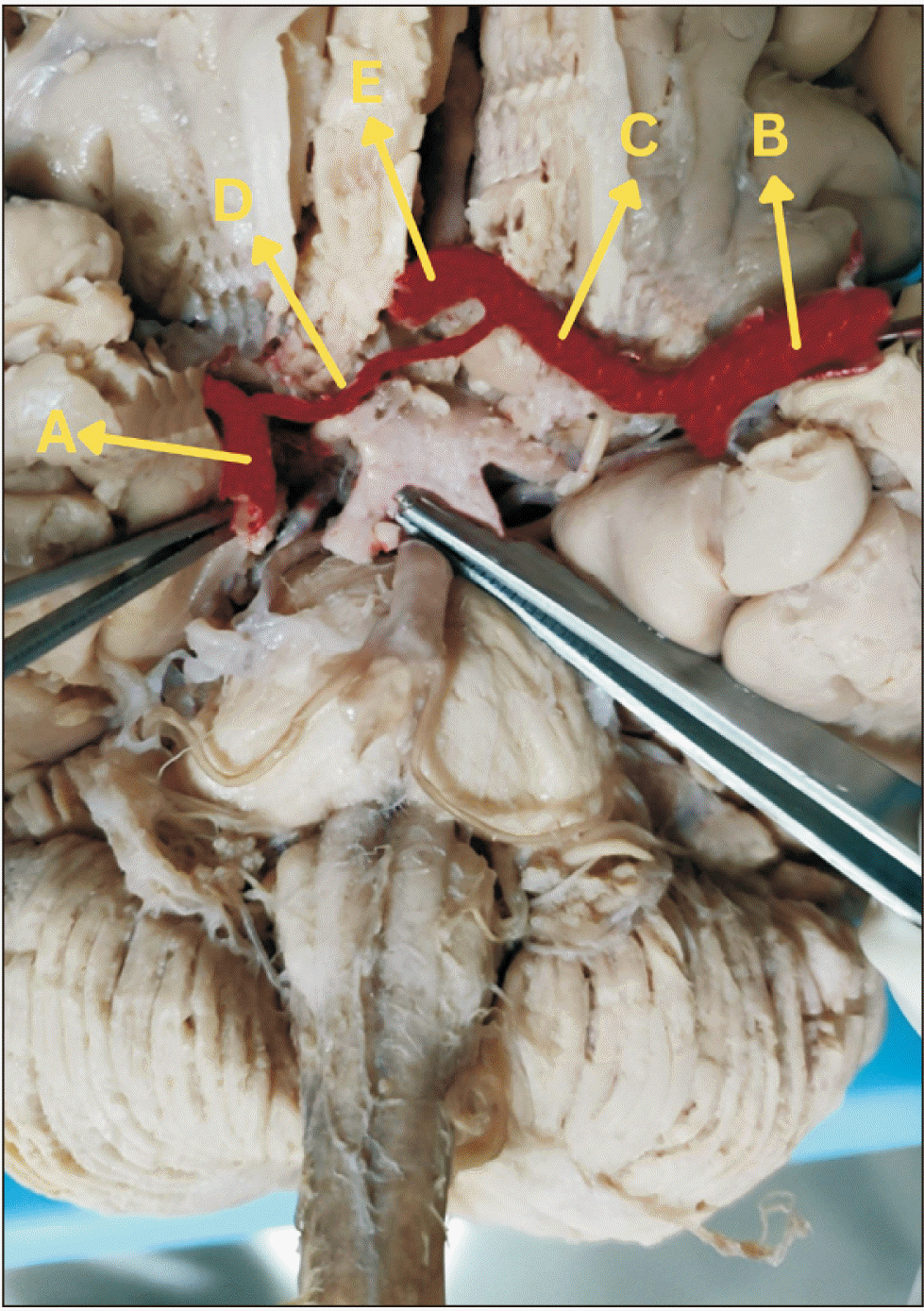
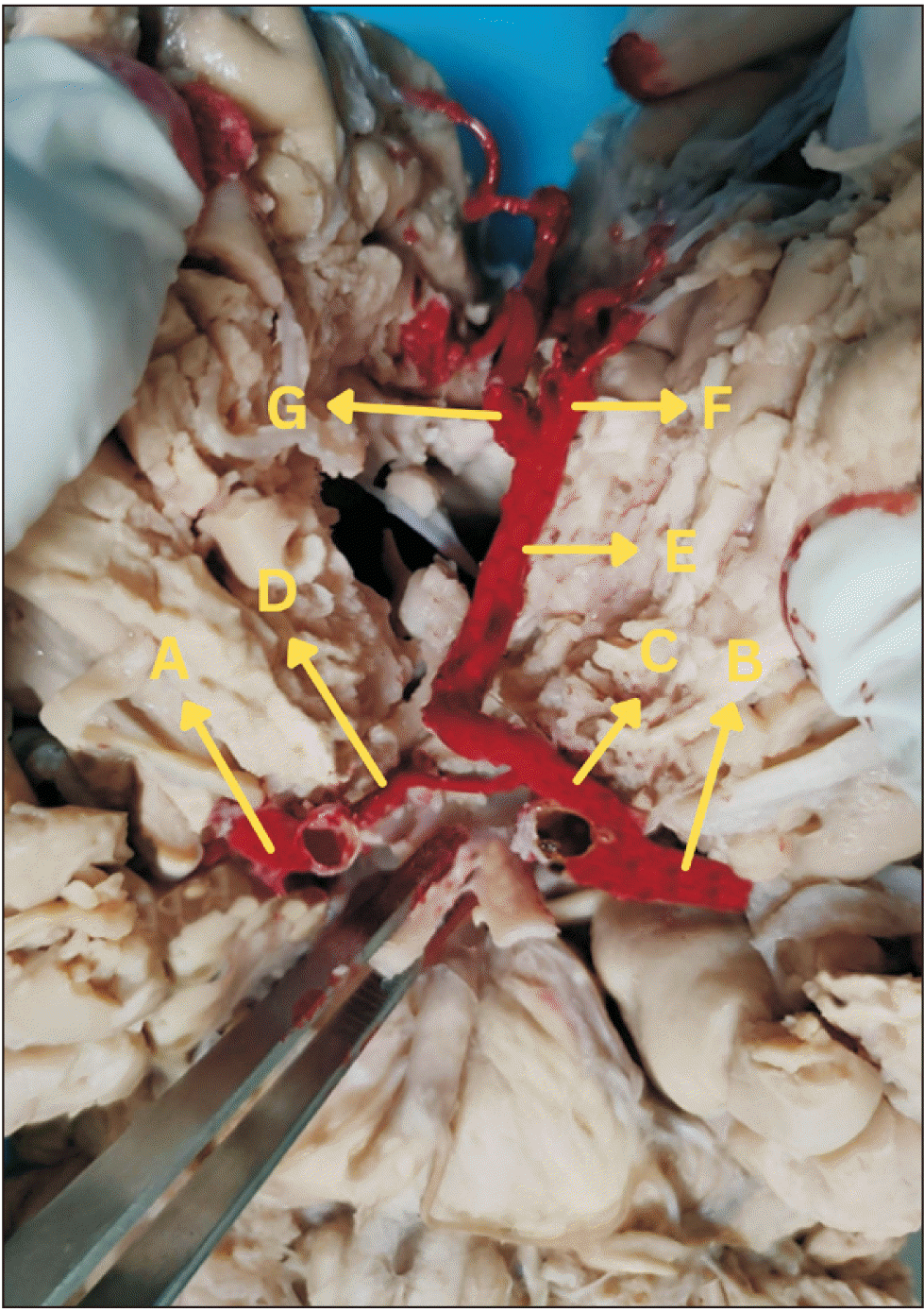
The AACA is running in the midline in the anterior median cerebral fissure transiently and then bifurcated into terminal branches to supply the respective hemispheres of the brain (Fig. 2).
This variation was found in routine cadaveric dissection so exempted from review by the ethical committee.
Go to :
Discussion
The brain receives its blood supply mostly from CW. Several variations in its formation and branching pattern are observed commonly. Most of these variations are either the absence of any vessel or the presence of any extra vessel or duplication of the vessel.
According to Gaillard et al. [3], the ACA is divided into five fragments. They are the A1 fragment (horizontal or pre-communicating fragment), A2 fragment (vertical or post-communicating fragment), A3 fragment (precallosal fragment), A4 fragment (supracallosal fragment), and A5 fragment (postcallosal fragment) [3].
According to Gunnal et al. [4], in their study, there are five types of AACA as shown in (Fig. 3).
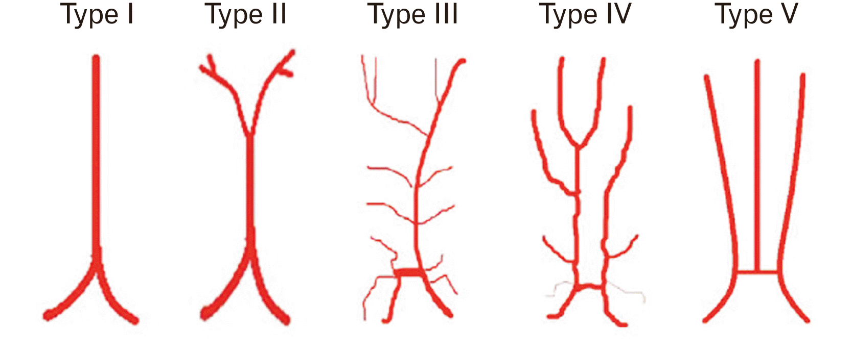 | Fig. 3Shows types of azygos anterior cerebral artery (AACA). Type I, classical AACA; Type II, short median stem of AACA with two terminal branches; Type III, two separate A2 fragments one short and one long; Type IV, azygos pericallosal artery; Type V, third azygos median A2 artery. Adapted from Gunnal et al. Neurol Asia 2013;18:249-59 [4].
|
Type I: Classical AACA, two A1 fragments fuse to form one A2 fragment.
Type II: Two A1 fragments fuse to form one short A2 fragment and terminate by dividing into two terminal branches.
Type III: Two separate A2 fragments, the right A2 fragment was very short and the left A2 fragment was long and showed an azygos course with branches to both hemispheres.
Type IV: Azygos pericallosal artery, Almost normal appearance, the right A2 fragment terminates as right and left pericallosal arteries and the left A2 fragment terminates as callosomarginal artery.
Type V: Third azygos median A2 fragment.
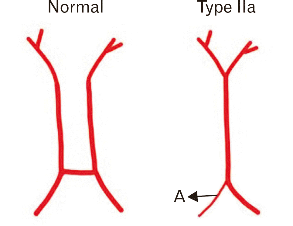
Among these five types of AACA, type II is described as the AACA is formed by two normal A1 fragments of ACA and the AACA runs as a short segment and terminates by dividing into two terminal branches which supply to the respective cerebral hemispheres [4], but in our case, the AACA is formed by hypoplastic A1 fragment of RACA and A1 fragment of normal LACA and the AACA runs as short segment and terminates by dividing into two terminal branches and can be described as type IIa (Fig. 4).
According to Pentyala et al. [5], in their study, the patients with hypoplastic A1 fragment of RACA show clinical features such as ipsilateral ischemic stroke, ipsilateral weakness of upper and lower extremities, loss of sensation, and slurred speech. The most common symptoms associated with this variation are chronic headache and dizziness, visual disturbances, nausea, weakness, and seizures [5], and these patients present with lower National Institutes of Health Stroke Scale (NIHSS) scores [6]. In Turner syndrome patients the most common intracranial vascular anomaly is an absence of the A1 fragment of ACA [7].
According to Pentyala et al. [5], the study was done in India, and the incidence of the hypoplastic A1 fragment of ACA is more predominant on the right side with an incidence of 25.4%, but according to Chuang et al. [6], the study was done in Taiwan, the incidence of hypoplastic A1 segment of ACA is more predominant on the left side with an incidence of 28%. As these two studies are done in different countries which explains the incidence of these anomalies will differ with race and region.
AACA is also linked to other midline anomalies such as corpus callosum agenesis, porencephalic cysts, holoprosencephaly, and saccular aneurysms. The most common anomaly is aneurysms because there is more hemodynamic stress [8].
This is the first cadaveric report of AACA associated with the hypoplastic A1 fragment of RACA which leads to more severe complications. The knowledge of these kinds of variations is important to anatomists, surgeons and radiologists during dissection, and surgical and radiological interventions.
Go to :
 XML Download