Abstract
Polymelia is an extremely rare congenital anomaly where an individual is born with an abnormally developed extra or supernumerary limb which is generally shrunken and functionless. A case of thoracomelia (a type of polymelia) was observed macroscopically and confirmed radiologically in 1.5 years old boy born in Nepal with an abnormal supernumerary upper limb attached to his back in the thoracic region. The limb was successfully amputated, and the boy had a favorable outcome after surgical treatment, without any adverse effects or impairment. Understanding the embryogenesis of thoracomelia is essential for unraveling the complex mechanisms underlying this condition and potentially aiding in early diagnosis and intervention. This case report and review aims to shed light on the intricate processes governing forelimb formation and their perturbations leading to thoracomelia.
Polymelia is an extremely rare congenital defect characterized by one or more supernumerary limbs which can further be categorized as pygomelia, thoracomelia, notomelia, and cephalomelia where the accessory limb is developed and attached at pelvis, thorax, back and head, respectively. Thoracomelia is a severe forelimb defect that occurs during embryonic development showing a significant challenge for affected individuals, as the forelimbs play a crucial role in various essential functions, including locomotion, dexterity, and communication. Despite its clinical importance, the embryological basis of thoracomelia remains poorly understood. By investigating the underlying mechanisms of forelimb development and the disruptions that lead to thoracomelia, we can provide valuable insights for improved prenatal diagnostics, counseling, and potential therapeutic interventions.
Scientific studies regarding the etiology of changes of this nature are limited in both human and veterinary medicine. So far, there is no consensus regarding if the origin of this congenital malformation is genetic or environmental [1, 2]. Limb malformations occur in approximately 6 per 10,000 live births and usually affect the upper limbs more than the lower limbs 3:1 [3].
Forelimb development in humans initiates during the fourth week of embryogenesis, with a complex interplay of genetic and molecular events. Although a few theories have been proposed, such as an insult during limb development or incomplete separation of monozygotic twins, perinatal injury, drugs and exposure to the teratogens [4]. Multiple signaling pathways, including the sonic hedgehog (SHH), fibroblast growth factor (FGF), and Wnt pathways, orchestrate the formation and patterning of forelimb buds. Perturbations in these pathways can disrupt the precise temporal and spatial regulation of limb development, leading to forelimb abnormalities [5].
Ethical approval was not required as this is a case report however the informed consent was obtained from the patient party for publication of patient information in a case report as the patient is minor.
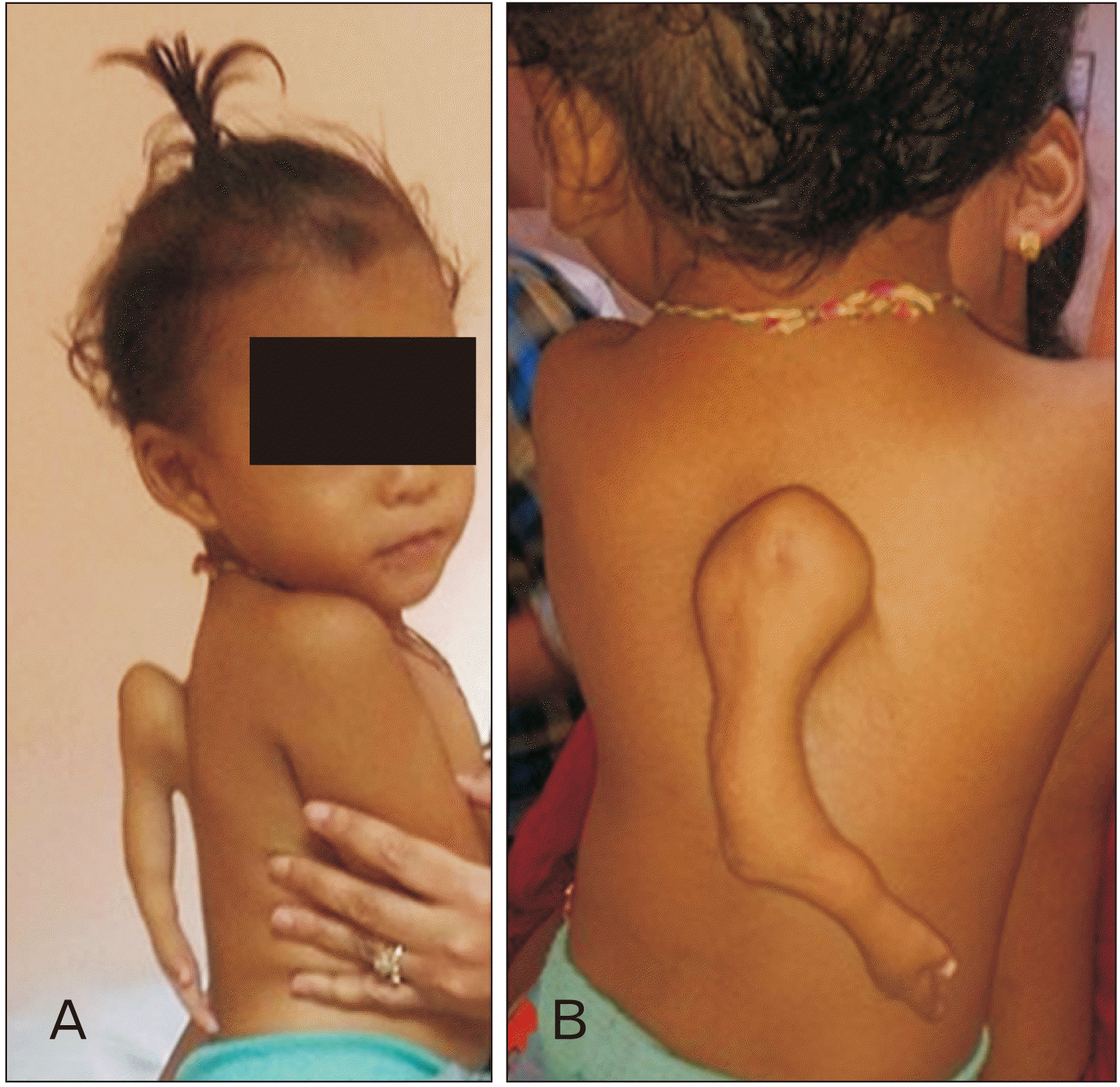
A 1.5-year-old boy was seen initially in a rural area of Nepal with an additional supernumerary upper limb attached to the back in the upper thoracic region. The presented case was the product of a full-term gestation without any evidence of complications due to ingestion of maternal drugs, infection, trauma, or teratogens. A written consent was obtained from the next of kin for publication of case findings. The patient was encountered at the primary care center in Nepal, where he was referred to the higher center for further consultation. The radiological diagnosis was also prescribed to confirm the case. The extra upper limb was successfully amputated without any post-operative complications (Fig. 1).
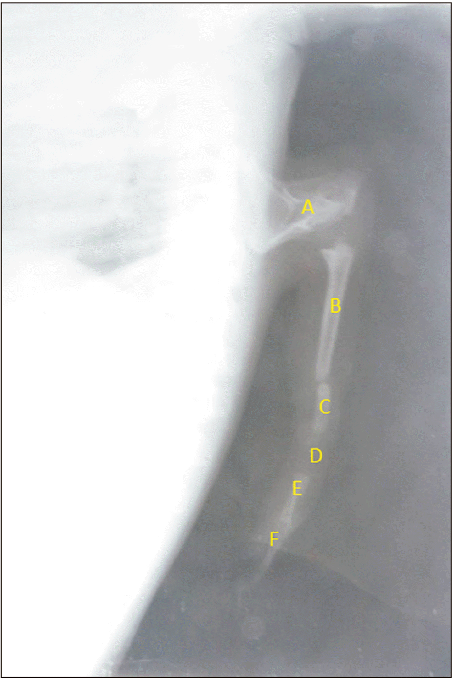
On the gross physical examination of the back, an ectopic accessory upper limb was observed attached to the area corresponding to the lower thoracic vertebrae. It was considered an accessory upper limb because the deltoid region of a shoulder with an arm, an elbow, a forearm, a hand and three digits were seen. The plain X-ray of the thoracic region with lateral view was recommended and performed to confirm the case radiologically. The conventional radiography revealed an ectopic bone resembling a scapula attached to the upper thoracic vertebrae with the other components of an upper limb. In addition, a few developing long bones resembling the humerus, radius, ulna, and phalanges were seen on the areas corresponding to the arm, forearm, and hand with incomplete ossification in the diaphyseal zone, confirming the case radiologically (Fig. 2).
The child was otherwise well-developed physically and mentally, with no other diagnosed malformations or congenital anomalies. After surgical intervention, the accessory limb was successfully amputated without any post-operative complications or deformities.
The histopathological diagnosis of the obtained specimen was confirmed as incomplete congenital duplication of the upper limb extremity (polymelia). Histologically, it was identified as diaphyseal endochondral ossification and cartilaginous epiphyseal plates matured according to the infant’s age.
The external appearance of the infant included two normal upper limbs, two normal lower limbs and an undeveloped extra forelimb attached to the posterior thoracic region and devoid of muscular tissues. In the extra forelimb, a scapula-like bone formed a joint with the incompletely duplicated humerus. The humerus fused with the incompletely duplicated radius. The ulna, carpal bones, metacarpal bones, and phalanges were completely duplicated.
The literature lacks the presence of forelimb polymelia, and therefore, our case can be considered the first human forelimb thoracomelia case reported in the literature. However, cases in the literature related to lower limb duplication in humans are available.
A neonate born in Niger with several congenital malformations on the posterior midline and the most rostral malformation was a polymelic limb at the level of the lumbar vertebrae composed of 2 long bones, a foot and 3 toes [6]. A case of dipygus with multiple extremities where the male fetus had 3 pelvises and 6 lower extremities, including feet and toes but no duplication anomalies or evidence of twinning was reported [7]. Another case of a newborn female with a tail-like extra right leg, which unfortunately was not detected during the routine antenatal ultrasonography, was reported [8]. Montalvo et al. [9] reported a case of a 6-month-old Hispanic boy who was born with a lower limb bud on the posteromedial face of the left thigh.
Another interesting case of an underdeveloped lower limb in a 6monthold female child was reported in India who presented with developed lower limbs and an additional underdeveloped left lower limb. A radiograph of the patient revealed a normal left hip joint with an accessory left underdeveloped limb. The accessory limb had a rudimentary femur falsely attached to normal acetabulum of the left hip joint. Both the rudimentary femur and the tibia formed a false knee joint. Distal most end of the accessory limb showed a single false digit with a single curved false metatarsal [10].
There are several cases of thoracomelia reported in the animal species. A rare case of thoracomelia has been reported in a female sheep due to an environmental cause [1]. In a male Korean native calf, polymelia (notomelia) was observed macroscopically and radiographically. External features included two normal forelimbs, two normal hindlimbs and two undeveloped extra forelimbs [11].
The development of the upper limb usually occurs during the fourth week of the gestational period by activating a group of mesenchymal cells in the somatopleuric layer of the lateral plate mesoderm. The upper limb bud develops opposite the caudal cervical segments, whereas the lower limb bud forms opposite the lumbar and upper sacral segments. At the apex of the limb bud, the ectoderm thickens to form the apical ectodermal ridge (AER), which is a specialized, multilayered epithelial structure that is induced by the paracrine factor, FGF10, from the underlying mesenchyme. Bone morphogenetic protein signaling is required for its formation. FGF8, secreted by the AER, exerts an inductive influence on the limb mesenchyme that initiates the growth and differentiation of the limbs in a proximodistal axis [12, 13]. Retinoic acid promotes the formation of the limb bud by inhibiting FGF signaling. Mesenchymal cells aggregate at the posterior margin of the limb bud to form the zone of polarizing activity, an important signaling center in limb development. FGFs from the AER activate the zone of polarizing activity, which causes the expression of the SHH genes. The AER itself is maintained by inductive signals from SHH and WNT7. As the AER grows more distal, the induced mesoderm cells, comprising rudimentary parts of the limb, can continue to grow without any developmental interference even if the AER is transplanted to the adjacent region. This leads to an assumption that duplication of the limb arises from the influence of the AER with abnormal splitting creating two sets of limbs [14].
One of the key genes associated with thoracomelia is the T-box transcription factor 5 (TBX5) gene. The TBX5 gene belongs to a family of transcription factors known as T-box genes, which play critical roles in regulating embryonic development. Mutations in genes such as WNT3A, FGF10, and SHH have been associated with limb malformations, including thoracomelia, in animal models and human studies. These genes are involved in signaling pathways critical for limb patterning and outgrowth during embryogenesis [15].
This case report highlights the need for continued research and collaboration to unravel the complexities of thoracomelia. Integrating molecular genetics, developmental biology, and clinical data will facilitate a deeper understanding of the etiology and pathogenesis of forelimb defects. Such knowledge will aid in developing early diagnostic tools, genetic counseling, and potential therapeutic interventions to mitigate the impact of thoracomelia on affected individuals and their families.
In conclusion, through this case report, we have provided an overview of the embryogenesis of thoracomelia, a rare forelimb defect in humans. By elucidating the intricate processes governing forelimb development and identifying potential genetic and environmental factors that contribute to thoracomelia, we hope to pave the way for improved diagnostic and therapeutic strategies. Early diagnosis of such cases can facilitate the parents’ management planning and psychological preparation.
Notes
References
1. Silva ES, Mustafa VS, Sá PA, Campebell RC. 2020; Thoracomelia in sheep: a case report. Braz J Vet Med. 42:e108220. DOI: 10.29374/2527-2179.bjvm108220. PMID: f4c7abab73664aafb03ac86fb1f05217.

2. Morath-Huss U, Drögemüller C, Stoffel M, Precht C, Zanolari P, Spadavecchia C. 2019; Polymelia in a chimeric Simmental calf: nociceptive withdrawal reflex, anaesthetic and analgesic management, anatomic and genetic analysis. BMC Vet Res. 15:102. DOI: 10.1186/s12917-019-1846-4. PMID: 30922306. PMCID: PMC6440010. PMID: 610edba4b47942f1a8828965b5482494.

3. Pitchaiah G, Pushpalatha E. 2017; Pathogenesis of horrifying rare genetic disorders in humans - a review. Pharma . Innov. 6:85–9.
4. Park KB, Kim YM, Park JY, Chung ML, Jung YJ, Nam SH. 2014; An accessory limb with an imperforate anus. Ann Surg Treat Res. 87:213–6. DOI: 10.4174/astr.2014.87.4.213. PMID: 25317418. PMCID: PMC4196430.

5. Munif MR, Safawat MS, Hannan A. 2023; Surgical correction of polymelia in the perineal region of a 2-day-old indigenous bovine calf: a case report from Bangladesh. Bull Natl Res Cent. 47:9. DOI: 10.1186/s42269-023-00988-0. PMID: 5d4a5a83dc494a748599c415ae249768.

6. Kelani AB, Moumouni H, Issa AW, Younsaa H, Fokou H, Sani R, Sanoussi S, Denholm LJ, Beever JE, Catala M. 2017; Notomelia and related neural tube defects in a baby born in Niger: case report and literature review. Childs Nerv Syst. 33:529–34. Erratum in: Childs Nerv Syst 2017;33:2065. DOI: 10.1007/s00381-017-3337-x. PMID: 28083641.

7. Scholl C, Thacker P. 2021; A rare sonographic case of caudal duplication versus polymelia. . J Diagn Med Sonogr. 37:180–3. DOI: 10.1177/8756479320975671.

8. Salameh K, Al-Bedaywi R, Elkabir NA, Valappil RP, Vellamgot A, Habboub L. 2020; Duplicate tail like right lower limb: a rare congenital malformation. J Pediatr Neonatal. 2:1–3. DOI: 10.33425/2689-1085.1015.

9. Montalvo N, Redrobán L, Espín VH. 2014; Incomplete duplication of a lower extremity (polymelia): a case report. J Med Case Rep. 8:184. DOI: 10.1186/1752-1947-8-184. PMID: 24920152. PMCID: PMC4077643.

10. Verma S, Khanna M, Tripathi VN, Yadav NC. 2013; Occurrence of polymelia in a female child. J Clin Imaging Sci. 3:18. DOI: 10.4103/2156-7514.111235. PMID: 23814690. PMCID: PMC3690670.

11. Kim C, Yeo S, Cho G, Lee J, Choi M, Won C, Kim J, Lee S. 2001; Polymelia with two extra forelimbs at the right scapular region in a male Korean native calf. J Vet Med Sci. 63:1161–4. DOI: 10.1292/jvms.63.1161. PMID: 11714039.

12. Roberts DJ, Tabin C. 1994; The genetics of human limb development. Am J Hum Genet. 55:1–6. PMID: 7912883. PMCID: PMC1918219.
13. Scherz PJ, Harfe BD, McMahon AP, Tabin CJ. 2004; The limb bud Shh-Fgf feedback loop is terminated by expansion of former ZPA cells. Science. 305:396–9. DOI: 10.1126/science.1096966. PMID: 15256670.

14. Zhao L, Li MQ, Sun XT, Ma ZS, Guo G, Huang YT. 2006; Congenital lumbosacral limb duplication: a case report. J Orthop Surg (Hong Kong). 14:187–91. DOI: 10.1177/230949900601400216. PMID: 16914786. PMID: 1b85f98dad22415c9a8c67db87fa805d.

15. Lovely AM, Duerr TJ, Qiu Q, Galvan S, Voss SR, Monaghan JR. 2022; Wnt signaling coordinates the expression of limb patterning genes during axolotl forelimb development and regeneration. Front Cell Dev Biol. 10:814250. DOI: 10.3389/fcell.2022.814250. PMID: 35531102. PMCID: PMC9068880. PMID: f207f57fd0104f598e67e66a502394ac.

Fig. 1
The lateral and posterior view of supernumerary forelimb attached to the posterior thoracic region.

Fig. 2
Lateral view of a plain radiograph of the accessory forelimb. A, an undeveloped scapula-like bone; B, an undeveloped humerus-like bone; C, an undeveloped ulna/radius-like bone; D, a wrist-like area without carpal bones; E, an undeveloped metacarpal-like bone; F, an undeveloped phalanx-like bone.


 PDF
PDF Citation
 Print
Print


 XML Download