This article has been
cited by other articles in ScienceCentral.
Abstract
In human fetuses, the left hepatic artery (LHA) issues the marginal artery that runs along the umbilical vein and, sometimes, reaches the umbilicus. The further observation demonstrated that, in 5 of 12 Japanese midterm fetuses (crown-rump length mm: 46, 50, 54, 59, 102), the marginal artery issued not only a thin umbilical branch but also a liver parenchymal branch that took a posterosuperior recurrent course in a peritoneal fold and supplied the anterior surface of the liver left lobe (segment III). However, in 22 Spanish fetuses of which gestational ages corresponded to the Japanese ones, we did not find the parenchymal branch. Therefore, between human populations, there seemed to be a considerable difference in the incidence as to whether or not the marginal artery issues the liver parenchymal branch. The parenchymal branch might be degenerated at the later stages due to friction between the liver free surface and growing diaphragm.
Go to :

Keywords: Left hepatic artery, Umbilical vein, Appendix fibrosa hepatis, Coronary ligament, Human fetus
Introduction
Detailed descriptions have been made about variant arteries at and around the human adult liver (
e.g., Michels [
1]), but to our knowledge, there was no descriptions of the variants to correspond to the fetal liver anatomy. Recently, we described that, in Spanish human fetuses, the umbilical cord contains not only a consistent arterial branch from the inferior epigastric artery but also, sometimes, that from the left hepatic artery (LHA) [
2]. The latter variation was seen at a relatively low incidence (4 of 25 midterm fetuses) and, we conducted a further observation using sagittal sections from 12 Japanese fetuses to understand the topographical relation between the LHA and umbilical vein. During the study, we incidentally found a novel arterial variant. The present study aimed to show a left marginal arterial branch to supply the anterior surface of the liver left lobe. The anatomy of the variant parenchymal branch will be compared with observations of 22 Spanish fetuses of which gestational ages corresponded to the Japanese ones.
Go to :

Case Report
The study was performed in accordance with the provisions of the Declaration of Helsinki, 1995 (as revised in 2013). We observed paraffin-embedded histological sections of 34 midterm fetal abdomen (approximately 10–16 weeks crown-rump length [CRL], 40–125 mm). Twelve Japanese specimens were part of the collection of the Department of Anatomy, Akita University, Akita, Japan and were donated by their families to the Department in 1975–1985 and preserved in 10% w/w neutral formalin solution for more than 30 years. Data on these specimens included the date of donation and the number of gestational weeks, but did not include the name of the family, obstetrician or hospital or the reason for abortion. The use of these specimens for research was approved by the Akita University Ethics Committee (No. 1428). Before a routine procedure for paraffin embedding, the trunks were decalcified by incubating them at room temperature in Plank-Rychlo solution (AlCl2/6H2O, 7.0 w/v%; HCl, 3.6; HCOOH, 4.6) for 3–7 days. The serial sections were stained with hematoxylin and eosin (HE). The sectional planes were sagittal.
To compare with Japanese fetuses, we examined 22 Spanish specimens those were part of the large collection at the Department of Anatomy of the Universidad Complutense, Madrid. These specimens were obtained from miscarriages and ectopic pregnancies from the Department of Obstetrics of the University. No information was available on the genetic background of the embryos or abortion. The serial sections were stained with HE, Azan or Goldner’s Masson trichrome stainings. The sectional planes were horizontal (12 fetuses) or sagittal (10 fetuses): the latter 10 specimens were overlapped with our recent study [
2]. The thickness of sections was 5–10 micron. This study was approved by the Ethics Committee of Complutense University (B08/374). All photographs of histology were taken with a Nikon Eclipse 80. Identification of the liver anatomy including the hepatic arteries was based on our previous topographical studies of the midterm liver [
3,
4].
General anatomy of the liver in fetuses
Japanese specimens are shown in
Figs. 1,
2, while a Spanish specimen in
Fig. 3. Topographical anatomy of the liver in fetuses showed a specific feature different from the adult liver. In sagittal sections of the midterm liver (
Figs. 1–
3), the caudate lobe is as large as, or even larger than, the quadrate lobe (segment IV). Immediately in front of the inferior vena cava, the liver is tightly attached to the membranous diaphragm at a small area of the top (
i.e., the bare area). The muscular diaphragm is restricted behind the vena cava. The LHA and umbilical vein are sandwiched by the quadrate lobe (segment IV) and the other parts of the liver left lobe (
Figs. 1B,
2A,
3A). These sections contain the gastric antrum, the transverse colon, the pancreatic head and the horizontal portion of the duodenum.
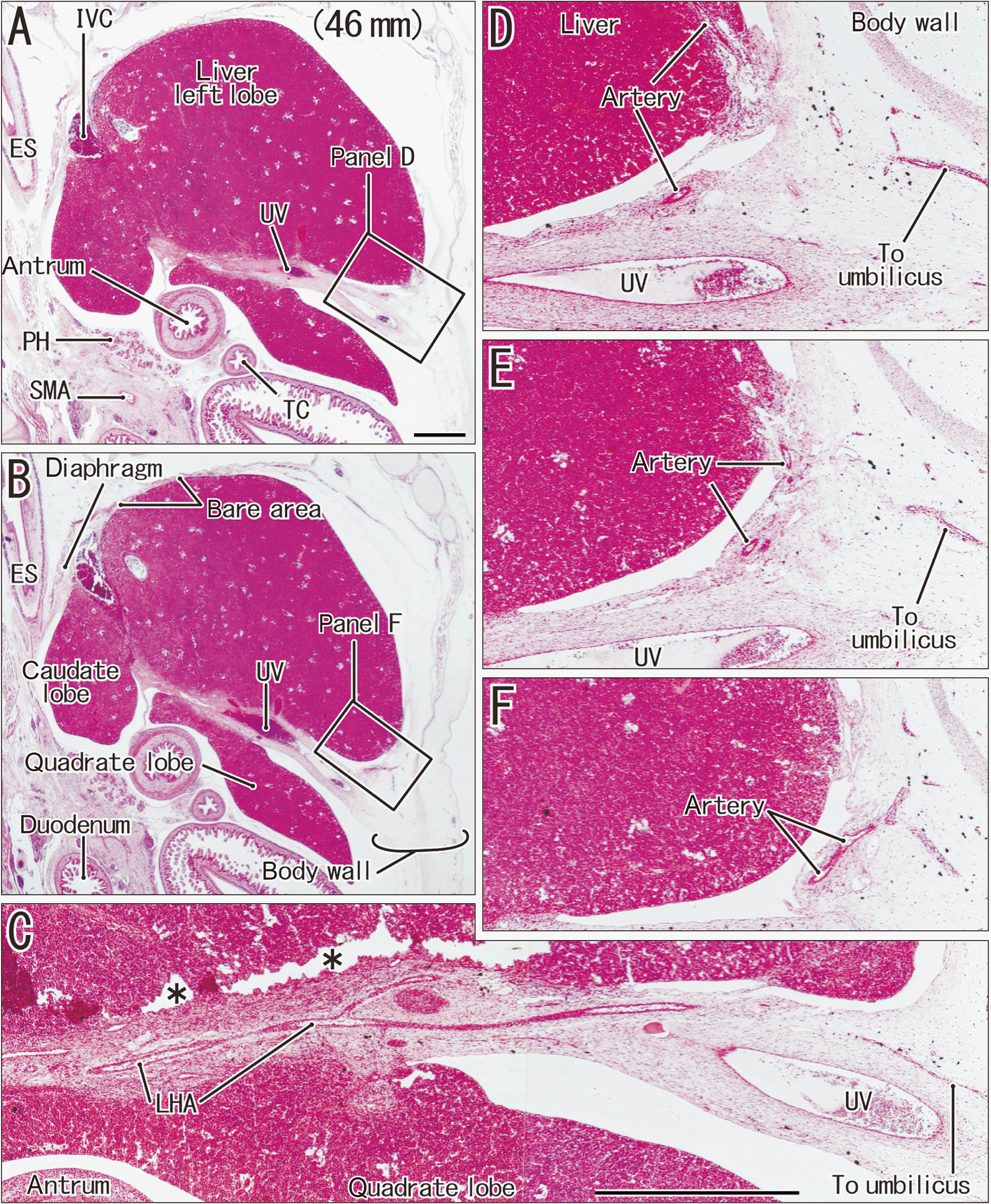
 | Fig. 1A marginal artery of the left hepatic artery to issue a parenchymal branch supplying the anterior surface of the liver left lobe. Sagittal sections from a Japanese fetus of 46 mm crown-rump length. Hematoxylin and eosin staining. (A) 0.6 mm left side of the midsagittal plane, shows a plane slightly medial to (B). (D, F) are higher magnification views of squares in (A, B), respectively. (E) displays an intermediate plane between panels (D, F). (C) 0.2 mm medial to (A), exhibits the longitudinal course of the marginal artery of the left hepatic artery (LHA) along the umbilical vein (UV). The LHA divides into the parenchymal branch (artery) and umbilical branch. The parenchymal branch takes a posterosuperior recurrent course beneath the peritoneum and enters the anterior liver surface (D–F). (A, B) or (C–F) were prepared at the same magnification. Scale bars in (A, C)=1 mm. Antrum, gastric antrum; ES, esophagus; IVC, inferior vena cava; PH, pancreatic head; SMA, superior mesenteric artery; TC, transverse colon. Asterisks (C) indicate artificial separation of tissues during the histological procedure. 
|
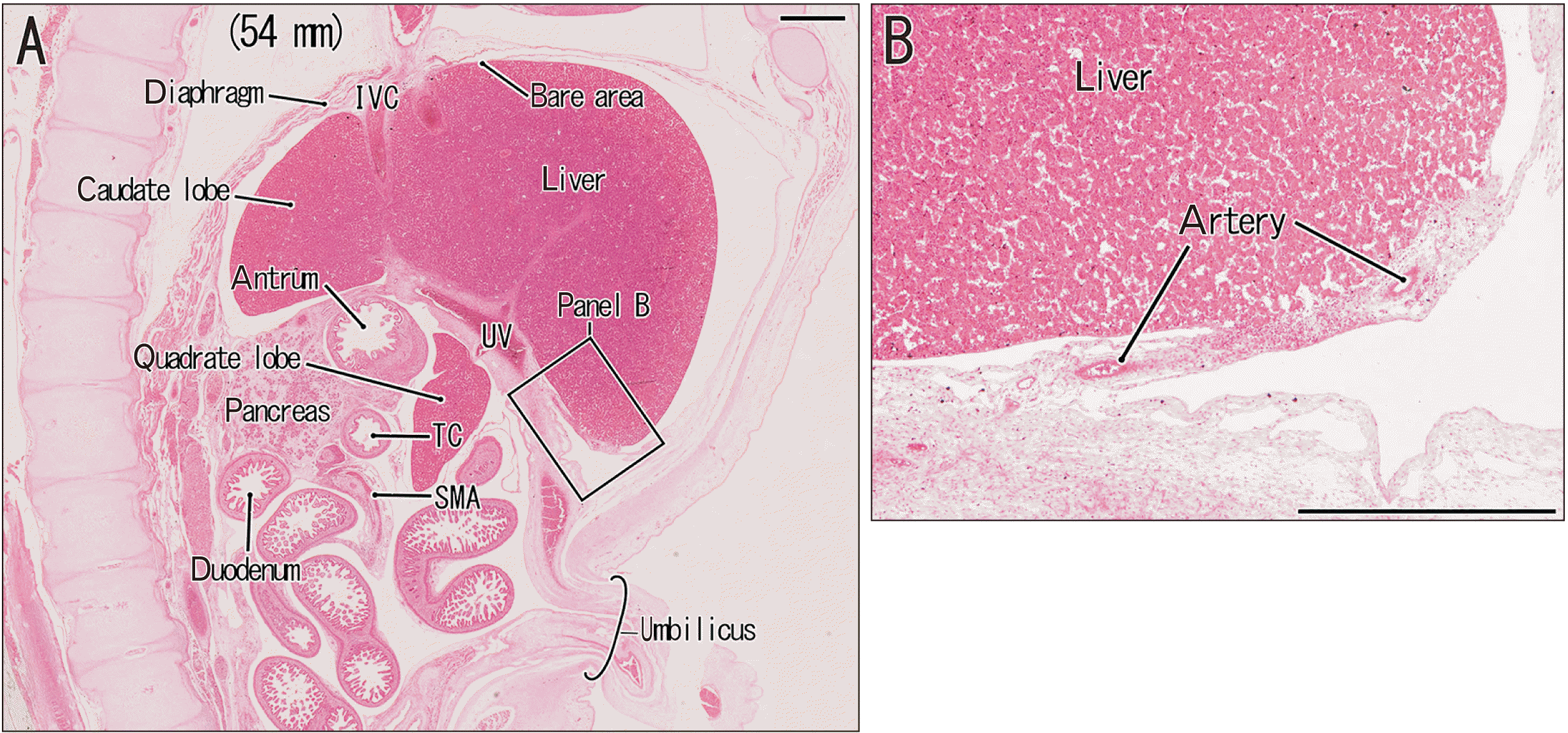
 | Fig. 2Another case of the parenchymal arterial branch supplying the anterior surface of the liver left lobe. Sagittal sections from a Japanese fetus of 54 mm crown-rump length. Hematoxylin and eosin staining. (A) displays the topographical anatomy. (B) corresponding to a square in (A), exhibits the parenchymal branch at higher magnification. Scale bars=1 mm. IVC, inferior vena cava; SMA, superior mesenteric artery; TC, transverse colon; UV, umbilical vein. 
|
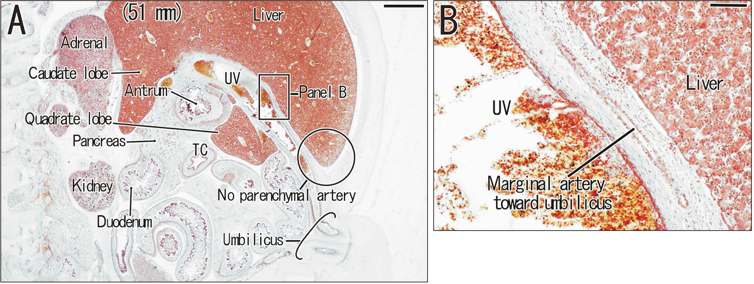
 | Fig. 3No parenchymal arterial branch supplying the anterior surface of the liver in a Spanish specimen. Sagittal sections from a Spanish fetus of 51 mm crown-rump length. Hematoxylin and eosin staining. (A) displays the topographical anatomy. (B) corresponding to a square in (A), exhibits the marginal artery along the umbilical vein (UV) at higher magnification. Scale bars=1 mm. TC, transverse colon. 
|
Case report of anomalous artery found in Japanese fetuses
Notably, in 5 of 12 Japanese midterm fetuses (CRL mm: 46, 50, 54, 59, 102), the LHA runs anteriorly along the umbilical vein (a marginal artery) and divides into a thick parenchymal branch and a thin umbilical branch (
Figs. 1C–F,
2B). The parenchymal branch takes a posterosuperior recurrent course beneath the peritoneum and supplies the anterior surface of the liver left lobe (segment III). The recurrent course of the artery provides a short peritoneal fold connecting between the liver left anterior end and the anterior body wall above the umbilicus. The umbilical branch reaches the umbilicus.
Spanish fetuses as a control
Although sagittal sections are limited in number, in 22 Spanish fetuses of which gestational ages corresponded to the Japanese ones, the LHA sometimes issues the marginal artery toward the umbilicus along the umbilical vein (5 specimens;
Fig. 3B). However, we do not find the parenchymal branch from the marginal artery. Rather than the simply attaching to the inferior aspect in Japanese fetuses, the quadrate lobe of the liver tends to completely surround the umbilical vein even at relatively early stages such as stages of 40–60 mm CRL. In addition, in contrast to its absence in Japanese fetuses, the inferior phrenic artery sometimes issues a branch to the liver parenchyma near the vena cava.
Go to :

Discussion
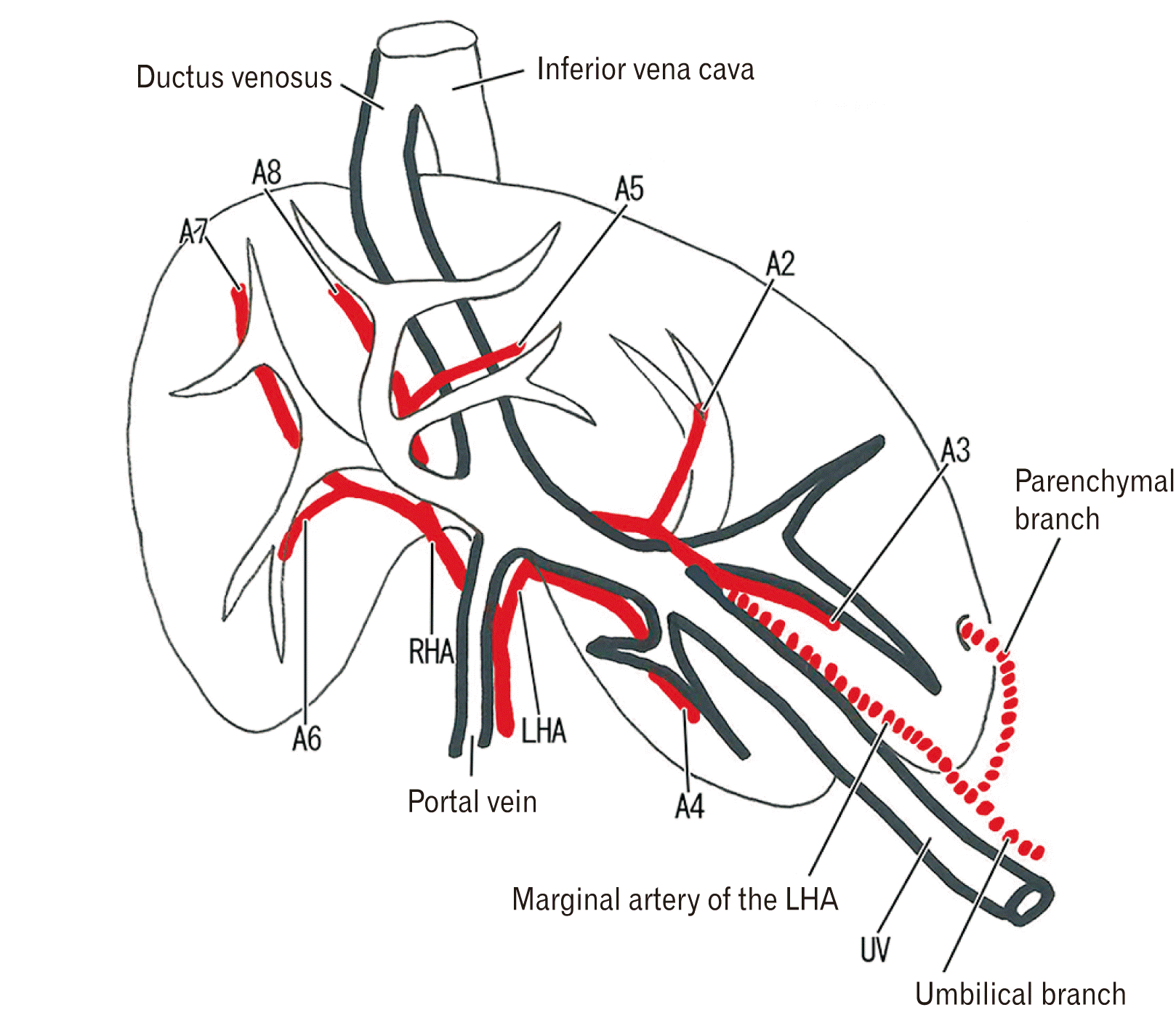
The present study demonstrated that, at the distal or anterior end of the course, the marginal artery of the LHA was likely to issue a parenchymal branch supplying the anterior liver surface.
Fig. 4 is a schematic representation of these uncommon arteries. In the figure, the arterial configuration in adults is based on Michels [
1] and Onishi et al. [
5] and it is modified according to the fetus-specific topographical anatomy of hepatic segments [
3] as well as the arterial anatomy at the hepatic hilum in fetuses [
4]. The marginal artery from the LHA was seen in both Japanese and Spanish specimens, but it did not issue a parenchymal branch in Spanish specimens. Our recent study demonstrated that, in Spanish specimens, the marginal artery sometimes reaches the abdominal wall at and around the umbilicus [
2].
 | Fig. 4Schematic representation of the marginal artery from the left hepatic artery: an anterolateral view of the liver corresponding to sagittal sections shown in the current study. A2–A8 indicates arteries to segments I-VIII, respectively. The caudate lobe (segments I and IX) is not drawn in the figure because of the posterior location along the inferior vena cava. The marginal artery originates from the left hepatic artery (LHA), runs anteriorly along the umbilical vein (UV) and divides into (1) the umbilical branch to the abdominal wall near the umbilicus and (2) the parenchymal branch to the anterior surface of the segment III. RHA, right hepatic artery. 
|
The surface-restricted distribution of parenchymal arteries seemed to be uncommon in the liver in contrast to well-known bile ducts exposing to the liver surface [
6]. Because of the long and straight course along the umbilical vein, the marginal artery of the LHA was most likely to correspond to thin arteries in and along the falciform ligament in adults: Michels [
1] demonstrated such arteries in his beautiful line-drawings of more than 30 cadaver dissections. However, to our understanding, none of them supplies the liver parenchyma but ends at the fibrous tissue of the anterior abdominal wall. The parenchymal branch of the marginal artery provided a peritoneal fold of which shape was similar to the appendix fibrosa hepatis in adults. The appendix fibrosa was situated in the left end of the bare area or the coronary ligament. In contrast, the present surface parenchymal branch was located near the umbilicus and distant from the bare area. Therefore, the present peritoneal fold was quite unlikely to develop into the appendix fibrosa. Macroscopic studies of the fetal liver also suggest a fact that, at midterm, the present parenchymal branch (near the umbilicus) is distant from the coronary ligament [
7-
9].
Although the specimens of sagittal sectionals were limited to in number (10/22), we did not find the parenchymal branch of the marginal artery in Spanish fetuses. We believed that, as in sagittal sections, serial horizontal sections also could demonstrate the artery if present. There seemed to be a considerable difference in incidence between human populations. The parenchymal branch of the marginal artery might be degenerated at the later stages due to friction between the liver free surface and growing diaphragm. Much or less, the left lobe surface parenchyma tended to be degenerate [
10,
11]. Actually, the appendix fibrosa contains degenerative liver parenchyma [
12,
13]. Degeneration of the liver parenchyma at the hepatic hilum is likely to be caused by smooth muscle contraction after birth [
13]. Overall, the present parenchymal artery seemed to be a transient variation seen in Japanese fetuses.
Go to :


 PDF
PDF Citation
 Print
Print


 XML Download