This article has been
cited by other articles in ScienceCentral.
Abstract
The muscles of the sole have been traditionally categorized into four layers, but it is more practical to divide them into peripheral and central groups. The peripheral groups include medial and lateral groups. The central plantar muscles are more numerous and divided into superficial and deep layers. During routine dissection in the Department of Anatomy, All India Institute of Medical Sciences Bibinagar, Hyderabad, variations are been observed in the plantar intrinsic muscle in the left foot & right foot of a 53-year-old male cadaver. This is the first cadaveric report of a combination of discrepancies especially the inter-tendinous connection between quadratus plantae and flexor digitorum brevis. Similar observations in the literature were not found by us. It is important to identify and study these dissimilarities of muscles of the sole for surgeons, anatomists, radiologists and orthopaedics as these muscles and tendons are used in foot reconstructive procedures, and for the treatment of some congenital anomalies.
Keywords: Quadratus plantae, Flexor digitorum brevis, Sole, Foot, Flexor digitorum accessories
Introduction
The muscles of the sole have been traditionally categorized into four layers, but it is more practical to divide them into peripheral and central groups. The peripheral groups include medial & lateral groups. The peripheral muscles, one long and one short, are located on each side of the foot and only reach the proximal phalanges of the digits. The central plantar muscles are more numerous and divided into superficial & deep layers. The superficial muscles are inserted into intermediate phalanges and deep muscles are inserted into distal phalanges. The central plantar muscles are flexor digitorum brevis (FDB), quadratus plantae (QP), lumbricals, plantar interossei, and dorsal interossei [
1].
The FDB is divided into four tendons for each of the four lateral toes. Each tendon divides at the base of the proximal phalanges into two fragments, which are inserted on eitherside of the shafts of the middle phalanx. The tendon divides into two fragments and forms a slot through which the flexor digitorum longus (FDL) tendon enters the distal phalanx. How FDB divides and is inserted into phalanges is similar to that of flexor digitorum superficialis in the hand [
2].
The other names of QP are “caro plantae pedis”, “flexor digitorum accessorius”, “Massae carnae”, “caro quadrata”, and “pronator pedis” [
3]. The origin of QP arises from two heads. The large fleshy medial head originates from the medial surface of the calcaneum. The small tendinous lateral head originates from the lateral tubercle of the calcaneum. It is inserted into the lateral part of the tendon of FDL [
2]. The QP has no homologous muscle in the hand.
The variations of the intrinsic muscles of the foot are uncommon. The Knowledge of exact variations of the intrinsic muscles of the foot is important to surgeons, ortho clinicians, and anatomists [
4].
In this case report, we are presenting a rare variation in the QP muscle that is associated with an unusual type of insertion, which is reported for the first time in India, to the best of our knowledge which is not mentioned in our literature search.
Case Report
Unusual variations in the intrinsic muscles of the foot were accidentally discovered in a 53-year-old male cadaver that had been embalmed in 10% formalin during practical sessions for undergraduate and postgraduate medical students at the Department of Anatomy at the All India Institute of Medical Sciences Bibinagar, Hyderabad, Telangana, India. This variation was found in routine cadaveric dissection so exempted from review by the ethical committee.
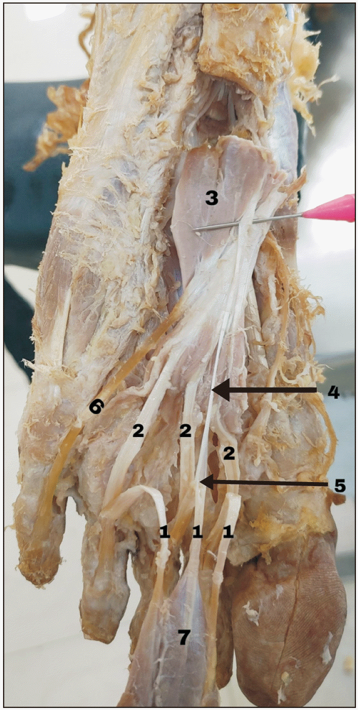
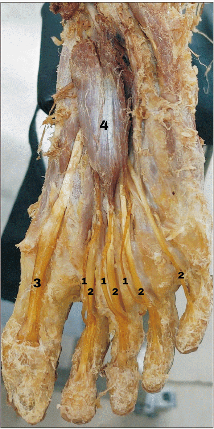
During the routine dissection of muscles of both feet, it was accidentally found that the fourth tendon of FDB was absent. There were only three tendons of FDB present and each tendon was divided into two fragments at the base of the proximal phalanx and was inserted into either side of the middle phalanx. The tendon of FDL passed through the slot between the two fragments (
Figs. 1,
2).
Later we detached the FDB from its origin and reflected it forward to see the second layer of muscles of both feet. We observed there were four tendons of FDL in which part of the fourth tendon of FDL was coming from QP in the left foot (
Fig. 1). On the right foot, there was no such variation found.
In the left foot, the flexor tendon of the third toe was coming from FDB and QP. A thin tendon of QP was inserted into the second tendon of FDB. The major part of the QP was inserted into the lateral side of the tendon of FDL.
Later we observed the tendons of FDL in the left foot. There were four tendons in which part of the fourth tendon was coming from QP. In the right foot, there was no such variation found.
Variations in the left foot
Absence of the fourth tendon of FDB.
A thin tendon of QP was inserted into the second tendon of FDB at the level of the head of the third metatarsal bone (Fig. 1).
The flexor tendon of the little toe was coming from both QP and FDL.
Variations in the right foot
Discussion
The most common variation of FDB is the absence of the fourth tendon to the little toe [
5]. The muscles of the fifth toe show gradual evolutionary changes because the utilization of the muscles of the fifth toe is very little and there is no opposition action [
6]. Clinically it is important to know the variations of the FDB because the tendons of FDB are used in the correction of claw and curly toe deformities which are congenital, musculocutaneous flaps for foot reconstruction procedures [
7].
Ilayperuma in their literary text mentioned that if the fourth tendon of FDB is absent mostly it is bilateral. Previously for the correction of claw toe deformities, the tendon of FDL transfer is the best method. In one study they compared the transfer of tendons of FDL and FDB using finite element simulation shows the transfer of tendons of FDB is the best modality for the treatment of claw toe deformities because it shows greater uniformity of stress distribution along the entire toe [
7].
Normally the medial plantar nerve passes between abductor hallucis and FDB but in some rare cases it passes superficial to the FDB. So, the variations of FDB may lead to the compression of medial plantar nerve [
8].
According to various authors, the incidence of the absence of the fourth tendon of FDB to the little toe varies among various populations as shown in
Table 1 [
9,
10].
The QP is unique to humans because it has two heads, medial and lateral head. In monkeys and apes, only a lateral head is present but in humans both heads especially the medial head is unique [
3]. Phylogenetically, the presence of QP in gorillas is 28%, orang-utans 48%, chimpanzees 50%, and humans 100% [
11].
According to the QP insertion pattern, it can be categorized into three types: muscular, tendinous, and aponeurotic [
12]. In the present case, it has both muscular which is inserted into the posterolateral part of FDL and a thin tendinous slip which is inserted into the second tendon of FDB.
According to Nayak and Vasudeva [
13] in their literary text, the accessory tendon slip of QP originated from the fascia of the abductor digiti minimi and was inserted into the FDL. The lateral plantar nerve is surrounded by the accessory tendon of QP and it may lead to compression of this nerve and mimics the symptoms of tarsal tunnel syndrome.
According to Coomar et al. [
14] the QP was inserted into the FHL and two tendinous slips arose from the medial head of QP & FHL and were inserted into the FDL at the level of metatarsophalangeal joints and the lateral head of QP was inserted into the second tendinous slip which changes the normal action of these muscles.
This is the first cadaveric report of a combination of all the above-mentioned variations especially the thin tendon of QP was inserted into the second tendon of FDB and it may lead to the compression of neurovascular structures and mimics the tarsal tunnel syndrome. Similar observations in the literature were not found by us. It is important to know the variations of muscles of the sole for surgeons, anatomists, and imaging studies as they are used in foot reconstructive procedures, and for the treatment of some congenital anomalies like club foot, claw toes, curling toes and for the treatment of diseases like the diabetic foot.
Acknowledgements
We would like to express our gratitude to the cadaver’s relatives for donating their relative’s body for education and research. Also we would like to acknowledge the efforts of anatomy laboratory attenders for regular maintenance of cadavers and the laboratory.
References
1. Ger R. 1986; The clinical anatomy of the intrinsic muscles of the sole of the foot. Am Surg. 52:284–5. PMID:
3706918.
2. Standring S. 2016. Gray's anatomy: the anatomical basis of clinical practice. 41st ed. Elsevier Limited;DOI:
10.1002/ca.22677.
3. Sooriakumaran P, Sivananthan S. 2005; Why does man have a quadratus plantae? A review of its comparative anatomy. Croat Med J. 46:30–5. PMID:
15726673.
4. Haratizadeh S, Seyyedin S, Nematollahi-mahani SN. 2023; Anatomical variation of quadratus plantae in relation to flexor hallucis longus and flexor digitorum longus: a rare case. Folia Morphol (Warsz). 82(2):412–5. DOI:
10.5603/FM.a2022.0034. PMID:
35380012.

5. Nathan H, Gloobe H. 1974; Flexor digitorum brevis--anatomical variations. Anat Anz. 135:295–301. PMID:
4413476.
6. Lobo SW, Menezes RG, Mamata S, Baral P, Hunnargi SA, Kanchan T, Bodhe AV, Bhat NB. 2008; Phylogenetic variation in flexor digitorum brevis: a Nepalese cadaveric study. Nepal Med Coll J. 10:230–2. PMID:
19558059.
7. Ilayperuma I. 2012; On the variations of the muscle flexor digitorum brevis: anatomical insight. Int J Morphol. 30:337–40. DOI:
10.4067/S0717-95022012000100059.

10. Bernhard A, Miller J, Keeler J, Siesel K, Bridges E. 2013; Absence of the fourth tendon of the flexor digitorum brevis muscle: a cadaveric study. Foot Ankle Spec. 6:286–9. DOI:
10.1177/1938640013489342. PMID:
23687345.
11. Talhar SS, Sontakke BR, Wankhede V, Shende MR, Tarnekar AM. 2017; A rare variation of flexor digitorum accessorius insole and its phylogenetic significance. J Mahatma Gandhi Inst Med Sci. 22:44–6. DOI:
10.4103/0971-9903.202017.

12. Pretterklieber B. 2018; Morphological characteristics and variations of the human quadratus plantae muscle. Ann Anat. 216:9–22. DOI:
10.1016/j.aanat.2017.10.006. PMID:
29166622.

13. Nayak SB, Vasudeva SK. 2022; Accessory slip of flexor digitorum accessorius (Quadratus plantae) muscle surrounding the lateral plantar nerve and vessels. Morphologie. 106:214–6. DOI:
10.1016/j.morpho.2021.06.004. PMID:
34247922.

Fig. 1
Variations in the left foot. 1: Three tendons of FDB, 2: tendons of FDL, 3: muscle belly of QP, 4: tendon slip of QP, 5: interconnection of tendon of QP with second tendon of FDB, 6: fourth tendon of FDL originates from both QP and FDL, and 7: reflected muscle belly of FDB. FDB, flexor digitorum brevis; FDL, flexor digitorum longus; QP, quadratus plantae.

Fig. 2
Variations in the right foot. 1: Three tendons of FDB, 2: four tendons of FDL, 3: tendon of Flexor hallucis longus, and 4: muscle belly of FDB. FDB, flexor digitorum brevis; FDL, flexor digitorum longus.

Table 1
Incidence of the absence of the fourth tendon of flexor digitorum brevis
|
Serial no. |
Author(s) |
Incidence (%) |
|
1 |
Nathan and Gloobe [5] |
63 |
|
2 |
Lobo et al. [6] |
100 |
|
3 |
Ilayperuma [7] |
72 |
|
4 |
Yalçin and Ozan [9] |
18 |
|
5 |
Bernhard et al. [10] |
48 |


 PDF
PDF Citation
 Print
Print


 XML Download