Journal List > Anat Cell Biol > v.56(4) > 1516085011

Priya, Philip, Jain, and Sikka: Variations of azygos vein: a cadaveric study with clinical relevance

Abstract

The azygos vein can be formed as a single root, two roots, and three roots, namely lateral, intermediate and the medial roots respectively. The hemiazygos vein and the accessory hemiazygos vein are the tributaries of azygos vein rather than its left side equivalents. Its variations, especially in young persons without any relevant risk factors, may result in thromboembolic illness. This study aimed to describe the morphological and morphometric variations of azygos system of veins. The present study was conducted on thirty formalin fixed adult human cadavers by dissecting azygos vein from formation to termination and variations were noted. The azygos vein was formed by a single root in 56.7%, by two roots: the lateral root and intermediate root in 36.7% cases and by the lateral root and medial root in 6.6%. The vertebral level of termination of azygos vein was seen at the level of T4 vertebrae in 70% cases, at the level of T3 vertebrae in 20% of cases and at the level of T5 vertebrae in 10% cases. The course of azygos vein was varying in 13.3%. These morphological variations can be useful while performing mediastinal surgery, mediastinoscopy, surgery of the deformations of the vertebral column, neurovascular surgeries of the retroperitoneal organs, disc herniation and fracture of thoracic vertebrae.

Introduction

The azygos vein (azygos=unpaired) is seen mostly on the right side of the vertebral column [1]. The azygos system consists of three interconnected major veins, the azygos vein (AV), hemiazygos vein (HAV) and accessory hemiazygos veins which drain the posterior wall of the thorax and abdomen [2, 3]. The formation of AV is variable due to the complexity in its embryological development [4, 5].
The AV can be formed as a single root, two roots, and three roots, namely lateral, intermediate and the medial roots respectively. Lateral root is the common trunk formed by the combination of right ascending lumbar vein and right subcostal vein in the thorax, formed at the level just below the head of twelfth rib. Intermediate root (lumbar azygos) arises from posterior aspect of the inferior vena cava and joins the lower end of AV [6]. The medial root is small plexiform of veins strongly associated with the ventral side of the lumbar vertebral bodies dorsal to the aorta and its lumbar segmental branches [5]. The AV typically leaves the abdomen through the aortic opening in the diaphragm at the level of T12 [7] and ascends vertically upwards lying in front of the vertebral column up to the level of T4, where it arches forward superior to the hilum of right lung to open into the posterior aspect of superior vena cava (SVC).
AV forms crucial cavo-caval and porto-caval interface, forming collateral circulation in case of portal hypertension and vena caval occlusion. Furthermore, the intercostal veins and vertebral venous plexuses link the azygos system to the cerebral venous system therefore forming potential metastatic pathway to brain in cases of breast and bronchial carcinoma. It also forms a significant communicating channel between superior and inferior vena cava, thus serving as an alternate route for venous return from the lower parts of the body if primary venous channel is obstructed [8-10].
Taking into consideration, all the aforementioned clinical prominences of AV, surgeons should be pre-acquainted about variations of AV while operating in the mediastinum (i.e., mediastinoscopy, surgery of the deformations of the vertebral column) to prevent accidental haemorrhage. Therefore, the aim of this study was to evaluate the morphological and morphometric variations in the formation, course, and termination of the AV to prevent iatrogenic complications.

Materials and Methods

Study design and sample size

This descriptive cross-sectional study was conducted in Department of Anatomy, Christian Medical College, Ludhiana from October 2017 to June 2019. This study was conducted on thirty formalin fixed adult human cadavers. The sample size was determined using convenience sampling.

Exclusion criteria

The cadavers in which AV were damaged due to any surgeries or trauma were excluded.

Data collection and analysis

Cadavers were dissected using Cunningham’s Manual for Dissection volume two to study AV [11]. The AV was dissected and traced from the formation to termination. Following parameters were studied and variations were noted:
  1. The formation, course, and termination of the AV.

  2. Vertebral level of the formation and termination of AV.

  3. External diameter of AV at the formation and termination.

Formation of the AV was noted as lateral root, intermediate root, medial root based on the way it is formed. The termination of AV was indicated by its drainage to the SVC, right brachiocephalic vein, or left brachiocephalic vein. Course of the AV in relation to vertebral column was evaluated with respect (in midline, right, or left side) to the vertebral column. Also, few very rare variations of AV were noted and mentioned separately under other variations. Vertebral levels of origin and termination of AV were determined by tracing the corresponding rib posteriorly up to the body of the thoracic vertebra. However, because the majority of thoracic ribs articulated with two vertebral bodies simultaneously, the upper one was considered. All observations and measurements were noted twice, independently by two observers. Mean value of the measurements by the two observers were considered as the exact value. External diameter was measured using Mitutoyo digital vernier calipers (precision 0.01 mm) in millimeters. All the data was analyzed using IBM SPSS Statistics 20.0 for Windows (IBM Co.). Descriptive analysis was done and categorical variables were presented as percentage (%).

Ethical approval

Ethical Clearance was obtained from the Institutional Ethics Committee, Christian Medical College and Hospital, Ludhiana, Punjab, India (IEC no: 2017/CMCL-1EC-445). Patient consent cannot be provided as this study is based on cadavers (which are donated cadavers) for which approval from ethics committee has already been taken and provided.

Results

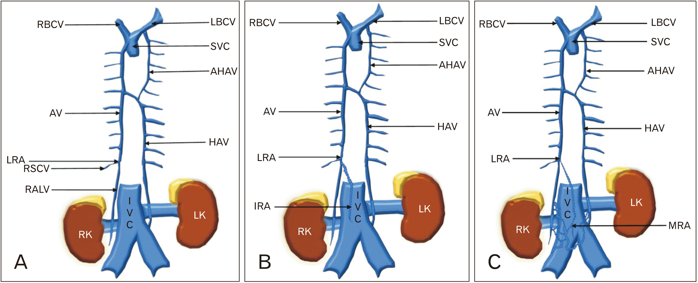
In the present study, the AV was formed by a single root (lateral root) in 17 (56.7%) of cases (Fig. 1A). In 11 (36.7%), the AV was formed by two roots (the lateral root and intermediate root) (Fig. 1B), wherein it was formed by two roots (the lateral root and medial root) in 2 (6.6%) (Fig. 1C). In none of the cases, AV was formed from all three roots i.e., lateral, intermediate root and medial root. It also exhibited typical termination (i.e., SVC) in all 30 cases. Although, the AV was formed at T12 vertebra in all 30 cases, it showed various vertebral levels of termination to SVC (Table 1). The mean external diameter of AV at the level of formation and termination was 3.18±1.85 mm and 7.44±2.84 mm, respectively. The maximum external diameter at termination was 13.97 mm and the minimum diameter was 1.28 mm. The course of AV in relation to vertebral column in shown in Table 2.

Other variations

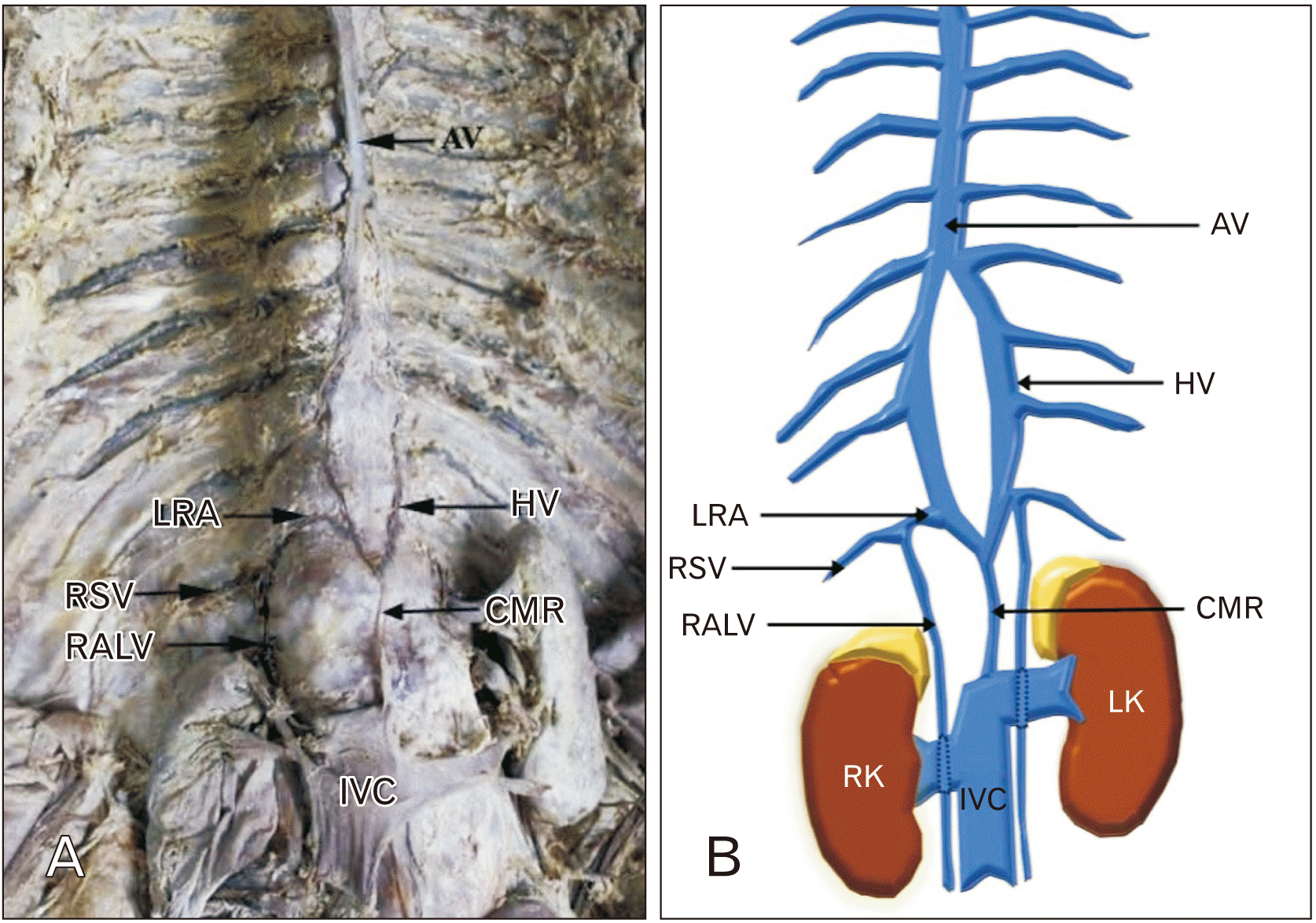
Variation 1: Both AV and HAV were seen arising as one common medial root from the postero-lateral aspect of inferior vena cava at L2 vertebrae. Then at L1 vertebrae this common medial root divided to form AV on right side and HAV on left side (Fig. 2).
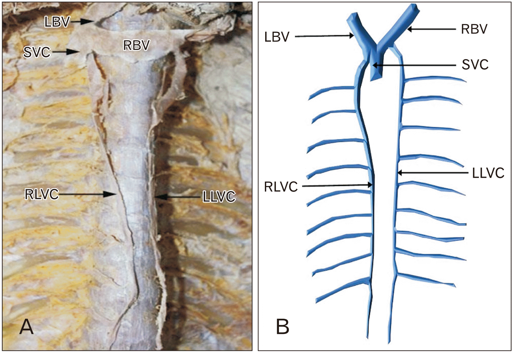
Variation 2: Two longitudinal venous channels were present on either side of vertebral column. Right longitudinal venous channel drained into SVC and left longitudinal venous channel drained into left brachiocephalic vein. Hemiazygos as well as accessory HAV were absent (Fig. 3).
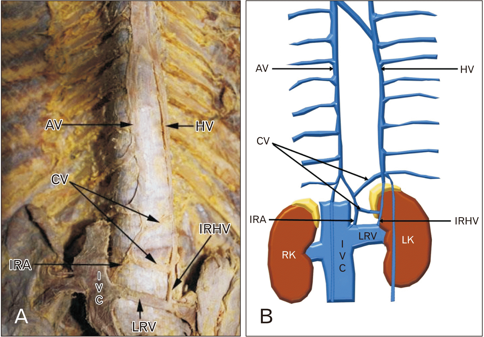
Variation 3: There were two communicating veins present between intermediate root of AV and intermediate root of HAV at the level of L1 vertebrae (Fig. 4).
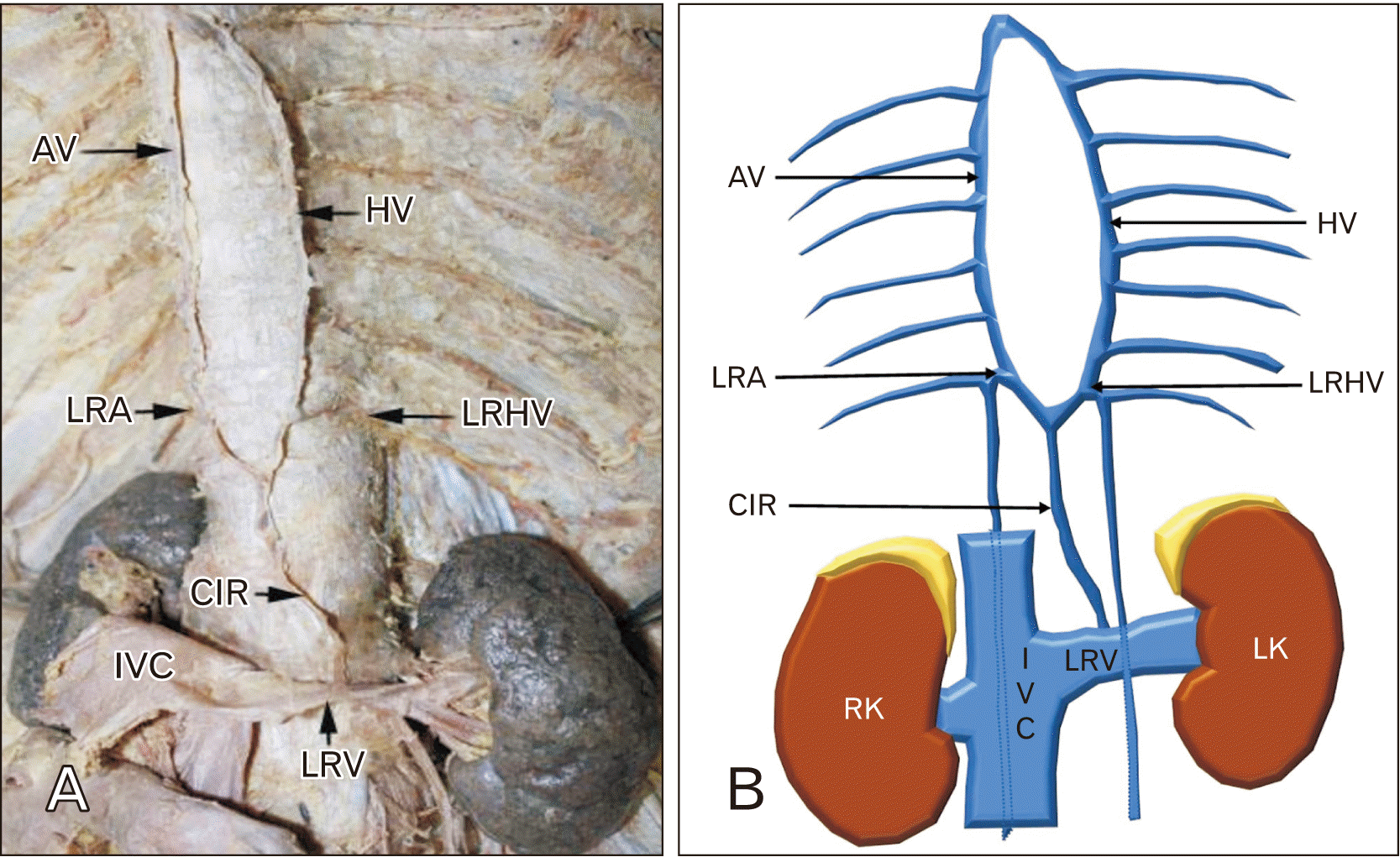
Variation 4: Both AV and HAV was seen arising as one common intermediate root from the left renal vein at the level of L2 vertebrae. At the level of L1 vertebrae this common intermediate root divided into two to form AV on right side and HAV on left side (Fig. 5).
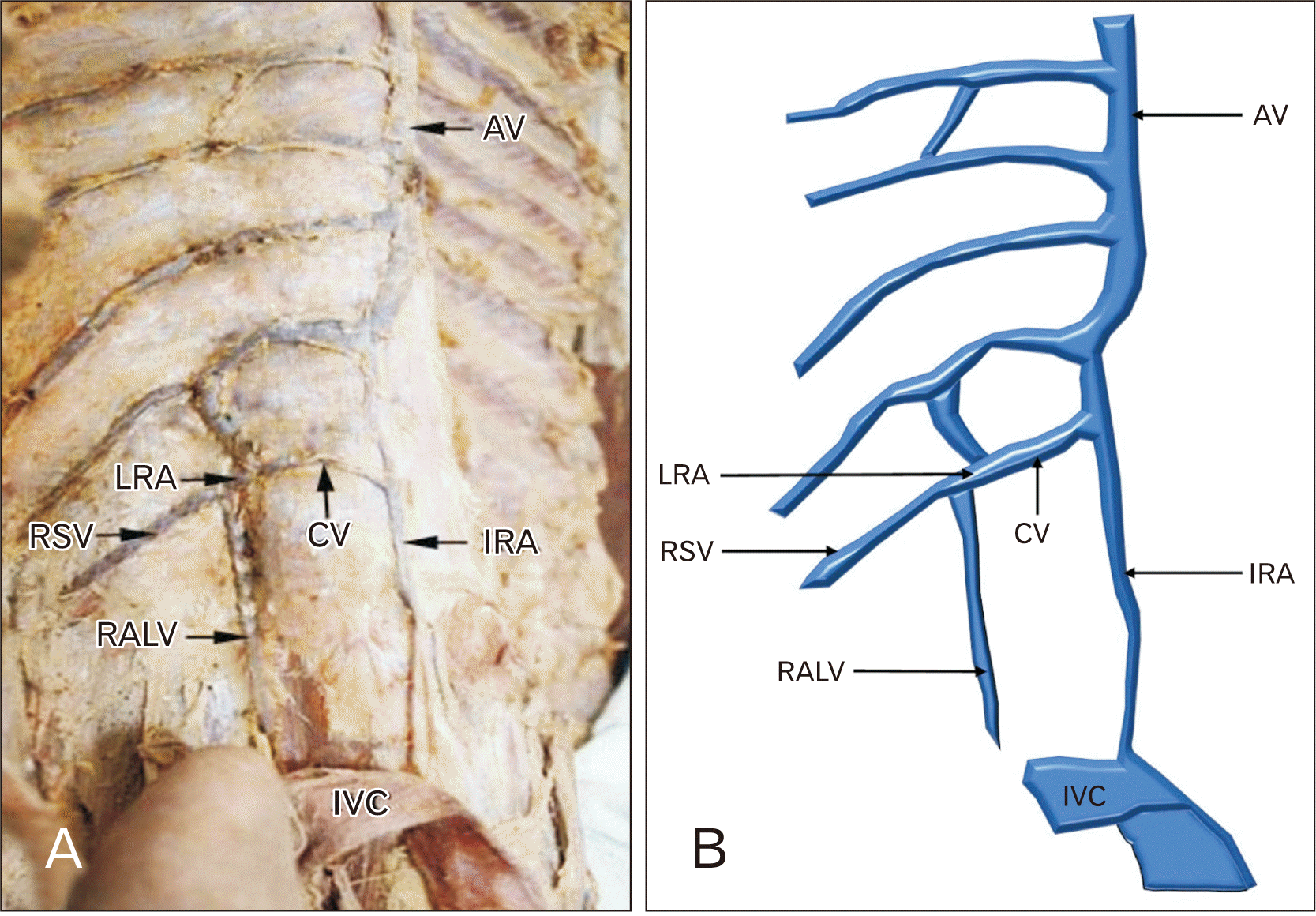
Variation 5: A rare variation, a communicating vein present between AV and intermediate root of AV at the level of L2 vertebrae (Fig. 6).

Discussion

Galen used the term “azygos vein” for the first time in literature, although not as an official anatomical name; rather, he used it as an adjective to describe the absence of a vein with a comparable pair on the left side [12]. The name “azygos” vein later saw a revival during Dubois’ [13] dissections and Vesalius’ [14] anatomical studies, where it obtained its final definition as an official anatomical term. Later, Vesalius [14] emphasized on course of AV in concordance with modern anatomical textbooks.
AV is seen to vary frequently in their mode of formation, course, and termination. These variations of the AV are commonly observed while performing radiological and surgical procedures of posterior mediastinum. They can simulate aneurysm, lymphadenopathy, and tumors, or might also lead to accidental intrathoracic haemorrhage during the operative procedures. Therefore, its relevant to know these variations for proper interpretation [4].
In the present study, the AV is formed either by single or double roots. The incidence of formation of AV from single root was higher in other studies [15-20] and from two roots were variable (Table 3) [15, 17, 19, 20].
Course of the AV in relation to vertebral column in the present study, in 40% cases AV was seen in the midline, in 33.3% cases it was noted to be at the right side and in 13.3% cases it was noted at the left side of the vertebral column (Table 2) [21, 22]. Osteophytosis of the vertebral column in old age is brought about by the mechanical stresses and strains acting on it. The osteophytes in the thoracic area grows asymmetrically, which is thought to be caused by the aorta along the left side of the vertebral column, which inhibits their growth. The growth of osteophytes, which mostly affect the right side of the thoracic vertebrae, can be reason for deviation of AV to the left side [23, 24]. On the contrary, Kagami and Sakai [25] in 1990 stated that the course of the AV is not affected by osteophytes, instead osteophytes can be influenced by the AV.
Termination of AV can be in SVC or brachiocephalic vein [1]. In the present study, termination of AV into SVC was noted in all 30 cases (100%), which is in accordance with the study done by Prasad and K [15] and Tatar et al. [26] but, Raghavendra and Bhosale [27] observed this in only 88.9% cases (Table 3).
Vertebral level of formation and termination of AV is shown in Table 1. Radhika et al. [16] observed the formation of AV at T12 vertebral level in 88% cases. In the present study it was observed in 100% cases, which is in accordance with study of Prasad and K [15]. The termination of AV was found to be at different vertebral levels by different authors. In majority of cases, Tatar et al. [26] and Prasad and K [15] observed it at T5 level, Kutoglu et al. [28] and Patra et al. [29] found it at T3 level. Nirmala and Teresa [17] observed it at T3–T4 level in all 100% cases. In the present study, in 70% cases it was seen to be at the level of T4 which was in agreement with Latha and Sugavasi [18] and Raghavendra and Bhosale [27].
External diameter at the level of formation and termination of AV is summarised in Table 4. The mean external diameter of AV at the level of formation and termination was similar to the study of Kutoglu et al. [28] and Tatar et al. [26], Raghavendra and Bhosale [27] respectively. Patra et al. [29] noted much larger diameter than the present study.
Normal diameter of AV at formation and termination should be known, to differentiate it from any pathological conditions such as mediastinal mass, enlarged lymph nodes and aneurysm. The increased size of AV in chest X-ray provides useful haemodynamic information [30, 31]. The AV diameter significantly correlate with the intravascular fluid circulation. Furthermore, increased capillary blood volume (CBV) following over-infusion as in cases of renal failure, with accompanying fluid overload , results in an associated increase in the width of the AV [10]. Therefore, increase in 5 mm of the vascular pedicle relates to 1 L increase in circulating fluid [32, 33].
Although, the width of the AV does not correlate substantially with the CBV, but it does with the right mean atrial pressure [10]. These relationships have been observed in patients with any degree of severity of cardiac failure and CBV values ranging from normal to very high [32, 33]. Thus, Milne [31] and Chait [32] reported that the main reason for increased width of AV is the increased mean right atrium pressure.
Increased width of the AV was correlated with a rising central venous pressure for an individual patient. When the width of the AV measured more than 15 mm it was associated with an abnormal central venous pressure (greater than 10 cm water). The linear relationship between the two was (AV width mm×1.4)–3=central venous pressure in cm water [34].

The other variations of AV

Variation 1: Both AV and HAV originated from one common medial root from the postero-lateral aspect of inferior vena cava. Similar finding was reported by Seema [35].
Variation 2: Two longitudinal venous channels (Right and left venous channels) were present on either side of vertebral column. Mustafa et al. [20], Dahran and Soames [36] and Patra et al. [29] observed exactly same finding. Rao and Banerjee [37] noted this finding in 10% cases which is higher than present study. Similar finding was observed by Keskin et al. [38] in computed tomography (CT) angiography of a 52-year-old female patient.
Variation 3, 4, and 5: To the best of our knowledge, no any similar case as these have been reported in the previous literature.

Embryology

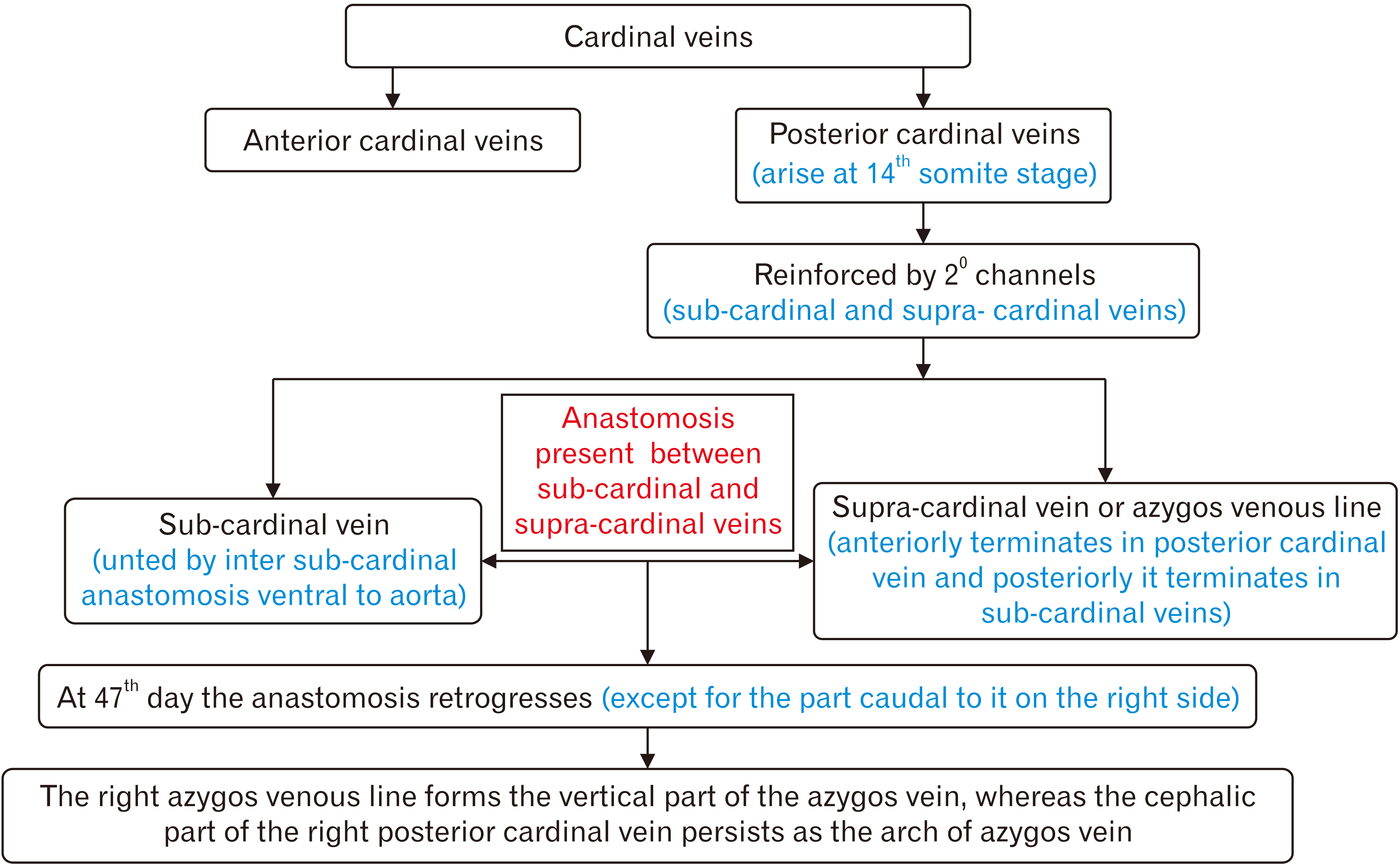
The entire course of AV is susceptible to variations due to its complex development (Fig. 7) [4, 5, 39]. If there is failure in this normal retrogression, this might lead to anomalies of variations in morphology of AV. Additionally, if the posterior cardinal vein fails to migrate supero-medially leading to a rare congenital anomaly called azygos lobe [40].

Clinical importance

The findings of this study can be applied to the treatment of disc herniation, thoracic vertebral fractures, deformities of the vertebral column, neurovascular surgery of the retroperitoneal organs, and mediastinal surgery. Surgeons should also keep in mind the varying course of AV before performing surgeries such as sympathectomy and repair of trachea-oesophageal fistula to prevent iatrogenic complications [24]. This study has brought out many facts about the variations of AV with respect to differentiating the normal anatomical variations from the pathological changes.

Limitations

The present study requires more data with respect to cadaveric as well as CT images/magnetic resonance imaging so as to compare with studies with such databases. The cadaveric data if compared with radiological entity would help the surgeons to understand the morphometry in a better way.

Acknowledgements

We are grateful to all the residents and faculty members of Christian medical college, Ludhiana and Rajendra Institute of medical sciences, Ranchi for their help and co-operation during the preparation of this manuscript.

Notes

Author Contributions

Conceptualization: AJ, AS, AP. Data acquisition: AP, AJ, AS. Data analysis or interpretation: AP, SEP, AJ, AS. Drafting of the manuscript: AP, SEP, AJ, AS. Critical revision of the manuscript: AP, SEP, AJ, AS. Approval of the final version of the manuscript: all authors.

Conflicts of Interest

No potential conflict of interest relevant to this article was reported.

References

1. Standring S. Standring S, editor. 2008. Mediastinum. Gray's Anatomy: The Anatomical Basis of Clinical Practice. 40th ed. Churchill Livingstone;p. 939–57. DOI: 10.1016/b978-0-443-06684-9.50063-9.
2. Snell RS. 2004. Clinical Anatomy. Lippincott Williams & Wilkins.
3. Drake RL, Vogl W, Mitchell AWM. 2015. Gray's Anatomy for Students. 3rd ed. Elsevier;p. 197.
4. Celik HH, Sargon MF, Aldur MM, Cumhur M. 1996; An anomalous course of the interazygos vein. Surg Radiol Anat. 18:61–2. DOI: 10.1007/BF03207766. PMID: 8685815.
crossref
5. Mezzogiorno A, Passiatore C. 1988; An atypic pattern of the azygos venous system in man. Anat Anz. 165:277–81. DOI: 10.53347/rid-26571. PMID: 3400891.
6. Casoni GL, Tomassetti S, Cavazza A, Colby TV, Dubini A, Ryu JH, Carretta E, Tantalocco P, Piciucchi S, Ravaglia C, Gurioli C, Romagnoli M, Gurioli C, Chilosi M, Poletti V. 2014; Transbronchial lung cryobiopsy in the diagnosis of fibrotic interstitial lung diseases. PLoS One. 9:e86716. DOI: 10.1371/journal.pone.0086716. PMID: 24586252. PMCID: PMC3938401. PMID: 79250b8f3a59492884f75f26253863fa.
crossref
7. Seib GA. 1934; The azygos system of veins in American whites and American negroes, including observations on the inferior caval venous system. Am J Phys Anthropol. 19:39–163. DOI: 10.1002/ajpa.1330190117.
crossref
8. Rosse C, Gaddum-Rosse P. 1997. Hollinshead's Textbook of Anatomy. 5th ed. Lippincott-Raven.
9. Shivanal U, HT G. 2015; Anomalous azygos veins - its embryological basis and clinical significance. Int J Res Med Sci. 3:2323–6. DOI: 10.18203/2320-6012.ijrms20150624.
crossref
10. Piciucchi S, Barone D, Sanna S, Dubini A, Goodman LR, Oboldi D, Bertocco M, Ciccotosto C, Gavelli G, Carloni A, Poletti V. 2014; The azygos vein pathway: an overview from anatomical variations to pathological changes. Insights Imaging. 5:619–28. DOI: 10.1007/s13244-014-0351-3. PMID: 25171956. PMCID: PMC4195836.
crossref
11. Romanes GJ. Romanes GJ, editor. 1986. The thorax. Cunningham's Manual of Practical Anatomy, Vol 2: Thorax and Abdomen. 15th ed. Oxford University Press;p. 1–82. DOI: 10.5005/jp/books/14241_6.
12. Paraskevas GK, Koutsouflianiotis KN, Patsikas M, Noussios G. 2019; What is the history of the term "azygos vein" in the anatomical terminology? Surg Radiol Anat. 41:1155–62. DOI: 10.1007/s00276-019-02238-3. PMID: 31028449.
crossref
13. Dubois J. 1555. In Hippocratis et Galeni Physiologiae Partem Anatomicam Isagoge. Apud Joannem Hulpeau;p. 18924. Latin.
14. Vesalius A. 1543. De Humani Corporis Fabrica Libri Septem. Ex Officina Joannis Oporini;p. 37. Latin. DOI: 10.5962/bhl.title.109299.
15. Prasad M, K J. 2018; Variations in formation and termination of azygos vein in South Indian population. Int J Anat Res. 6:5384–9. DOI: 10.16965/ijar.2018.212.
crossref
16. Radhika D, Nirmala BV, Latha GK. 2016; Tributaries of azygos vein. J Evid Based Med Healthc. 3:49–51. DOI: 10.18410/jebmh/2016/11. PMID: 27871101.
crossref
17. Nirmala BV, Teresa RS. 2015; Study of azygos system and its variations. J Evol Med Dent Sci. 4:5652–7. DOI: 10.14260/jemds/2015/827.
crossref
18. Latha K, Sugavasi R. 2013; Study of the azygos system of veins in human cadaver. Int J Curr Res Rev. 5:113–7.
19. Alves EC, Porciúncula Junior RW, Monte Bispo RF, de Sousa-Rodrigues CF, da Rocha AC. 2011; Formation of the azygos vein. Int J Morphol. 29:140–3. DOI: 10.4067/S0717-95022011000100024.
20. Mustafa SIA, Ali TO, Ahmed AA. 2016; Anatomical variation of the azygos vein in human cadavers. Int J Innov Sci Res. 25:368–72.
21. Krakowiak-Sarnowska E, Wiśniewski M, Szpinda M, Krakowiak H. 2003; Variability of the azygos vein system in human foetuses. Folia Morphol (Warsz). 62:427–30. PMID: 14655133.
22. Vedapriya KA, Priyanka D. 2019; Morphological variation in azygos vein. Int J Sci Res. 8:1396–9. DOI: 10.32388/rqli8f.
23. Nathan H. 1960; Anatomical observations on the course of the azygos vein (vena azygos major). Thorax. 15:229–32. DOI: 10.1136/thx.15.3.229. PMID: 13727912. PMCID: PMC1018568.
crossref
24. Saito A, Murakami M, Tomioka K, Ezure H, Moriyama H, Mori R, Nakajima K, Nakamura M, Otsuka N. 2015; The impact of aging on the course of the azygos vein. Okajimas Folia Anat Jpn. 92:7–10. DOI: 10.2535/ofaj.92.7. PMID: 26448373.
crossref
25. Kagami H, Sakai H. 1990; The problems in the arrangement of the azygos vein. Okajimas Folia Anat Jpn. 67:111–4. DOI: 10.2535/ofaj1936.67.2-3_111. PMID: 2216303.
crossref
26. Tatar I, Denk CC, Celik HH, Oto A, Karaosmanoglu DA, Ozdemir BM, Surucu SH. 2008; Anatomy of the azygos vein examined by computerized tomography imaging. Saudi Med J. 29:1585–8. DOI: 10.53347/rid-955. PMID: 18998005.
27. Raghavendra AY, Bhosale SM. 2017; Variations of arch of azygos vein: an anatomical overview with clinical importance. Int J Anat Res. 5:4251–6. DOI: 10.16965/ijar.2017.299.
crossref
28. Kutoglu T, Turut M, Kocabiyik N, Ozan H, Yildirim M. 2012; Anatomical analysis of azygos vein system in human cadavers. Rom J Morphol Embryol. 53:1051–6. PMID: 23303031.
29. Patra A, Singla RK, Kaur H, Malhotra V. 2019; Analysis of multiple variations in azygos venous system anatomy with its classification: a cadaveric study. Eur J Anat. 23:9–15.
30. Bass JE, Redwine MD, Kramer LA, Huynh PT, Harris JH Jr. 2000; Spectrum of congenital anomalies of the inferior vena cava: cross-sectional imaging findings. Radiographics. 20:639–52. DOI: 10.1148/radiographics.20.3.g00ma09639. PMID: 10835118.
crossref
31. Milne EN. 1978; Some new concepts of pulmonary blood flow and volume. Radiol Clin North Am. 16:515–36. PMID: 746145.
32. Chait A. 1972; Interstitial pulmonary edema. Circulation. 45:1323–30. DOI: 10.1161/01.CIR.45.6.1323. PMID: 5032828.
crossref
33. Rossi SE, McAdams HP, Rosado-de-Christenson ML, Franks TJ, Galvin JR. 2001; Fibrosing mediastinitis. Radiographics. 21:737–57. DOI: 10.1148/radiographics.21.3.g01ma17737. PMID: 11353121.
crossref
34. Preger L, Hooper TI, Steinbach HL, Hoffman JI. 1969; Width of azygos vein related to central venous pressure. Radiology. 93:521–3. DOI: 10.1148/93.3.521. PMID: 4898452.
crossref
35. Seema MA. 2012; A rare anatomical variation in medial root of azygos vein with its embryological aspect. Int J Anat Sci. 3:14–6.
36. Dahran N, Soames R. 2016; Anatomical variationos of the azygos venous system: classification and clinical relevance. Int J Morphol. 34:1128–36. DOI: 10.4067/S0717-95022016000300051.
37. Rao YS, Banerjee A. 2014; Anatomical variations of the azygos venous system. Int J Biol Med Res. 5:4535–8. DOI: 10.1142/9781783269150_0048.
38. Keskin S, Keskin Z, Sekmenli N. 2013; The independent right and left azygos veins with hemiazygos absence: a rare case presentation. Case Rep Vasc Med. 2013:282416. DOI: 10.1155/2013/282416. PMID: 23841014. PMCID: PMC3690739. PMID: d9d21dc797b94231824f5d09ab5f988c.
crossref
39. Moore KL, Persaud TVN, Torchia MG. Torchia MG, Persaud TVN, editors. 2016. Cardiovascular system. The Developing Human: Clinically Oriented Embryology. 10th ed. Elsevier;p. 283–334.
40. Kc S, Shrestha P, Shah AK, Jha AK. 2018; Variations in human pulmonary fissures and lobes: a study conducted in Nepalese cadavers. Anat Cell Biol. 51:85–92. DOI: 10.5115/acb.2018.51.2.85. PMID: 29984052. PMCID: PMC6026823.
crossref

Fig. 1
Schematic representation of normal roots of origin of azygos vein (AV). (A) Origin only by lateral root. (B) Origin by lateral and intermediate root. (C) Origin by lateral and medial root. RBCV, right brachiocephalic vein; LBCV, left brachiocephalic vein; SVC, superior vena cava; AHAV, accessory hemiazygos vein; HAV, hemiazygos vein; LRA, lateral root of AV; RSCV, right sub costal vein; RALV, right ascending lumbar vein; RK, right kidney; IVC, inferior vena cava; LK, left kidney; IRA, intermediate root of AV; MRA, medial root of AV.
acb-56-4-448-f1.tif
Fig. 2
Showing both azygos vein (AV) and hemiazygos vein (HV) arising from common medial root (CMR). It also shows AV arising from both lateral root and CMR. (A) Cadaveric image. (B) Schematic representation. LRA, lateral root of AV; RSV, right subcostal vein; RALV, right ascending lumbar vein; IVC, inferior vena cava; RK, right kidney; LK, left kidney.
acb-56-4-448-f2.tif
Fig. 3
Showing two longitudinal venous channels on either side of vertebral column. (A) Cadaveric image. (B) Schematic representation. LBV, left brachiocephalic vein; RBV, right brachiocephalic vein; SVC, superior vena cava; RLVC, right longitudinal venous channel; LLVC, left longitudinal venous channel.
acb-56-4-448-f3.tif
Fig. 4
Showing two communicating veins (CVs) present between intermediate root of azygos vein (IRA) and intermediate root of hemiazygos vein (IRHV). (A) Cadaveric image. (B) Schematic representation. AV, azygos vein; HV, hemiazygos vein; IVC, inferior vena cava; LRV, left renal vein; RK, right kidney; LK, left kidney.
acb-56-4-448-f4.tif
Fig. 5
Showing a communicating vein (CV) present between azygos vein (AV) and intermediate root of azygos vein (IRA). (A) Cadaveric image. (B) Schematic representation. LRA, lateral root of azygos vein; RSV, right subcostal vein; RALV, right ascending lumbar vein; IVC, inferior vena cava.
acb-56-4-448-f5.tif
Fig. 6
Showing both azygos vein (AV) and hemiazygos vein (HV) formed from lateral root and common intermediate root (CIR) arising from left renal vein (LRV). (A) Cadaveric image. (B) Schematic representation. LRA, lateral root of azygos vein; LRHV, lateral root of hemiazygos vein; IVC, inferior vena cava; RK, right kidney; LK, left kidney.
acb-56-4-448-f6.tif
Fig. 7
Flow diagram of the development of azygos vein.
acb-56-4-448-f7.tif
Table 1
Showing comparison of vertebral level of termination of AV
References n Findings (%) at different vertebral level
T2 T2–T3 T3 T3–T4 T4 T4–T5 T5 T6 Absent AV
Tatar et al., 2008 [26] 103 - - - - 40 - 55 5 -
Kutoglu et al., 2012 [28] 48 12.5 12.5 62.5 6.2 - - - - 6.2
Latha and Sugavasi, 2013 [18] 100 - - 8 - 85 - 7 - -
Nirmala and Teresa, 2015 [17] 50 - - - 100 - - - - -
Raghavendra and Bhosale, 2017 [27] 36 - 2.8 11.1 13.9 52.8 8.3 11.1 - -
Prasad and K, 2018 [15] 50 - - - - 6 4 90 - -
Patra et al., 2019 [29] 30 20 16.6 53.3 6.6 3.3 - - - -
Present study, 2019 30 - - 20 - 70 - 10 - -

AV, azygos vein.

Table 2
Showing comparison of course of azygos vein
References Type of study n Findings (%)
Right Midline Left Others
Krakowiak-Sarnowska et al., 2003 [21] Human fetuses 32 90.6 9.4 - -
Tatar et al., 2008 [26] Chest computed tomography 103 37.9 39.8 22.3 -
Saito et al., 2015 [24] Cadaveric 47 - 6 94 -
Vedpriya and Priyanka, 2019 [22] Cadaveric 22 73 - 27 -
Patra et al., 2019 [29] Cadaveric 30 66.7 - - 33.3
Present study, 2019 Cadaveric 30 33.3 40.1 13.3 13.3
Table 3
Showing comparison of mode of formation and termination of azygos vein
References Type of study n Formation (%) Termination (%)
Single root Two roots Three roots SVC RBCV LBCV
Tatar et al., 2008 [26] Chest computed tomography 103 - - - 100 - -
Alves et al., 2011 [19] Cadaveric 30 50 30 20 - - -
Latha and Sugavasi, 2013 [18] Cadaveric 100 100 - - - - -
Nirmala and Teresa, 2015 [17] Cadaveric 50 98 2 - - - -
Radhika et al., 2016 [16] Cadaveric 100 100 - - - - -
Mustafa et al., 2016 [20] Cadaveric 30 80 20 - - - -
Raghavendra and Bhosale, 2017 [27] Cadaveric 36 - - - 88.9 8.3 2.8
Prasad and K, 2018 [15] Cadaveric 50 84 10 6 100 - -
Present study, 2019 Cadaveric 30 56.7 43.3 - 100 - -

SVC, superior vena cava; RBCV, right brachiocephalic vein; LBCV, left brachiocephalic vein.

Table 4
Showing comparison of external diameter of azygos vein at the level of formation and termination
References Type of study No. of cases Formation (mm) Termination (mm)
Tatar et al., 2008 [26] Chest computed tomography 103 - 8.11±2.02
Kutoglu et al., 2012 [28] Cadaveric 48 4.05±1.03 8.55±1.25
Dahran and Soames, 2016 [36] Cadaveric 30 2.14±0.39 6.21±1.36
Raghavendra and Bhosale, 2017 [27] Cadaveric 36 - 8.19±2.71
Patra et al., 2019 [29] Cadaveric 30 6.08±1.46 12.21±1.58
Present study, 2019 Cadaveric 30 3.18±1.85 7.44±2.84

Values are presented as mean±SD.

TOOLS
Similar articles