

 PDF
PDF Citation
 Print
Print


Introduction
The malignant potential of thyroid macrocalcifications remains controversial, despite efforts to stratify risks in terms of macrocalcification patterns.1-3) The recently revised Korean Thyroid Imaging Reporting and Data System (K-TIRADS) categorized the entirely calcified nodules as an intermediate suspicion nodules.4) The problem with the macrocalcifications (defined as calcification thickness exceeding 1 mm on ultrasound) arises from their dense calcification, causing difficulty in performing fine-needle aspiration biopsy (FNAB) because of restricted needle motion and difficulty in penetrating dense macrocalcifications.3,5) To that end, core needle biopsy (CNB) may be a better solution than FNAB for obtaining sufficient amount of specimens of thyroid nodules with sclerosis or dense calcifications.3,6-8) Nevertheless, CNB presents undue difficulties in performing biopsy because of poor visualization of the cutting cannula and hesitance in performing the biopsy when the target is near the important anatomical structures.
With hydrodissection techniques and utilization of conventional mammography, CNB of thyroid nodules with macrocalcification can be safely and effectively performed.9) The case report aimed to evaluate the feasibility of hydrodissection-assisted CNB of thyroid nodules with macrocalcification and the utility of mammography to confirm calcification in the CNB specimens.
Go to : 
Case Report
The case report was approved by the Institutional Review Board (IRB registration: VC23ZASI0184), and the requirement for informed consent was waived. In the same month of 2023, three patients (mean age: 69.3 years, 100% female) underwent CNB due to thyroid nodules with macrocalcification. In accordance with the 2021 Korean Thyroid Imaging Reporting and Data System (K-TIRADS), the entirely calcified nodules were categorized as intermediate suspicion nodules (K-TIRADS C4, 10-40% malignancy risk), and the nodule with rim macrocalcification was categorized as a low suspicion nodule (K-TIRADS C3, 3-10% malignancy risk).10)
The first patient (a 68-year-old female) presented with entirely calcified thyroid nodule measured 6 mm (anteroposterior)×7 mm (transverse)×11 mm (craniocaudal) in the vicinity of the left common carotid artery (Fig. 1). Anterolateral hydrodissection was performed to create a safe margin between the entirely calcified nodule and the left common carotid artery. Two CNB specimens were acquired and underwent subsequent mammography magnification examination, which demonstrated conspicuous clustered punctate calcifications. The normalized signal intensity (SI) values of calcifications were 2.1 and non-calcified portion were 1.5. Pathological examination confirmed that the CNB specimen contained adenomatous hyperplasia with microfollicular and macrofollicular proliferation.
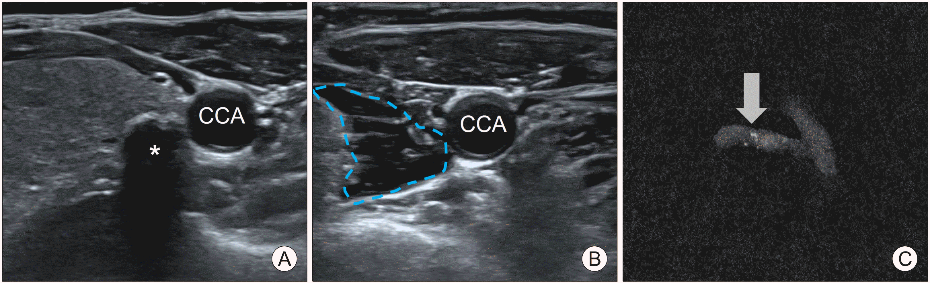 | Fig. 1Anterolateral hydrodissection-assisted core needle biopsy (CNB) of entirely calcified nodule. (A) A 68-year-old female patient presented with an entirely calcified nodule (*) with dimension of 6×7×11 mm on the left side of the thyroid gland with prominent posterior acoustic shadowing. (B) Anterolateral hydrodissection was performed to create a safe margin (dotted lines). CCA: common carotid artery. (C) Two CNB specimens were acquired and underwent subsequent mammography magnification examination, which demonstrated clustered punctate calcifications (arrow). The normalized SI values of calcifications (arrow) were 2.1 and non-calcified portions were 1.5. The pathology confirmed it as adenomatous hyperplasia with microfollicular and macrofollicular proliferation.
|
The second patient (a 57-year-old female) presented with an entirely calcified thyroid nodule on the left side of thyroid gland near the trachea. The calcified nodule had dimensions of 7 mm (anteroposterior)×12 mm (transverse)×13 mm (craniocaudal) with posterior acoustic shadowing (Fig. 2). Successful hydrodissection was performed using a pre-tracheal approach to create a safety margin.9) The trachea comprises several layers of echogenic arcs that may hinder the visualization of the CNB needle. The anechoic background after injection of the lidocaine and saline solution may improve the visibility and maneuverability of the CNB needle for the operator. The mammography evaluation demonstrated conspicuous visibility of the calcification (normalized SI of calcification: 2.2 vs. normalized SI of non-calcified specimen: 1.4). Pathological examination confirmed it to be a probable benign nodule with microfollicular and macrofollicular proliferation, lymphoid aggregates, and calcification.
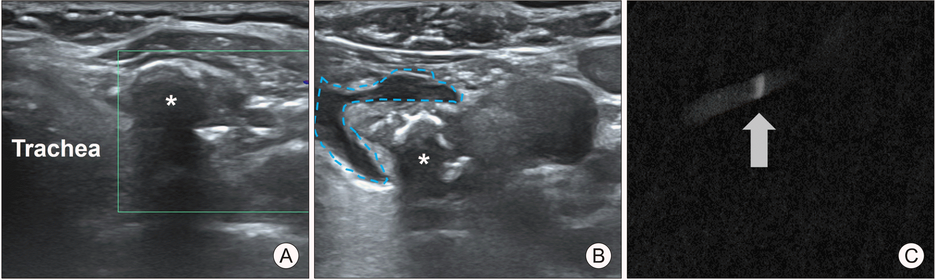 | Fig. 2Pre-tracheal hydrodissection-assisted core needle biopsy (CNB) of entirely calcified nodule. (A) A 57-year-old female patient presented with an entirely calcified nodule (*) with dimension of 7×12×13 mm on the left side of the thyroid gland near the trachea. (B) Pre-tracheal hydrodissection was performed to create safe margin (dotted lines) between the trachea and the target nodule (*). (C) Two CNB specimen were acquired and underwent subsequent mammography magnification examination, which demonstrated a thick calcified line (arrow). The normalized SI values of calcification (arrow) was 2.2 and non-calcified portion was 1.4.
|
The third patient (a 83-year-old female) presented with a large isoechoic nodule with thick rim macrocalcification on the left side of the thyroid gland (Fig. 3). The isoechoic nodule had dimensions of 21 mm (anteroposterior)×24 mm (transverse)×25 mm (craniocaudal) with thick rim macrocalcification. Hydrodissection was not performed in this case because of sufficient clearance from the adjacent anatomical structures. Mammography demonstrated a normalized SI of calcification of 2.0, while that of the non-calcified portion was 1.3. Pathological examination confirmed it to be a probable benign nodule with hyalinized stroma, calcification, and a focal area of macrofollicular proliferation. All three patients underwent successful specimen sampling without immediate CNB-related complications.
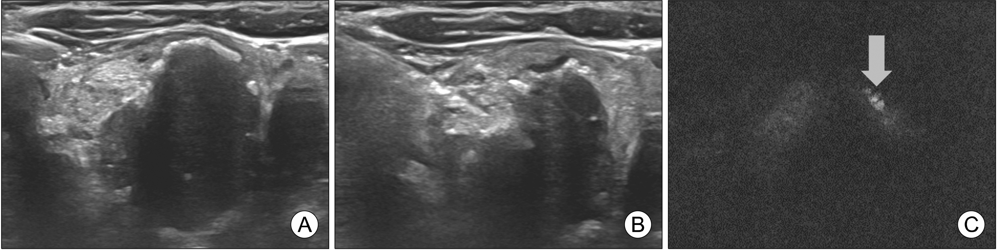 | Fig. 3Core needle biopsy (CNB) of a large isoechoic nodule with thick rim macrocalcification. (A) A 83-year-old female patient presented with a large isoechoic nodule (21×24×25 mm) on the left side of the thyroid with thick rim macrocalcification with prominent posterior acoustic shadowing. (B) Since macrocalcification had enough clearance from the left common carotid artery and trachea, hydrodissection was not performed. Trans-isthmic approach of CNB needle was performed. (C) The mammography demonstrated normalized SI of calcification (arrow) as 2.0, while non-calcified portion as 1.3. The pathology confirmed it as probable benign nodule with hyalinized stroma with calcification and focal area of macrofollicular proliferation.
|
CNB Procedure
All CNBs were performed by a single neuroradiologist with more than five years of biopsy experience. The CNB device used in this study was an 18-gauge automatic spring-powered device with an 11 mm cutting cannula (ACECUT, TSK Laboratory, Tochigi, Japan), and ultrasound device was Canon Aplio i800 (Canon Medical System, Otawara, Japan) with a high-frequency linear transducer.
The procedure began with a careful surveillance of important anatomical structures. Vascular mapping was also conducted using Doppler for safe CNB needle trajectory and planning for hydrodissection. For anesthesia and hydrodissection, 2-3 mL of 1% lidocaine was mixed with 9-10 mL of normal saline solution into a 10 mL syringe.11) After administration of diluted lidocaine at the puncture site, subsequent hydrodissection was performed. Similar to continuous hydrodissection during the radiofrequency ablation, the single-shot (discrete) hydrodissection was intended for three reasons: 1) to alleviate the pain associated with the CNB, 2) to ensure safety margin between important anatomical structures such as common carotid artery, and 3) to enhance visibility of CNB needle during maneuvering and positioning of the cutting canulae. Hydrodissection with little over 10 mL was performed for the first case, while hydrodissection with approximately 7 mL was performed for the second case. The injected solution for hydrodissection drained relatively quickly.
All CNBs were conducted via transisthmic approach, and no incisions were made at the puncture site.8) As the CNB needle approached the target calcified nodule, the cutting cannula was fired using a spring-power mechanism. If the cutting cannula did not penetrate the calcified portion of the lesion, gentle thrust and twisting movements were additionally performed to avoid ricocheting off the calcified portion. Because prominent posterior acoustic shadowing impedes the visualization of the needle, it is necessary to estimate the length of the needle trajectory and the position of the fired cutting cannula. The immediate minor and major complication criteria were evaluated in accordance with the Society of Interventional Radiology Clinical Practice Guidelines.12)
For mammography evaluation, the biopsy specimen was placed on a Petri dish with wet gauge to prevent tissues drying. Mammography was performed using Selenia Dimensions (Hologic, Marlborough, USA). A mammography magnified view with a radiation dose of 26 kVp and 18 mAs was acquired. Pixel SI was measured at the calcification portion and at the non-calcified nodular portion. The SI was then compared, after normalization by dividing it by the background SI.
Go to :
Discussion
This is the first case report describing the hydrodissection-assisted CNB and use of mammography in the biopsy of thyroid nodules with macrocalcifications. Hydrodissection-assisted CNB effectively created a safe distance from important anatomical structures and enhanced the visibility of the CNB needle with requirement of little over 10 mL of lidocaine and saline solution. The merits of mammography for confirming a biopsy specimen are that it is a fast, simple, cost-effective, and definite means to reduce unnecessary rebiopsies and false-negative results. In some institutions, on-site pathologists provide immediate feedback on the biopsy specimen.13) However, for patients without on-site pathologists, alternative confirmation with mammography may be an efficient way to provide feedback on the adequacy of CNB specimens.
Thyroid nodules with macrocalcifications are challenging in terms of performing effective CNB. In the three cases presented here, two were easily penetrated by the CNB device, whereas the first case was partially penetrated. Gentle thrust and twisting motions were applied until a slight advancement of the CNB needle was observed. Even if the CNB needle appears to penetrate the macrocalcification, it is unclear whether correct targeting and proper acquisition of specimens have been performed, partly because of thick posterior acoustic shadowing. For that reason, mammography evaluation of the CNB specimen may be a rapid, efficient means to confirm the correct targeting of the CNB specimen.
For the prospect, a randomized performance of hydrodissection-assisted CNB with mammography confirmation in a large sample population may be necessary to validate the technical efficacy. In conclusion, the case report presented three successful cases of US-guided CNB with mammographic confirmation, in which two cases were performed with hydrodissection. Not only these techniques may offer safe and effective methods to obtain the specimen of thyroid nodules with macrocalcification, but also instill confidence in the biopsy specimen.
Go to :
 XML Download