

 PDF
PDF Citation
 Print
Print


INTRODUCTION
Choledochal cysts (CDC) are congenital conditions involving cystic or fusiform dilatations of the biliary system. They are classified into 5 types by Todani et al. [1]. Multiple intrahepatic and extrahepatic biliary tree dilatations characterize type IV-A CDC. CDC are treated by surgical excision of the extrahepatic bile duct with formation of a Roux-en-Y anastomosis hepaticojejunostomy (HJ). This operation aims to prevent bile stasis and reflux of pancreatic juice into the bile duct by abnormal long pancreaticobiliary channels and remove the common bile duct and gallbladder where malignancy arises [2,3]. This surgery works well in type I CDC, but the persistence of intrahepatic cysts in type IV-A CDC may lead to long-term complications [4]. There is a lack of literature on the long-term outcome of type IV-A CDC after extrahepatic cyst excision. We report a 10-year follow-up of 45 postoperative cases of type IV-A CDC.
CASES
This was a descriptive case series of a prospectively maintained database of patients with type IV-A CDC who underwent resection of the extrahepatic cyst between January 2013 and December 2021 Specific indications for surgery in patients with type IV-A CDC at our center were pain in the abdomen, jaundice, and cholangitis, apart from the development of malignancy. The follow-up period ranged from 2 to 10 years (median, 25 months). Cases with acquired dilatation of the intra and extrahepatic biliary tree induced by a benign or malignant biliary obstruction were excluded. Demographic data were collected from the hospital database. Supplementary information was obtained from patients during follow-up or through telephonic conversation. Institutional clearance was waived because of the retrospective nature of the study. At postoperative follow-up, all patients were assessed by clinical examination, liver function tests (LFTs), and ultrasound to investigate the changes in intrahepatic biliary tracts over 6 and 12 months initially and yearly later. Additional investigations were conducted to detect stenosis and stone, including a computed tomography scan (CT) and/or magnetic resonance cholangiopancreatography (MRCP). Patients with anastomotic stenosis initially underwent percutaneous transhepatic biliary drainage (PTBD) and dilatation. A definitive reoperation was performed if a patient with severe stenosis or hepatolithiasis did not respond to the initial conservative treatment.
Results
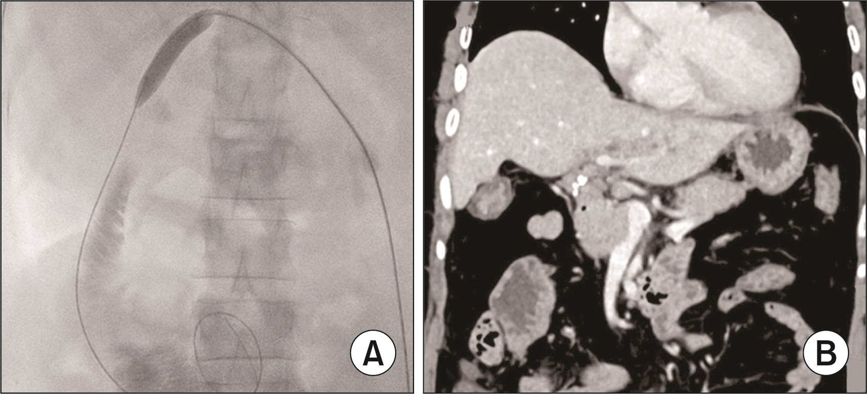
Forty-five patients with type IV-A CDC were included in the final analysis. Their median age was 29 years (range, 15–40 years). Thirty patients remained asymptomatic; however, five had abnormal LFTs, requiring regular monitoring. Late complications in varying combinations were seen in 10 patients, which included cholangitis and/or intrahepatic-hepatic stones in 9 patients, intrahepatic bile duct stenosis with stones in 2 patients, anastomotic stricture with or without stone formation in 6 patients, and left lobar atrophy with intrahepatic stones in 3 patients (Table 1). MRCP and/or CT scans were performed to evaluate the cause of the stricture, which revealed an anastomotic stricture in 6 patients and web-like stenosis of the left intrahepatic bile duct in 2 patients. Percutaneous transhepatic biliary dilatation was performed in 2 patients with anastomotic stricture without cystolithiasis (Fig. 1A). Re-do HJ was required in the remaining four patients. The stenotic web was incised, and HJ was performed with the left intrahepatic duct in both patients. The median time interval from primary surgery to reintervention was 24 months. The median follow-up period after reoperation was five years. Out of 6 patients who required a re-do HJ, three patients had left lobe atrophy with patent HJ anastomosis with a recurrent attack of cholangitis on follow-up at 3, 8, and 10 years (Fig. 1B). Two of them underwent left hepatectomy and refashioning of anastomosis, and the other patient received conservative management. Two patients experienced recurrent episodes of pain in the abdomen with raised amylase and lipase levels. These patients were diagnosed with a residual intrapancreatic cyst on an ultrasonography examination and CT scan of the abdomen. Intrapancreatic cyst excision was performed in both patients. None of the patients developed carcinoma in the intrahepatic cyst during the follow-up period. No mortality was documented.
DISCUSSION
In adults, extrahepatic cyst excision for type IV-A CDC may lead to recurrent complications or malignant transformation in the remaining intrahepatic cyst [5]. Wide anastomosis and smooth bile flow may provide adequate drainage resulting in excellent long-term outcomes in children and infants, and intrahepatic dilatation often regresses with time [6]. Unrecognized stenosis or web in biliary confluence, intrahepatic stones, and anastomotic stricture were the leading causes of postoperative complications in this series.
Anastomotic strictures after cyst excision are more common when the anastomosis is performed below the confluence of the bile duct, especially in type IV-A CDC. This may be due to resistance to the free flow of bile from the dilated intrahepatic cyst and inflammation of the cyst wall above the confluence, leading to frequent attacks of cholangitis [7]. Recurrent cholangitis leads to epithelial mucosal damage in adults causing anastomotic stricture. Therefore, construction of wide HJ (larger than 1 cm) at the confluence with extension to the left duct for free flow of bile, preservation of blood supply of bile ducts, and tension-free anastomosis are critical factors in reducing anastomosis-related complications. An urgent surgical or radiological intervention is mandatory once signs of cholangitis develop in the postoperative period.
PTBD and dilatation of anastomotic strictures are the preferred modes of treatment for anastomotic strictures. However, there are chances of failure of PTBD and dilatation in postoperative strictures associated with CDC. This may be due to persistent inflammation of the remaining intrahepatic cyst and associated intrahepatic membranous web or stenosis. In our series, PTBD and dilatation were possible in 2 cases; the remaining cases required revision surgery.
Congenital stenosis of the hepatic ducts near the confluence is common in patients with type IV-A CDC. If left untreated, even after creation of a wide anastomosis at the confluence after excision of the cyst, bile stasis occurs, leading to frequent attacks of cholangitis and intrahepatic stone formation. Stenosis at the hilum may be membranous or septal. Histological examination of these two types of stenosis showed no difference with the cyst wall. One should be vigilant about this stenosis in intrahepatic ducts during surgery and it should be excised before creating HJ at the confluence [8]. In our series, two patients with cholangitis were found to have intrahepatic stenosis, and both required revision surgery.
A retained intrapancreatic cyst causes secretion of mucin, which may lead to formation of protein plugs at the junction of the biliary and pancreatic ducts, resulting in recurrent attacks of acute pancreatitis. Complete excision of the intrapancreatic cyst is necessary to prevent attacks of recurrent acute pancreatitis, which may ultimately lead to chronic pancreatitis [9]. In our series, 2 patients had recurrent pain in the abdomen during the postoperative follow-up; and on investigations, they were found to have persistence of the intrapancreatic cyst with biochemical evidence of raised amylase and lipase levels.
The incidence of bile duct carcinoma is reported to be 0.7%–5.4% and it is independent of the extent of excision of a CDC [10]. Fortunately, we have not detected intrahepatic cholangiocarcinoma in our series to date.
In our series of 45 patients with type IV-A CDC, 10 (22.2%) had severe complications requiring interventions. Five patients had minor symptoms, such as pain in the abdomen and fluctuating jaundice, which resolved after treatment with antibiotics. The evaluation revealed abnormal LFTs, and therefore, follow-up studies were performed. The remaining 30 patients have been asymptomatic to date. The most likely explanation for this occurrence may be limited involvement of the intrahepatic duct in these patients, and development of symptoms primarily due to the extrahepatic cysts.
All patients with type IV-A CDC do not respond to extrahepatic cyst excision. A tailored surgical approach is necessary. Extrahepatic cyst excision is adequate in cases with minimal involvement of intrahepatic ducts. The risk of cholangiocarcinoma in the remaining ductal system is minimal after complete resection of the extrahepatic cyst. Partial hepatectomy for localized intrahepatic disease, and in rare circumstances, liver transplantation in patients with diffuse involvement and secondary biliary cirrhosis are required for type IV-A CDC [11].
A close follow-up and early intervention are necessary to detect residual intrahepatic cyst complications. Awareness of the possibility of a web at the confluence and wide anastomosis in the hilum extending into the left duct are essential for preventing long-term complications.
 XML Download