Abstract
Backgrounds/Aims
Pancreatoduodenectomy (PD) is recommended in fit patients with a carcinoma (PDAC) of the pancreatic head, and a delayed resection may affect survival. This study aimed to correlate the time from staging to PD with long-term survival, and study the impact of preoperative investigations (if any) on the timing of surgery.
Methods
Data were extracted from the Recurrence After Whipple’s (RAW) study, a multicentre retrospective study of PD outcomes. Only PDAC patients who underwent an upfront resection were included. Patients who received neoadjuvant chemo-/radiotherapy were excluded. Group A (PD within 28 days of most recent preoperative computed tomography [CT]) was compared to group B (> 28 days).
Results
A total of 595 patents were included. Compared to group A (median CT-PD time: 12.5 days, interquartile range: 6–21), group B (49 days, 39–64.5) had similar one-year survival (73% vs. 75%, p = 0.6), five-year survival (23% vs. 21%, p = 0.6) and median time-to-death (17 vs. 18 months, p = 0.8). Staging laparoscopy (43 vs. 29.5 days, p = 0.009) and preoperative biliary stenting (39 vs. 20 days, p < 0.001) were associated with a delay to PD, but magnetic resonance imaging (32 vs. 32 days, p = 0.5), positron emission tomography (40 vs. 31 days, p > 0.99) and endoscopic ultrasonography (28 vs. 32 days, p > 0.99) were not.
Patients with resectable pancreatic head malignancy without metastases may be offered pancreatoduodenectomy (PD) with curative intent. However, only one in seven PD patients with a pancreatic ductal adenocarcinoma (PDAC) is alive five years after their resection [1]. The standard preoperative workup includes a computed tomography (CT) scan of the chest, abdomen and pelvis to accurately stage the disease [2], but some patients require additional preoperative investigations. If distant metastases cannot be ruled out by CT alone, positron emission tomography (PET-CT) is indicated [3]. Further, patients with indeterminate liver lesions may require magnetic resonance imaging (MRI) [4] and those with suspected peritoneal metastases may undergo staging laparoscopy (SL) [5]. In addition, if CT is unable to characterize a pancreatic lesion, or if further information is required from a biopsy or cytology, endoscopic endoscopic ultrasound (EUS) can provide clarity [6]. While preoperative biliary stenting (PBS) is no longer recommended in patients with uncomplicated obstructive jaundice, it is necessary in some patients and PBS complications can delay PD [7]. In some instances, serious complications can result in a surgical resection being canceled altogether.
Since PDAC is an aggressive malignancy [8], there is only a narrow window of opportunity for treatment before the tumor becomes unresectable or metastases develop [9]. Therefore, patients should undergo a timely PD once the decision to offer a resection has been made. However, current British guidelines do not specify the ideal timing of PD [10]. All of the investigations mentioned above have the potential to cause a delay, and patients who experience a significant delay should arguably undergo a repeat preoperative CT to ensure that their disease remains surgically resectable [9], and ensure they do not undergo major surgery which is unlikely to provide a survival benefit [11]. Using a large multicentre cohort of PD patients with PDAC, this study aimed to investigate if a delay in the time from staging CT to resection, as dictated by required further investigations, affected survival. We also aimed to determine if the investigations mentioned above were associated with a delayed resection, and compare radiological and histological staging to investigate concordance.
Patients were included if they underwent PD for histologically-confirmed PDAC at one of twenty-nine participating centers between June 1st, 2012 and May 31st, 2015. The study involved nineteen centers from the UK, three from Spain, two from Italy, and one from Australia, Austria, Mexico, Pakistan, and Sudan (Appendix 1). The end date of May 31st, 2015 was selected so that five-year follow-up data was available for all the included patients (except those who died within five years of PD). The data was collected from physical and electronic patient records at each participating unit and uploaded onto an electronic REDCap (v11.0.3; Nashville, TN, USA) database. The following information was collected: demographics, preoperative investigations, histology results, adjuvant treatment (if any), and five-year outcomes (actual recurrence and survival rates). Data on patient signs/symptoms and preoperative assessment (excluding staging) were unavailable. As the research period was from 2012–2015, both radiological and histological staging were performed using the seventh edition of the TNM classification system [12]. For this sub-study, patients who received neoadjuvant chemotherapy and/or neoadjuvant radiotherapy were removed. Also excluded were those in whom radiological staging was not performed for PDAC (either not recorded or another malignancy was suspected) and those in whom the exact date of the preoperative staging CT was unknown/not recorded.
This study was approved by Greater Manchester South Research Ethics Committee as part of the Recurrence After Whipple’s (RAW) study and University Hospitals Plymouth NHS Trust Research and Development Department (IRAS ID: 280423; REC reference: 20/NW/0397). The study was also supported by the collaborating centers’ research and development departments and adhered to the standards laid down in the Declaration of Helsinki (revised 2013).
The patients were divided into those who underwent PD within 28 days of their most recent preoperative CT (group A) and those who did not (group B). The figure of 28 days was chosen arbitrarily as this seemed reasonable (British guidelines do not advise on the exact timing of PD) [10]. Univariable tests were performed to compare the groups. Distributions were compared using the Mann–Whitney U test, and proportions were compared using Fisher’s exact test. Among those who did not achieve five-year survival, times to death (both time from PD to death, and time from staging CT to death) were compared using the Kaplan-Meier method. The patients were categorized into those who underwent preoperative MRI/PET-CT/EUS/SL/PBS, and those who did not. The groups were compared by median time from CT to PD using the Mann–Whitney U test. Following this, radiological staging results were compared to those obtained after histological staging using the Mann–Whitney U test (distributions) and Fisher’s exact test (all other comparisons). Categorical data are presented as frequency counts, with associated percentages, and continuous data are presented as medians, with interquartile range (IQR). A p-value < 0.05 was considered significant. Analyses were performed using Microsoft Excel (v2013; Microsoft), GraphPad Prism (v9.3.1; GraphPad Software), and IBM SPSS Statistics (v25; IBM Corp.). See table legends for specific details.
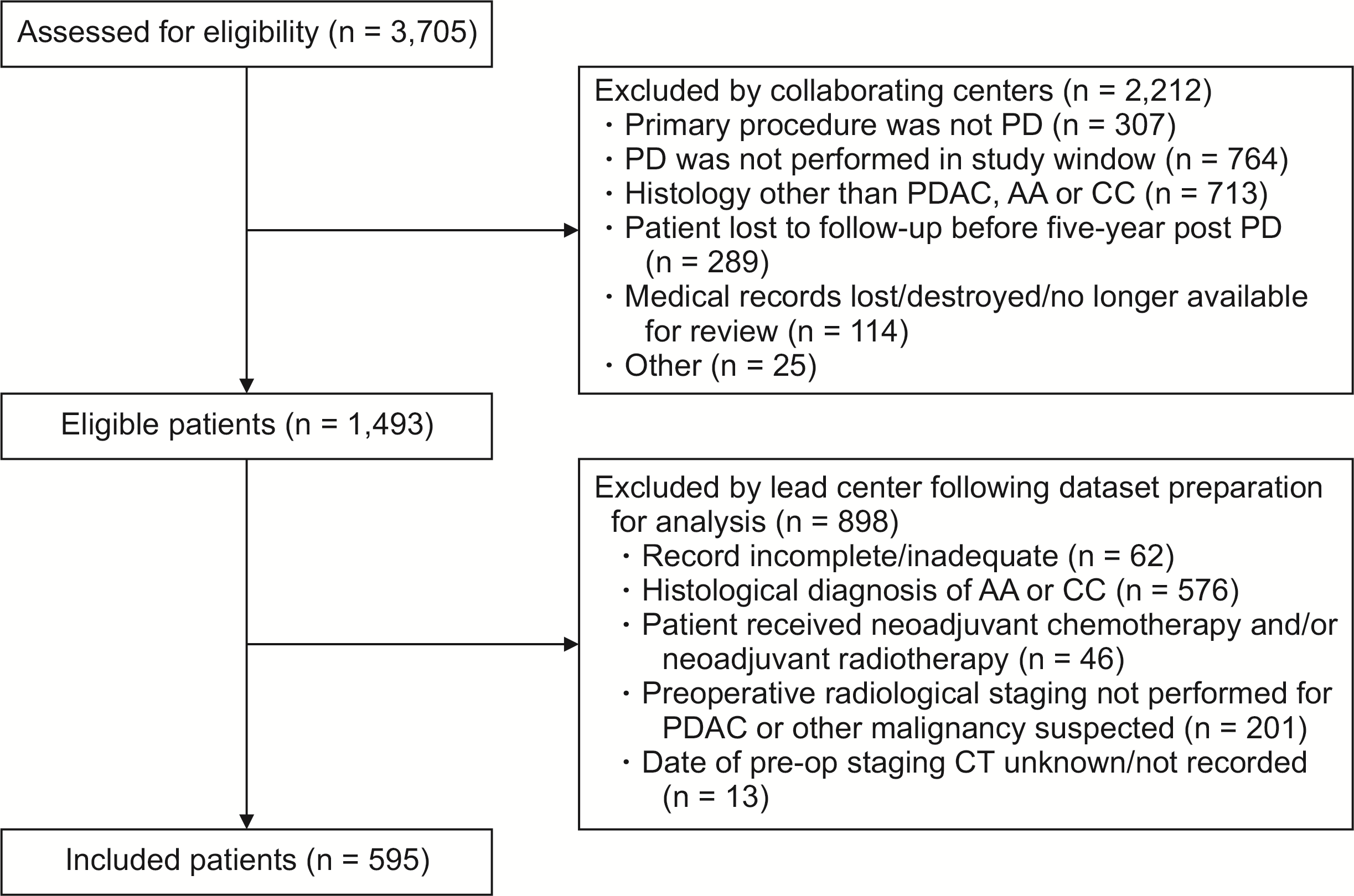
Of the 1,493 potentially eligible patients (Fig. 1), 62 patients were excluded as their records were incomplete, 46 were excluded as they received neoadjuvant chemotherapy and/or radiotherapy, and 201 were excluded because radiological staging was not performed for PDAC. A further thirteen patients were excluded as the date of their most recent preoperative CT was unknown/not recorded. In total, 595 patients were included in the final analysis. The median patient age was 68 years (IQR: 13 years) and 47.7% were female (Table 1). The median tumor size on staging CT was 27 mm (IQR: 14 mm). Concerning radiological staging, 20.2%, 39.5%, 26.2%, 2.1%, and 12.1% were T1, T2, T3, T4, and Tx, respectively, and 65.7%, 27.2%, and 7.1% were N0, N1, and Nx, respectively. The median time from CT to PD was 32 days (IQR: 13–50 days). In total, 25.0%, 4.5%, 33.6%, 10.4%, and 58.3% of patients underwent preoperative MRI (type not specified), PET-CT, EUS, SL, and PBS, respectively. Table 1 outlines the median time from CT to these investigations, and from these investigations to PD. Of those who underwent EUS, 65.6% had a biopsy/cytology sample which confirmed malignancy, 19.4% had a sample that could not confirm a malignancy, and 15.1% had no sample taken. Regarding histological staging, 6.9%, 10.8%, 79.3%, and 2.5% of patients were T1, T2, T3, and T4, respectively, and 22.7% and 76.9% were N0 and N1, respectively. Actual five-year cancer recurrence was 69.9% and actual five-year survival was 21.9%.
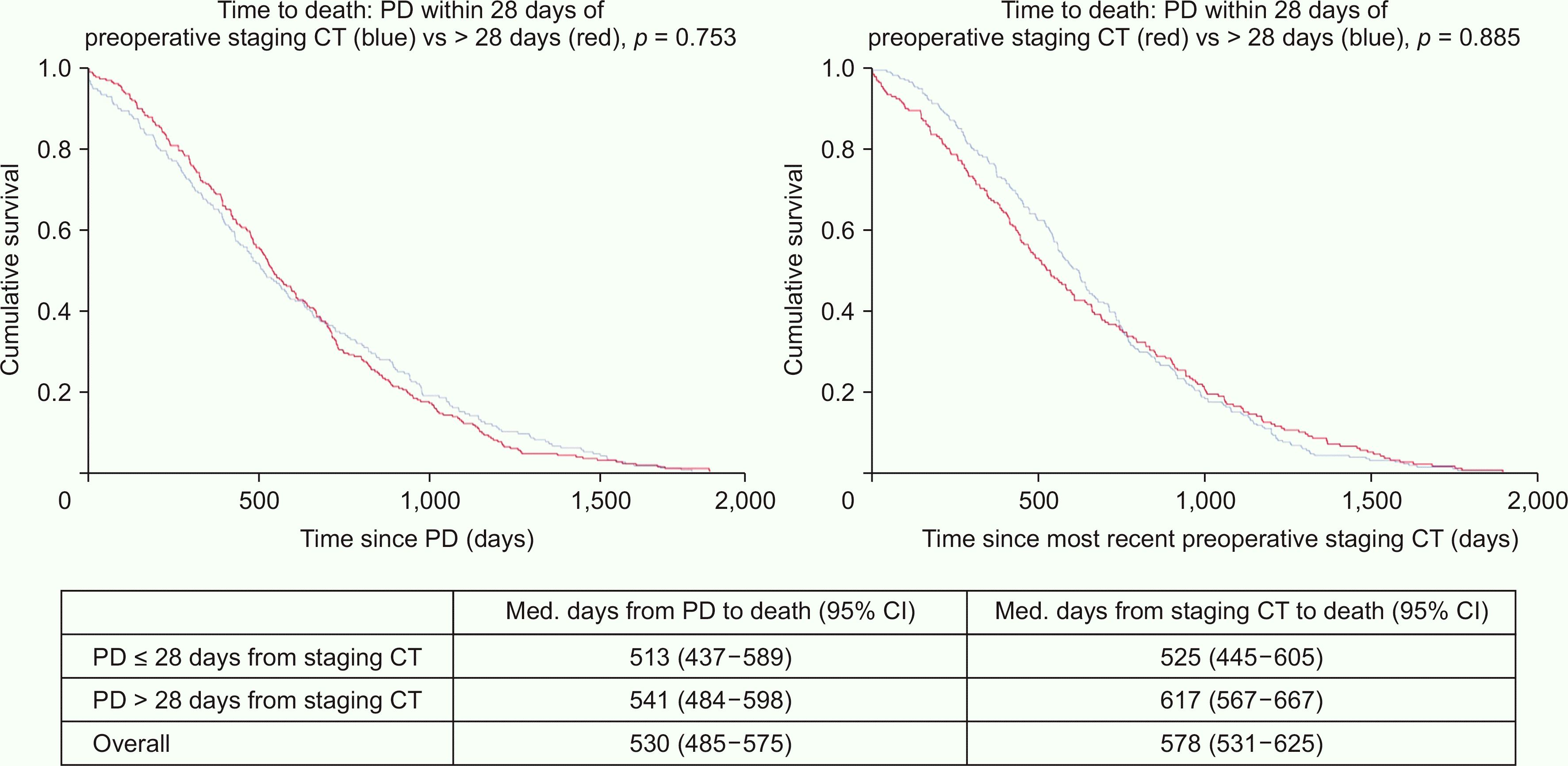
In total, 46% of patients (n = 270) underwent PD within 28 days of their most recent preoperative CT (group A), and 54% (n = 316) did not (group B). These groups exclude nine cases where the exact date of the most recent preoperative CT and PD were not specified (these included only the month and year). The two groups were similar in age, sex, radiological tumor size, and radiological staging (Table 2). Comparable numbers in each group underwent preoperative MRI, PET-CT, and EUS. However, SL (5.9% vs. 13.6%, p = 0.0023) and PBS (69.3% vs. 45.6%, p < 0.001) were significantly more common in group B. Postoperatively, patients in group A were more often T1–2 (25.3% vs. 10.7%, p < 0.001) and had more often undergone a complete (R0) resection (49.2% vs. 38.5%, p = 0.0095). However, actual one-year survival, five-year recurrence and five-year survival rates were similar, as was the median time from PD to death (17 vs. 18 months, p = 0.8) and the median time from the most recent preoperative CT to death (18 vs. 21 months, p = 0.9) (Fig. 2). In addition, the rate of PDAC recurrence was 70% in both groups (p = 0.09).
The patients who underwent preoperative MRI or EUS had similar median times from CT to PD as those who did not (Table 3). Having a PET-CT delayed surgery by a median of nine days, but this was not significant (p = 0.08). SL and PBS were associated with a median increase of 14 (p = 0.009) and 19 days (p < 0.001) to PD, respectively. Table 4 compares the included patients’ preoperative radiological staging to their postoperative histological staging. Following histological staging (vs. radiological), patients were significantly less likely to be T1–2 (17.7% vs. 59.7%) or N0 (22.7% vs. 65.7%, both p < 0.001).
In our multicentre study, patients with PDAC who underwent resection within 28 days of their latest preoperative CT had similar survival to those who did not. This was although an R0 resection was more common among the former. While a complete resection correlates with improved survival, patients who undergo an incomplete resection have similar survival to those with metastatic disease [4]. Hence, patients should be spared PD if a complete resection is unlikely. Patients must be staged promptly so those who might benefit from PD are identified as soon as possible. However, our study suggests that the time from CT to PD is unimportant for long-term outcomes.
While those in the delayed group had more advanced histologic disease and higher rates of incomplete resection (which we would hypothesize was due to the delay), surprisingly, this did not correlate with reduced one- or five-year survival or reduced time to death. Therefore, our data would not support the re-scanning of patients who do not undergo surgery within 28 days of CT. We were surprised by the findings, so we repeated our study using a threshold of 56 days. Again, patients in the “delayed PD” group had similar long-term outcomes to those in the “timely PD” group. It is possible that patients who undergo a timely PD might be at greater risk of very early recurrence if they only undergo CT and not more sensitive modalities. This might help to explain our findings. Future studies which are specifically designed to investigate this phenomenon are required.
British guidelines do not specify when PD should be performed relative to the most recent preoperative CT (hence, we used an arbitrary cut-off of 28 days) [13]. Dutch national guidelines suggest that treatment should occur within three weeks of the multidisciplinary team (MDT) meeting where the decision to offer PD is made. While this is not evidence-based, it would seem a common-sense approach. Having said this, Steen et al. [14], using Dutch national data (n = 2,027), grouped patients (all periampullary tumors) into those who underwent PD within 18 days (33%) of the MDT meeting, those who underwent PD within 19–32 days (33%), and those who waited 33 days or more (34%). The three groups’ overall survival and complete resection rates were similar [14]. The authors concluded that a longer interval between the last MDT meeting and PD did not decrease overall survival. Unfortunately, our dataset did not include MDT meeting dates so we could not make comparisons. In addition, we could not consider the timing of patient’s signs/symptoms as the relevant data were not collected as part of the RAW study.
Since it is less readily available, most patients do not undergo MRI preoperatively. MRI tends to be used as a supplementary modality [15]. A quarter of the patients in our study underwent preoperative MRI. A recent study by Deng et al. [16] (n = 132) showed that the accuracy of MRI for the evaluation of T and N stages was 83% and 74%, respectively, and the sensitivity and specificity of MRI in assessing resectability were 94% and 71%, respectively. Using the eighth edition of the TNM staging classification, no significant differences were observed between the preoperative MRI and postoperative staging findings.
In our study, preoperative MRI did not correlate with a delay to PD. In addition, patients undergoing delayed PD were no more likely to have undergone MRI, if there is diagnostic uncertainty surrounding resectability status, MRI should be considered as this modality can detect liver metastases not seen by conventional CT. As well as characterizing pancreatic lesions with greater detail compared with CT, MRI (along with PET-CT and/or EUS) may have a role for reducing the number of patients diagnosed with very early recurrence following PD for PDAC.
PET-CT can be useful for ruling out metastases before resection [3]. PET-CT is now recommended in those with resectable disease. It has a similar sensitivity to CT and MRI, and significantly higher specificity and accuracy [15]. Prior authors have argued that combining PET-CT with CA19-9 could further enhance its diagnostic efficiency [15]. In a multi-center Chinese study (n = 467), the joint application of PET-CT and CA19-9 significantly enhanced diagnostic efficiency compared with PET-CT alone (sensitivity: 97% vs. 91%, p = 0.0003; specificity: 100% vs. 96%, p = 0.005) [15]. Other recent studies have demonstrated PET-CTs ability to stop patients undergoing PD unnecessarily [3]. In our study, just 5% of patients underwent preoperative PET-CT, and the time from staging CT to PD was a median of nine days longer (median) in this group. Although this was not significant, the small sample size may explain this. Additionally, twice as many patients in group B had undergone preoperative PET-CT. Again, this was not significant, likely due to the small numbers involved.
EUS is an endoscopic imaging modality which can provide high resolution spatial images. It is particularly useful for evaluating small pancreatic lesions because the proximity of the transducer and the pancreas enables magnified imaging, which minimizes the influence of bowel gas and adipose tissue [6]. EUS is more sensitive, specific and accurate than CT in detecting pancreatic lesions [17], particularly small diameter lesions [18]. EUS also provides the option of fine needle aspiration and fine needle biopsy. These techniques can help confirm a diagnosis and rule out malignancy e.g., if there are suspicious liver lesions [6]. Further, in selected patients who become unwell secondary to biliary obstruction, EUS can assist with PBS when conventional techniques have failed [6]. However, EUS is operator-dependent and resource-intensive. In addition, it can also cause complications (e.g., pancreatitis), which might delay PD. Therefore, it should only be carried out when there is a clear indication.
In our study, a third of the included patients underwent EUS before PD. Of these, 77% had a biopsy/cytology sample which confirmed malignancy. The patients with a positive biopsy had a shorter median time from CT to PD (16 vs. 55 days, p < 0.001) and a shorter median time from EUS to PD (20.5 vs. 35 days, p = 0.0003) than those with a negative biopsy/one which could not confirm malignancy. However, actual five-year survival was similar in the two groups (28.1% vs. 25.0%, p = 0.8). EUS was not associated with a delay to PD, and group B patients were no more likely to have undergone EUS.
SL can help to identify hepatic or peritoneal metastases that are not obvious following conventional imaging. This can prevent unnecessary PD and assist with the early initiation of palliative therapy. Suspicious lesions can also be biopsied [5]. Ashraf [5] studied 120 patients who underwent SL before a planned PD and found that the plan for a curative resection changed in five patients. In our study, SL correlated with a median increase in time from CT to PD of 14 days. In addition, a higher proportion of patients in group B had undergone SL. The patients who underwent SL were no more likely to have undergone preoperative MRI (32% vs. 24%, p = 0.2) or EUS (24% vs. 35%, p = 0.1) than those who did not. However, this group had more often undergone PET-CT (10% vs. 4%, p = 0.052) and/or PBS (79% vs. 56%, p = 0.0004). Although the former was not significant, this likely reflects the small sample size. Therefore, the delay in patients who underwent SL can partly be explained by the fact that these patients were more likely to have undergone other preoperative investigations. The SL patients may represent a more complex group. This may partly explain the association with a delay to PD. Unfortunately, we do not know whether frozen section was performed during SL, as our dataset did not include this information.
PBS is now only recommended in patients with a clear indication e.g., those with biliary sepsis or an acute kidney injury secondary to biliary obstruction. Multiple studies have shown that routine PBS is not associated with improved short- or long-term outcomes [7]. In contrast, it correlates with a higher incidence of infectious complications and higher treatment costs [19]. As a result, an upfront surgical approach is preferred. In our study, the patients who underwent PBS had their operation 19 days later (median) than those who did not. In addition, PBS was more common in group B. Those who underwent PBS were no more likely to have undergone MRI (26% vs. 24%, p = 0.7) or PET-CT (5% vs. 4%, p = 0.8), and this group underwent EUS less frequently (28% vs. 41%, p = 0.001). Therefore, other preoperative investigations did not account for the delay associated with PBS.
Our study found a low degree of concordance between preoperative radiological and postoperative histological staging. Patients were more likely to have early disease on the former and more advanced disease on the latter. While the reasoning behind this is unclear, our data might suggest that the radiological staging system used tended to understage PDAC. This highlights the difficulties radiologists face when attempting to characterize lesions using CT alone and underlines the importance of other preoperative investigations which can provide further information and allow a more complete assessment. Indeed, the radiological difficulties associated with TNM staging in pancreatic cancer are well recognized [20]. The TNM classification system has been updated since the research period so the figures obtained are now of limited relevance.
The radiological stage was similar in groups A and B. However, patients in the latter less often had T1 tumors (4% vs. 10%, p = 0.02) and more often had T3 tumors (85% vs. 73%, p = 0.0001). This did not affect long-term outcomes as the actual one- and five-year survival rates were similar. It is unclear why patients who waited longer for their PD had similar survival despite more often having an incomplete resection or a more advanced tumor (both of these are well recognized poor prognostic factors [1]). One potential explanation is that a preoperative delay allows those with low volume metastatic disease to be identified and removed from the resected population. Since our dataset only contained patients who underwent PD, we are not able to explore this further.
Our study has several limitations, as it was originally set up to investigate cancer recurrence patients after PD. Firstly, it has the inherent weaknesses and biases associated with a retrospective study, and there were no controls or comparators. Secondly, a not insignificant proportion of patients had to be excluded for practical reasons (Fig. 1); this is another potential source of bias. Of those excluded after the initial screen, just 18% were removed as they were lost to follow-up or because their clinical records were unavailable (it was impossible to include these records as this data was not collected as part of the RAW study). Although this is a considerable proportion, we have no reason to believe that including these records would have significantly affected our findings. Thirdly, the RAW study was designed to study PD outcomes and the timing of surgery relative to radiological staging was not a primary outcome measure. To perform similar comparisons, we had to exclude many patients form the dataset, e.g., those who received neoadjuvant therapy, those in whom the exact staging CT or PD date was unknown, and those who were initially diagnosed with a suspected ampullary adenocarcinoma or cholangiocarcinoma. However, of the 808 PDAC patients eligible for inclusion, 595 (74%) were included.
Fifth, while we have demonstrated a relationship between SL/PBS and delayed PD, we accept that there are potential confounders. Sixth, since our study was set up to investigate recurrence patterns, all of the included patients underwent PD. Therefore, we could not determine the number of patients who avoided an unnecessary resection. Seventh, our data is historic and practice has evolved since the research period, e.g., staging is now performed using the eighth edition of the TNM classification system and some of the studied investigations, particularly EUS and PET-CT, are now more readily available. However, this does not detract from our core message and we feel our conclusions remain valid. Finally, our dataset did not include the indication for the preoperative investigations performed or whether a frozen section was performed at the time of SL. This was entirely at the discretion of the treating team.
In our multicentre study, PDAC patients who received timely PD (relative to their most recent preoperative CT) had similar survival to those who did not. Our dataset would not support the re-scanning of patients who do not undergo PD within 28 days of CT. SL and PBS both correlated with a delay to PD, but the other studied investigations did not. Preoperative MRI, PET-CT and EUS should be performed whenever there is diagnostic uncertainty to ensure that PD is not carried out inappropriately. The additional information obtained from these investigations may reduce the number of patients diagnosed with very early recurrence following PD.
REFERENCES
1. Russell TB, Labib PLZ, Aroori S. 2023; Five-year follow-up after pancreatoduodenectomy performed for malignancy: a single-centre study. Ann Hepatobiliary Pancreat Surg. 27:76–86. DOI: 10.14701/ahbps.22-039. PMID: 36168824. PMCID: PMC9947371.
2. Tamm EP, Balachandran A, Bhosale PR, Katz MH, Fleming JB, Lee JH, et al. 2012; Imaging of pancreatic adenocarcinoma: update on staging/resectability. Radiol Clin North Am. 50:407–428. DOI: 10.1016/j.rcl.2012.03.008. PMID: 22560689.
3. Ghaneh P, Hanson R, Titman A, Lancaster G, Plumpton C, Lloyd- Williams H, et al. 2018; PET-PANC: multicentre prospective diagnostic accuracy and health economic analysis study of the impact of combined modality 18fluorine-2-fluoro-2-deoxy-d-glucose positron emission tomography with computed tomography scanning in the diagnosis and management of pancreatic cancer. Health Technol Assess. 22:1–114. DOI: 10.3310/hta22070. PMID: 29402376. PMCID: PMC5817411.
4. Pietryga JA, Morgan DE. 2015; Imaging preoperatively for pancreatic adenocarcinoma. J Gastrointest Oncol. 6:343–357. DOI: 10.3978/j.issn.2078-6891.2015.024. PMID: 26261722. PMCID: PMC4502157.
5. Ashraf MI. 2019; Role of staging laparoscopy in patients undergoing pancreaticoduodenectomy. Cureus. 11:e5906. DOI: 10.7759/cureus.5906. PMID: 31777693. PMCID: PMC6853269.
6. Yousaf MN, Chaudhary FS, Ehsan A, Suarez AL, Muniraj T, Jamidar P, et al. 2020; Endoscopic ultrasound (EUS) and the management of pancreatic cancer. BMJ Open Gastroenterol. 7:e000408. DOI: 10.1136/bmjgast-2020-000408. PMID: 32414753. PMCID: PMC7232396.
7. Lee PJ, Podugu A, Wu D, Lee AC, Stevens T, Windsor JA. 2018; Preoperative biliary drainage in resectable pancreatic cancer: a systematic review and network meta-analysis. HPB (Oxford). 20:477–486. DOI: 10.1016/j.hpb.2017.12.007. PMID: 29526466.
8. Arnold M, Abnet CC, Neale RE, Vignat J, Giovannucci EL, McGlynn KA, et al. 2020; Global burden of 5 major types of gastrointestinal cancer. Gastroenterology. 159:335–349.e15. DOI: 10.1053/j.gastro.2020.02.068. PMID: 32247694. PMCID: PMC8630546.
9. Raman SP, Reddy S, Weiss MJ, Manos LL, Cameron JL, Zheng L, et al. 2015; Impact of the time interval between MDCT imaging and surgery on the accuracy of identifying metastatic disease in patients with pancreatic cancer. AJR Am J Roentgenol. 204:W37–42. DOI: 10.2214/AJR.13.12439. PMID: 25539271. PMCID: PMC4872713.
10. O'Reilly D, Fou L, Hasler E, Hawkins J, O'Connell S, Pelone F, et al. 2018; Diagnosis and management of pancreatic cancer in adults: a summary of guidelines from the UK National Institute for Health and Care Excellence. Pancreatology. 18:962–970. DOI: 10.1016/j.pan.2018.09.012. PMID: 30292643.
11. Niesen W, Hank T, Büchler M, Strobel O. 2019; Local radicality and survival outcome of pancreatic cancer surgery. Ann Gastroenterol Surg. 3:464–475. DOI: 10.1002/ags3.12273. PMID: 31549006. PMCID: PMC6749949.
12. Cong L, Liu Q, Zhang R, Cui M, Zhang X, Gao X, et al. 2018; Tumor size classification of the 8th edition of TNM staging system is superior to that of the 7th edition in predicting the survival outcome of pancreatic cancer patients after radical resection and adjuvant chemotherapy. Sci Rep. 8:10383. DOI: 10.1038/s41598-018-28193-4. PMID: 29991730. PMCID: PMC6039534.
13. National Institute for Health and Care Excellence. Pancreatic cancer in adults: diagnosis and management. National Institute for Health and Care Excellence (NICE);2018. DOI: 10.1016/j.pan.2018.09.012.
14. Steen MW, van Rijssen LB, Festen S, Busch OR, Groot Koerkamp B, van der Geest LG, et al. 2020; Impact of time interval between multidisciplinary team meeting and intended pancreatoduodenectomy on oncological outcomes. BJS Open. 4:884–892. DOI: 10.1002/bjs5.50319. PMID: 32841533. PMCID: PMC7528524.
15. Huang S, Chong H, Sun X, Wu Z, Jia Q, Zhang Y, et al. 2021; The Value of 18F-FDG PET/CT in diagnosing pancreatic lesions: comparison With CA19-9, enhanced CT or enhanced MR. Front Med (Lausanne). 8:668697. DOI: 10.3389/fmed.2021.668697. PMID: 34692714. PMCID: PMC8531126.
16. Deng Y, Ming B, Wu JL, Zhou T, Zhang SY, Chen Y, et al. 2020; Magnetic resonance imaging for preoperative staging of pancreatic cancer based on the 8(th) edition of AJCC guidelines. J Gastrointest Oncol. 11:329–336. DOI: 10.21037/jgo.2020.03.06. PMID: 32399274. PMCID: PMC7212112.
17. Kamata K, Kitano M, Kudo M, Sakamoto H, Kadosaka K, Miyata T, et al. 2014; Value of EUS in early detection of pancreatic ductal adenocarcinomas in patients with intraductal papillary mucinous neoplasms. Endoscopy. 46:22–29. DOI: 10.1055/s-0034-1367539. PMID: 24218310.
18. Sakamoto H, Kitano M, Suetomi Y, Maekawa K, Takeyama Y, Kudo M. 2008; Utility of contrast-enhanced endoscopic ultrasonography for diagnosis of small pancreatic carcinomas. Ultrasound Med Biol. 34:525–532. DOI: 10.1016/j.ultrasmedbio.2007.09.018. PMID: 18045768.
19. Morris S, Gurusamy KS, Sheringham J, Davidson BR. 2015; Cost-effectiveness of preoperative biliary drainage for obstructive jaundice in pancreatic and periampullary cancer. J Surg Res. 193:202–209. DOI: 10.1016/j.jss.2014.07.060. PMID: 25172090. PMCID: PMC4274324.
20. Sahani DV, Shah ZK, Catalano OA, Boland GW, Brugge WR. 2008; Radiology of pancreatic adenocarcinoma: current status of imaging. J Gastroenterol Hepatol. 23:23–33. DOI: 10.1111/j.1440-1746.2007.05117.x. PMID: 18171340.
Table 1
Variable | Value |
---|---|
Median age in years (IQR) | 68 (13) |
Female sex | 284 (47.7) |
Median tumor size on pre-op CT in mm (IQR) | 27 (14) |
Unknown/not recorded | 162 (27.2) |
Radiological T stage on pre-op CT (unknown/not recorded in 15 cases*) | |
T1 | 117 (20.2) |
T2 | 229 (39.5) |
T3 | 152 (26.2) |
T4 | 12 (2.1) |
Tx | 70 (12.1) |
Radiological N stage on pre-op CT (unknown/not recorded in 15 cases*) | |
N0 | 381 (65.7) |
N1 | 158 (27.2) |
Nx | 41 (7.1) |
Median time from CT to PD in days (IQR) | 32 (38) |
Underwent pre-op MRI | 149 (25.0) |
Median time from CT to MRI in days (IQR) | 4 (15) |
Median time from MRI to PD in days (IQR) | 34 (40.5) |
Underwent pre-op PET-CT | 27 (4.5) |
Median time from CT to PET-CT in days (IQR) | 20 (13) |
Median time from PET-CT to PD in days (IQR) | 27.5 (36) |
Underwent pre-op EUS | 200 (33.6) |
Did the EUS biopsy/cytology sample confirm malignancy? (n = 200; unknown: 14*) | |
Yes | 122 (65.6) |
No | 36 (19.4) |
No biopsy/cytology sample taken | 28 (15.1) |
Median time from CT to EUS in days (IQR) | 13 (27) |
Median time from EUS to PD in days (IQR) | 27 (36) |
Underwent pre-op staging laparoscopy | 62 (10.4) |
Median time from CT to staging lap. in days (IQR) | 29.5 (20) |
Median time from staging lap. to PD in days (IQR) | 9.5 (17) |
Underwent PBS | 347 (58.3) |
Median tumor size in mm (IQR) | 30 (13) |
Histological T stage (unknown/not recorded in 2 cases*) | |
T1 | 41 (6.9) |
T2 | 64 (10.8) |
T3 | 470 (79.3) |
T4 | 15 (2.5) |
Tx | 3 (0.5) |
Histological N stage (unknown/not recorded in 1 case*) | |
N0 | 135 (22.7) |
N1 | 457 (76.9) |
Nx | 2 (0.3) |
Resection margin (R) status (unknown/not recorded in 45 cases*) | |
R0 | 241 (43.8) |
R1 | 289 (52.5) |
R2 | 20 (3.6) |
Actual five-year cancer recurrence (unknown/not recorded in 1 case*) | 415 (69.9) |
Actual five-year survival (unknown/not recorded in 1 case*) | 130 (21.9) |
Table 2
Variable | Timely PD | Delayed PD | p-value |
---|---|---|---|
Number of patients | 270 | 316 | - |
Median age in years (IQR) | 68 (12) | 68 (13) | 0.476 |
Female sex | 139 (51.5) | 140 (44.3) | 0.097 |
Radiological tumor size in mm (IQR) | 27 (15) | 26 (14) | 0.472 |
Radiological T stage | |||
T1 | 58 (21.6) | 58 (19.1) | 0.468 |
T2 | 108 (40.1) | 115 (38.0) | 0.606 |
T3 | 71 (26.4) | 81 (26.7) | > 0.999 |
T4 | 9 (3.3) | 3 (1.0) | 0.076 |
Tx | 23 (8.6) | 46 (15.2) | - |
Unknown | 1a) | 13a) | - |
Radiological N stage | |||
N0 | 179 (66.8) | 194 (64.0) | 0.538 |
N1 | 71 (26.5) | 87 (28.7) | 0.575 |
Nx | 18 (6.7) | 22 (7.3) | - |
Unknown | 2a) | 13a) | - |
Underwent pre-op MRI | 66 (24.4) | 79 (25.0) | 0.924 |
Underwent pre-op PET-CT | 8 (3.0) | 19 (6.0) | 0.112 |
Underwent pre-op EUS | 99 (36.7) | 98 (31.0) | 0.161 |
Underwent pre-op staging laparoscopy | 16 (5.9) | 43 (13.6) | 0.0023* |
Underwent PBS | 123 (45.6) | 219 (69.3) | < 0.001* |
Histological tumor size in mm (IQR) | 30 (13) | 30 (15) | 0.0101* |
Histological T stage | |||
T1 | 26 (9.7) | 14 (4.4) | 0.0139* |
T2 | 42 (15.6) | 20 (6.3) | 0.0004* |
T3 | 195 (72.5) | 269 (85.4) | 0.0001* |
T4 | 5 (1.9) | 10 (3.2) | 0.433 |
Tx | 1 (0.4) | 2 (0.6) | - |
Unknown | 1a) | 1a) | - |
Histological N stage | |||
N0 | 70 (25.9) | 63 (20.0) | 0.093 |
N1 | 199 (73.7) | 251 (79.7) | 0.095 |
Nx | 1 (0.4) | 1 (0.3) | - |
Unknown | 0 (0) | 1a) | - |
Resection margin (R) status | |||
R0 | 127 (49.2) | 110 (38.5) | 0.0095* |
R1 | 120 (46.5) | 167 (58.4) | 0.010* |
R2 | 11 (4.3) | 9 (3.1) | 0.502 |
Unknown | 12a) | 30a) | - |
Actual one-year survival | 197 (73.0) | 238 (75.3) | 0.570 |
Actual five-year recurrence | 190 (70.4) | 219 (69.3) | 0.857 |
Actual five-year survival | 63 (23.3) | 65 (20.6) | 0.558 |
Nine patients had to be excluded from this sub-analysis as their exact CT date was unknown (only the month of the scan was known). Where data were missing, cases were excluded from the relevant sub-analyses (see Table 1). Statistics: Mann–Whitney U test: medians/distributions, Fisher’s exact test: proportions.
Table 3
Investigation/intervention | Median time (days) from CT to PD: investigation vs. none (IQR) | p-value |
---|---|---|
MRI | 32 (37) vs. 31 (37) | 0.548 |
PET-CT | 40 (27) vs. 31 (37) | 0.084 |
Endoscopic ultrasound | 28 (38) vs. 32 (37.5) | 0.989 |
Staging laparoscopy | 43 (27) vs. 29.5 (36.5) | 0.00896* |
Pre-op biliary stenting | 39 (33) vs. 20 (32) | < 0.001* |