Abstract
Background
Paradoxical responses (PR) occur more frequently in lymph node tuberculosis (LNTB) than in pulmonary tuberculosis and present difficulties in differential diagnosis of drug resistance, new infection, poor patient compliance, and adverse drug reactions. Although diagnosis of mediastinal LNTB has become much easier with the development of endosonography, limited information is available. The aim of this study was to investigate the clinical course of mediastinal LNTB and the risk factors associated with PR.
Methods
Patients diagnosed with mediastinal LNTB via endosonography were evaluated retrospectively between October 2009 and December 2019. Multivariable logistic regression was applied to evaluate the risk factors associated with PR.
Results
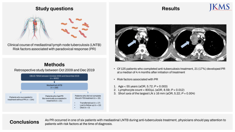
Of 9,052 patients who underwent endosonography during the study period, 158 were diagnosed with mediastinal LNTB. Of these, 55 (35%) and 41 (26%) concurrently had pulmonary tuberculosis and extrapulmonary tuberculosis other than mediastinal LNTB, respectively. Of 125 patients who completed anti-tuberculosis treatment, 21 (17%) developed PR at a median of 4.4 months after initiation of anti-tuberculosis treatment. The median duration of anti-tuberculosis treatment was 6.3 and 10.4 months in patients without and with PR, respectively. Development of PR was independently associated with age < 55 years (adjusted odds ratio [aOR], 5.72; 95% confidence interval [CI], 1.81–18.14; P = 0.003), lymphocyte count < 800/μL (aOR, 8.59; 95% CI, 1.60–46.20; P = 0.012), and short axis diameter of the largest lymph node (LN) ≥ 16 mm (aOR, 5.22; 95% CI, 1.70–16.00; P = 0.004) at the time of diagnosis of mediastinal LNTB.
Graphical Abstract
Extrapulmonary tuberculosis (EPTB) is an infectious disease in which Mycobacterium tuberculosis (MTB) affects organs other than the lungs. EPTB accounted for 17% of the 6.4 million reported cases in 2021, ranging from 8% in the Western Pacific Region to 23% in the Eastern Mediterranean Region.1 Lymph node tuberculosis (LNTB) is one of the most common presentations of EPTB, accounting for approximately 20–40% of all cases.23 The majority of previous studies on LNTB analyzed peripheral LNTB rather than mediastinal LNTB because peripheral lymph nodes (LNs) are easier to recognize and access.2
A paradoxical response (PR) during anti-TB treatment refers to worsening of pre-existing disease or development of new lesions in patients who have been on anti-tuberculosis (TB) treatment for at least 2 weeks, excluding documented relapse or presence of another disease.4 Most cases of PR in patients with peripheral LNTB manifest as enlarged LNs, new affected nodes, pain, and sinus draining between three weeks and four months after anti-TB treatment begins.56 This phenomenon demonstrates the importance of differential diagnosis from drug resistance, new infection, poor patient compliance, and adverse drug reactions. Moreover, a PR occurs relatively commonly, in approximately 20% of patients with peripheral LNTB during anti-TB treatment, compared with less than 5% of cases of pulmonary TB in human immunodeficiency virus (HIV)-negative patients.4567
Although diagnosis of mediastinal LNTB has become much easier with the development of endobronchial ultrasound-guided transbronchial needle aspiration (EBUS-TBNA), limited information is available.8 The aim of this study was to investigate the clinical course of mediastinal LNTB and the risk factors associated with PR.
This was a retrospective observational study of patients with mediastinal LNTB. We reviewed EBUS-TBNA registry data collected prospectively between October 2009 and December 2019 at Samsung Medical Center, a tertiary hospital in the Republic of Korea. This study included all patients diagnosed with mediastinal LNTB via EBUS-TBNA.
Mediastinal LNTB was diagnosed when a patient met one or more of the following: (a) a positive culture result for MTB from the rinse fluid of the EBUS needle and/or core tissue of the EBUS-TBNA, (b) a positive polymerase chain reaction (PCR) result for MTB from the rinse fluid and/or core tissue, or (c) histologically confirmed LNTB with granulomatous inflammation with caseating necrosis with or without a positive result of nested PCR.
Details of the EBUS-TBNA procedure were described in our previous reports.910 We selected the target LNs based on imaging studies, including chest computed tomography (CT) and integrated positron emission tomography–CT (PET), if available. Nodal sampling was performed on the short diameter of LNs ≥ 5 mm in EBUS after systematic inspection of mediastinal and hilar LNs. We conducted three passes per node when possible or at least two passes when core tissue was obtained. During the study period, rapid on-site cytology and elastography were not available. If necessary, bronchial washing was performed during the EBUS session.
Core tissue was spread on filter paper to absorb excess blood, fixed in 10% formalin, and sent for histopathologic examination. If TB was suspected while pathologists were examining the tissue, they performed nested PCR analysis (Seeplex MTB Nested ACE Detection; Seegene, Seoul, Korea) using formalin-fixed, paraffin-embedded tissue specimens.11 Immediately after obtaining the core tissue, we collected one bottle of rinse fluid of the EBUS needle for each patient by flushing 1 cc of sterile normal saline through the needle. The rinse fluid was sent for acid-fast bacilli (AFB) staining, culturing, and an MTB PCR assay.12 If the operator strongly suspected mediastinal LNTB, some core tissue was immersed in normal saline and subjected to AFB staining, culturing, and MTB PCR assay.
The AFB smears were examined using an auramine-rhodamine fluorescent stain, followed by confirmation with Ziehl-Neelsen staining. The results were graded according to guidelines, and more than 1+ (1 to 9 bacilli in 100 fields) were defined as smear-positive.1314 The AFB were cultured with both 3% Ogawa solid medium (Shinyang, Seoul, Korea) and liquid broth medium (mycobacterial growth indicator tube [MGIT] 960 system; Becton Dickinson, Sparks, MD, USA) at our institution beginning in January 2009.15 To confirm MTB in samples with positive culture, MPT64 antigen testing (SD BIOLINE TB Ag MPT64 Rapid; Standard Diagnostics Inc., Yongin, Korea) and/or a PCR test using the MTB-ID V3 kit (YD Diagnostics, Yongin, Korea) or the GENEDIA MTB/NTM Detection Kit (GENEDIA MTB/NTM; Green Cross Medical Science Corp., Eumseong, Korea) were carried out. MTB PCR assays were performed using the COBAS TaqMans MTB test (Roche Diagnostics, Basel, Switzerland) or the GENEDIA MTB Detection Kit (GENEDIA assay; Green Cross Medical Science Corp.). The first identified MTB isolate for each patient was subjected to a drug susceptibility test (DST) based on the absolute concentration method using a solid Löwenstein-Jensen medium and on liquid media using an MGIT 960 system.16
Initial anti-TB treatment began with a combination of isoniazid, rifampin, ethambutol, and pyrazinamide. When the DST results were confirmed, anti-TB regimens were adjusted accordingly. All patients underwent initial plain chest radiography and chest CT. We counted the number of LNs sampled, suspected of TB, and confirmed of TB. The definitions for these are in the footnotes of Supplementary Table 1. The largest LN was determined based on the short axis diameter in the CT scan conducted at the time of diagnosis.17 Both the short and long axis diameters of these nodes were measured. A plain chest radiograph was obtained monthly, and chest CT scans were performed 2–3 months after treatment initiation to evaluate treatment response. A chest CT was performed when symptoms of suspected PR or deterioration findings on plain chest radiography were observed.
Treatment success for mediastinal LNTB was evaluated by the clinical judgment of the attending physician based on improvement in symptoms and reduction in LN size.18 In the case of coexisting pulmonary TB, treatment success was defined as negative results of sputum smear and culture at 5 months after treatment.19 In addition, treatment success was defined as completion of the prescribed course of treatment and no signs of failure.20
PR was defined as worsening of a pre-existing disease or development of new lesions in a patient who has been on appropriate anti-TB treatment for at least 2 weeks with exclusion of a documented relapse or presence of another disease.46 Worsening of LNs was defined as a 20% or greater increase in short axis diameter on the contrast-enhanced CT compared to the baseline measurement.17 The time to onset of PR was defined as the interval between initiation of anti-TB treatment and detection of PR. Treatment for PR included adding anti-TB drugs, extending the treatment period, and adding steroids according to the attending physicians.
To analyze the risk factors of PR in patients with mediastinal LNTB, we classified patients by clinical outcome: (a) patients who succeeded in treatment without PR, (b) patients who had PR but eventually succeeded in treatment, and (c) patients who did not complete anti-TB treatment.
Data are reported as number (percentage) for categorical variables and as median with interquartile range (IQR) for continuous variables. Data were compared using the χ2 test or Fisher’s exact test for categorical variables and the Mann–Whitney U test for continuous variables. After excluding patients who did not complete anti-TB treatment, multivariable logistic regression analysis with a backward stepwise selection with P < 0.05 for entry and P > 0.10 for removal was used to identify the risk factors for PR. In logistic regression analysis, continuous variables were entered after converting to binary variables using the highest Youden’s index (optimal cut-off values) or clinically meaningful cut-off values to provide more useful information to clinicians.21 All tests were two-sided, and a P-value < 0.05 was considered significant. All statistical analyses were performed using SPSS software (IBM SPSS Statistics ver. 27; IBM Corp., Chicago, IL, USA).
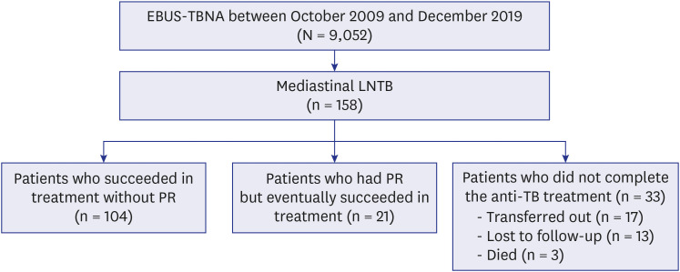
Of the 9,052 patients who underwent EBUS-TBNA during the study period, 158 were diagnosed with mediastinal LNTB, and all were negative for HIV (Fig. 1). Of these, 33 patients (21%) did not complete anti-TB treatment because they transferred to another hospital (n = 17), were lost to follow-up (n = 13), or died (n = 3). Of 125 patients who completed anti-TB treatment, PR occurred in 21.
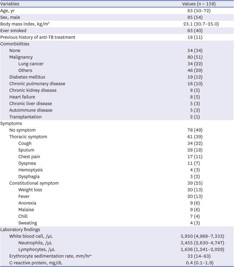
The baseline characteristics of patients diagnosed with mediastinal LNTB are presented in Table 1. The median age was 63 years (IQR, 50–72) and 85 (54%) were male. There were 18 patients (11%) with a history of previous anti-TB treatment. Malignancy (51%) was the most common comorbidity, and only 54 (34%) patients had no comorbidities. At the time of diagnosis, 78 (49%) had no symptom, 61 (39%) had thoracic symptoms including cough and sputum, and 39 (25%) had constitutional symptoms such as weight loss or fever. In laboratory results, the median counts of leukocytes and lymphocytes were 5,950/μL (IQR, 4,968–7,333) and 1,636/μL (IQR, 1,241–2,029), respectively.
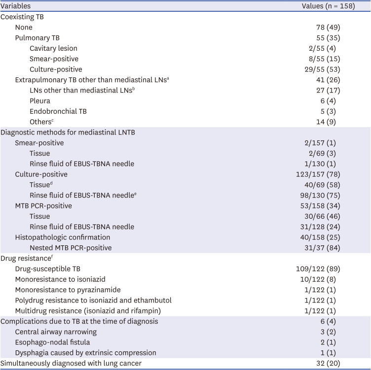
The baseline disease status of patients with mediastinal LNTB is listed in Table 2. Of the 158 patients, 55 (33%) and 41 (26%) concurrently had pulmonary TB and EPTB other than mediastinal LNTB, respectively. The most common sites of EPTB were LNs other than mediastinal LNs (n = 27, 17%). Of 157 patients tested with AFB stain and culture, only two (1%) had positive results on AFB stain and 123 (78%) had positive results on AFB culture. Of 158 patients tested with MTB PCR and histopathologic examination, 53 (34%) and 40 (25%) had positive results, respectively. Eventually, 78 patients (49%) could only be diagnosed with tests of rinse fluid for an EBUS needle. DST was performed on all culture-positive patients, except one whose MTB isolates were contaminated. Of these 122 patients, 109 (89%) were identified as having drug-susceptible TB, 10 (8%) were monoresistant to isoniazid, and 1 (1%) was multidrug resistant. There were 6 local complications (4%) at the time of diagnosis. All three patients with central airway narrowing presented with cough and dyspnea, however their symptoms and CT findings improved solely with anti-TB treatment. One of the two patients who had a esophago-nodal fistula at the time of diagnosis was asymptomatic and was transferred immediately after diagnosis. The other had dysphagia, however it improved solely with anti-TB treatment. One patient with dysphagia caused by extrinsic compression died of pneumonia on the 123rd day of anti-TB treatment. There were two more deaths, all of which had hematological malignancies, and died of pneumonia and cancer progression, respectively, within one month of treatment. Lung cancer was diagnosed simultaneously in 20% of patients.
Data are reported as number (%).
LNTB = lymph node tuberculosis, TB = tuberculosis, LN = lymph node, EBUS-TBNA = endobronchial ultrasound-guided transbronchial needle aspiration, MTB = Mycobacterium tuberculosis, PCR = polymerase chain reaction.
aOf 52 extrapulmonary manifestations in 41 patients, 26 (50%) were diagnosed by imaging and the remaining half were diagnosed by invasive procedures (body fluid aspiration = 11, LN biopsy = 10, and bronchoscopy = 5).
bCervical (n = 27), abdominal (n = 4), and axillary (n = 1) LNs.
cPericardium (n = 4), bone (n = 3), liver (n = 2), spleen (n = 2), soft tissue (n = 2), peritoneum (n = 1), bone marrow (n = 1), prostate (n = 1), and meninges (n = 1).
dPositive results from liquid medium (n = 36) and solid medium (n = 23).
ePositive results from liquid medium (n = 87) and solid medium (n = 57).
fDrug susceptibility testing was not available in one patient whose MTB isolates were contaminated.
Most commonly sampled LNs were 4R (right lower paratracheal, n = 107) and 7 (subcarinal, n = 100) (Supplementary Table 1). Most commonly TB-suspected and confirmed LNs were also 4R and 7.
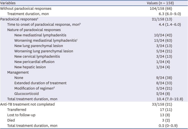
Table 3 lists the clinical outcomes. For the 104 patients who succeeded in treatment without PR, the median duration of anti-TB treatment was 6.3 months (IQR, 6.0–9.0). Of the 125 patients who were followed until the end of anti-TB treatment, 21 (17%) experienced a total of 24 PR events. Considering only the first PR event in each patient, the median onset timing of PR was 4.4 months (IQR, 1.4–6.0) after initiation of anti-TB treatment. The most commonly observed nature associated with PR was worsening mediastinal lymphadenitis (15/24, 63%). As management for these PR events, attending physicians extended the treatment period in 8 (33%) patients, modified the regimen in 5 (21%), and added oral glucocorticoid in 2 (8%). Eventually, the median duration of anti-TB treatment in these 21 patients with PR events was 10.4 months (IQR, 7.0–12.8). Of 33 patients who did not complete the anti-TB treatment, 15 patients were transferred or lost to follow-up at the initiation of treatment. The remaining 18 patients were treated for median 0.9 months (IQR, 0.5–1.9) at our hospital, and no PR was identified during this period.
Data are reported as median (interquartile range) and number (%).
TB = tuberculosis.
aThree patients experienced two separate paradoxical responses. A total of 24 paradoxical responses was analyzed and presented.
bA total of 21 cases were analyzed, excluding the three cases of recurrent paradoxical response.
cWorsening lymphadenitis was defined as a 20% or greater increase in short axis diameter compared to the baseline value.
dPyrazinamide (n = 4) or moxifloxacin (n = 1) was added to maintenance regimen.
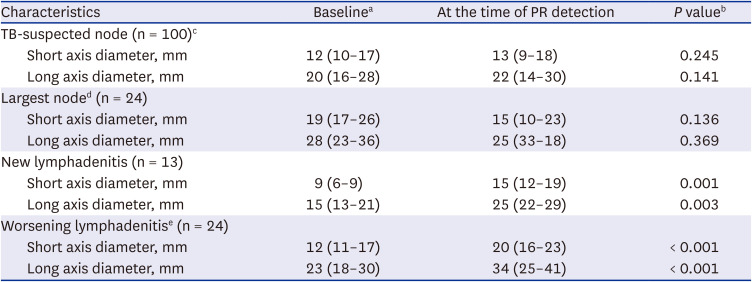
Table 4 presents a comparative analysis of TB-suspected LNs at the baseline and at the time of PR detection. Notably, no statistically significant changes were observed in the short and long axis diameters of TB-suspected LNs or the largest LNs between the baseline and the PR. However, new lymphadenitis that occurred during PR exhibited significant increases in both short (9 to 15 mm, P = 0.001) and long axis diameters (15 to 25 mm, P = 0.003) of the LNs compared to the baseline. Similarly, worsening LNs demonstrated notable increases in short (12 to 20 mm, P < 0.001) and long axis diameters (23 to 34 mm, P < 0.001) between the two phases. In only 9 of a total of 24 PR events, invasive procedures were performed to rule out drug resistance and other diseases (EBUS-TBNA to mediastinal LN = 4, fine needle aspiration to neck LN = 2, bronchoscopic biopsy to endobronchial lesion = 1, liver biopsy = 1, and surgical lung biopsy = 1). None of these 9 events were diagnosed with drug resistance or other diseases. Moreover, all patients with PR events demonstrated clinical and radiological improvements while maintaining anti-TB treatment, conclusively confirming the diagnosis of PR. Fig. 2 illustrates a representative case of a PR.
Data are reported as median (interquartile range) number (%).
LN = lymph node, PR = paradoxical response, TB = tuberculosis, CT = computed tomography, LNTB = lymph node tuberculosis.
aCT images at the time of mediastinal LNTB diagnosis were analyzed as baseline. For the second PR of three patients who had two PRs, the CT immediately before the second episode of PR was analyzed as baseline.
bData were compared using the Wilcoxon signed-rank test.
cThis includes (1) cases in which necrosis was seen inside the LN on contrast-enhanced CT, or (2) the short diameter of the LN was 10 mm or more, even though no biopsy was performed, and (3) cases confirmed as TB as a result of the biopsy.
dThe largest LN was determined based on the short axis diameter.
eWorsening lymphadenitis was defined as a 20% or greater increase in short axis diameter compared to the baseline value.
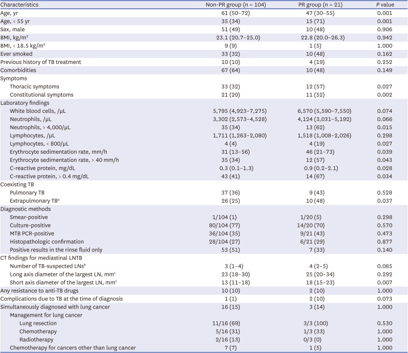
Compared with patients without PR, patients with PR were younger (47 vs. 61 years, P = 0.001), had more frequent thoracic (57% vs. 32%, P = 0.027) and constitutional (52% vs. 20%, P = 0.002) symptoms, lower level of lymphocytes (< 800/µL; 19% vs. 4%, P = 0.027), higher neutrophil level (> 4,000/µL; 62% vs. 34%, P = 0.015), higher erythrocyte sedimentation rate (> 40 mm/h; 57% vs. 34%, P = 0.043), higher C-reactive protein level (> 0.4 mg/dL; 67% vs. 41%, P = 0.034), more frequent EPTB other than mediastinal LNTB (48% vs. 25%, P = 0.037), and greater short axis diameter of the largest LN (18 vs. 13 mm, P = 0.007) at initial diagnosis (Table 5). However, there were no statistical differences in body mass index, previous history of anti-TB treatment, diagnostic methods, DST results, and proportions of patients simultaneously diagnosed with lung cancer.
Data are reported as median (interquartile range) and number (%). There was no recurrence of TB during the follow-up period up to the time of data collection (median 20.9 months after the treatment initiation).
TB = tuberculosis, PR = paradoxical response, BMI = body mass index, MTB = Mycobacterium tuberculosis, PCR = polymerase chain reaction, CT = computed tomography, LNTB = lymph node tuberculosis, LN = lymph node.
aOther than mediastinal LNs.
bThis includes (1) cases in which necrosis was seen inside the LN on contrast-enhanced CT, or (2) the short diameter of the LN was 10 mm or more, even though no biopsy was performed, and (3) cases confirmed as TB as a result of the biopsy.
cThe largest LN was determined based on the short axis diameter.
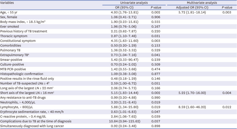
In multivariable logistic analysis, PR was independently associated with age < 55 years (adjusted odds ratio [aOR], 5.72; 95% confidential interval [CI], 1.81–18.14; P = 0.003), short axis diameter of the largest LN ≥ 16 mm (aOR, 5.22; 95% CI, 1.70–16.00; P = 0.004), and lymphocytes < 800/μL (aOR, 8.59; 95% CI, 1.60–46.20; P = 0.012) at initial diagnosis (Table 6).
All variables are values at the time of diagnosis of mediastinal LN TB.
OR = odd ratio, CI = confidence interval, TB = tuberculosis, MTB = Mycobacterium tuberculosis, PCR = polymerase chain reaction, LN = lymph node.
aOther than mediastinal LNs.
bThis includes (1) cases in which necrosis was seen inside the LN on contrast-enhanced CT, or (2) the short diameter of the LN was 10 mm or more, even though no biopsy was performed, and (3) cases confirmed as TB as a result of the biopsy.
cThe largest LN was determined based on the short axis diameter.
This retrospective observational study investigated the clinical course of mediastinal LNTB and the frequency of PR in patients with mediastinal LNTB and risk factors associated with PR. In our study, PR occurred in 21 (17%) of 125 patients at a median of 4.4 months after initiation of anti-TB treatment. We found that PR was independently associated with younger age, larger LN, and lymphocytopenia using multivariable logistic regression analysis. To our knowledge, this is the first study to describe the clinical course of mediastinal LNTB and the characteristics of PR in mediastinal LNTB.
Before the development of EBUS-TBNA, a variety of techniques, including transesophageal ultrasonography-guided needle aspiration, CT-guided percutaneous needle aspiration, conventional transbronchial needle aspiration, and mediastinoscopy, were available for sampling mediastinal LNs.2223 Mediastinoscopy has long been the preferred method for obtaining specimens from mediastinal LNs, but it has some limitations. It is an invasive, expensive surgical procedure that requires general anesthesia and has a 1–2% morbidity rate. In addition, the posterior subcarinal and hilar nodes are generally inaccessible.2425 Now widely used, EBUS-TBNA is a minimally invasive technique that allows access to nodes commonly affected by TB. Previous studies concluded that EBUS-TBNA is a safe and effective modality for mediastinal LNTB.8 However, the clinical course of mediastinal LNTB has been poorly reported, as it was before the advent of EBUS-TBNA. The present study analyzed the largest number of patients with mediastinal LNTB to date and reported detailed clinical information, including symptoms, coexisting TB, drug resistance, mechanical complications, and PR.
The peak age of onset for LNTB is 30 to 40 years. Some recent studies of mediastinal LNTB reported that 5–22% of patients were asymptomatic at the time of diagnosis.262728 Compared with these studies, the patients in our study were older, with a median age of 63 years, and half as many were asymptomatic. One possible explanation for this observation is that many patients were diagnosed with mediastinal LNTB while undergoing evaluation for mediastinal metastases associated with pre-existing or newly identified malignancies. Lung cancer and mediastinal LNTB were simultaneously diagnosed in 20% of patients in our study, and the diagnosis of mediastinal LNTB was facilitated by routine AFB culture for the rinse fluid of EBUS-TBNA needle.12
In our previous study, we demonstrated that routine AFB culture for rinse fluid of the EBUS needle increased the diagnostic yield of LNTB for all patients undergoing EBUS-TBNA, irrespective of pre-procedural diagnosis.12 Similarly, Lucey et al.28 concluded that an AFB culture of core tissue should be performed regardless of pre-test TB probability for patients undergoing EBUS-TBNA. In our study, none of the patients who were simultaneously diagnosed with lung cancer were initially considered to have mediastinal LNTB in the pretest diagnosis. Early detection in this manner would prevent reactivation of TB in cancer patients undergoing chemotherapy.29 Compared with a tissue culture, rinse fluid analysis does not require additional procedural time or punctures. Additionally, half of the patients could only be diagnosed with a culture of the needle rinse fluid in this study.
We described mediastinal LNTB, including coexisting TB and complications. In non-endemic regions, patients with LNTB rarely present with pulmonary TB. In contrast, 20–40% of patients with LNTB exhibit pulmonary involvement in TB-endemic regions.3031 As in previous studies, mediastinal LNTB in our study was accompanied by pulmonary TB in 35% of patients. There are some case reports of complications of mediastinal LNTB, such as esophageal dysphagia, tracheobronchial stenosis, vocal cord paralysis, esophago-mediastinal fistula, and broncho-esophageal fistula.32333435 Because of the small number of case reports, the incidence of these cases in patients with mediastinal LNTB is not well known. In our study, complications of mediastinal LNTB occurred in 4% of patients, which means that attending physicians should be mindful of such serious complications during the course of the disease.
Diagnosis of PR requires evaluation of whether adequate anti-TB treatment has been administered and whether other diseases or drug resistance has been ruled out.46 When PRs are suspected in patients with EPTB, invasive procedures are often required, and DST is relatively difficult compared to pulmonary TB.236 Therefore, it is often challenging to diagnose PRs in patients with EPTB in actual clinical practice, so it is usually made based on clinical and radiological improvements while maintaining anti-TB treatment. Our study revealed CT findings and risk factors that help clinicians recognize the PR. We found that in PR situations, only some LNs were significantly enlarged and not all LNs deteriorated together. In addition, the PR was more likely to occur with larger LN size, consistent with previous reports on peripheral LNTB.3738
A PR occurs frequently in HIV-positive patients who receive highly active antiretroviral therapy (HAART).3940 However, PRs are not uncommon, even in HIV-negative patients, and particularly in EPTB.41 Cheng et al.41 reported that PRs occurred in 15.4% of patients with culture-confirmed TB infection regardless of the organ involved, and the median time to development of PR after initiating anti-TB treatment was 56 days. In another study on pleural TB, the incidence of PR of pleural TB was 16–23%, and the median time to onset of PR was 51 days.4243 Cho et al.6 reported that PR occurred in 23% of patients with peripheral LNTB, and the median time to onset of PR after initiation of anti-TB treatment was 8 weeks. In our study, the incidence of PR in mediastinal LNTB was 17%, which was similar to those of other types of EPTB. However, the median time to onset of PR was 4.4 months, which was longer than those of other types of EPTB. This result is thought to be due to delayed detection because of the location of mediastinal LNs. However, the number of patients who developed PR within 30 days, between 30–60 days, 60–90 days, 90–120 days, 120–150 days, 150–180, and after 180 days was 3, 4, 1, 2, 4, 3, and 4, respectively. The timing of PR detection was fairly evenly distributed, so PR detection timing may not simply depend on CT date.
The pathogenesis of PR remains uncertain. However, the known risk factors for PR suggest that high mycobacterial bacillary and/or antigen load and immune system dysfunction are associated with PR.44 In a study of HIV-positive patients, low lymphocyte count and enhanced tuberculin skin test were related to PR after starting HAART.40 Cheng et al.41 reported that lower baseline lymphocyte count and a greater change in lymphocyte count in HIV-negative patients were risk factors for PR. Another study found that baseline anemia, hypoalbuminemia, and lymphocytopenia were independently associated with PR in patients with pulmonary TB.7 These previous studies found that immunodeficiencies such as lymphocytopenia were associated with development of PR. A strong inflammatory response by the normalized immune system and many microbial components killed by anti-TB drugs may be associated with PR.44
Our study had some limitations. First, it was conducted retrospectively at a single center, which poses difficulties in generalization and increases the risk of selection bias. Second, 33 of 158 patients did not complete anti-TB treatment and were excluded from the analysis of risk factors for PR. In addition, LN characteristics such as findings of ultrasonography and PET could not be identified as risk factors for PRs in this study. Therefore, this study’s ability to evaluate the exact incidence of and risk factors for mediastinal LNTB is limited. Third, our study does not provide adequate management of PR. Since there is no standard treatment guideline for PRs, various treatments have been applied by each attending physician. In addition, the number of PRs was not large enough to analyze which treatment was more useful. Further research is needed on appropriate treatment for PR.
In conclusion, we found that the risk factors for development of PR in mediastinal LNTB were age < 55 years, short axis diameter of the largest LN ≥ 16 mm, and lymphocyte count < 800/μL at the time of diagnosis. As one in six patients with mediastinal LNTB can be expected to experience PR during anti-TB treatment, clinicians should pay attention to patients with risk factors.
Notes
Author Contributions:
Conceptualization: Jeong BH.
Data curation: Choe J, Han A, Jeong BH.
Formal analysis: Choe J, Han A, Jeong BH.
Investigation: Choe J, Han A, Jeong BH.
Methodology: Kim TY, Huh HJ, Choi YL, Han J, Jeong BH.
Resources: Shin SH, Lee K, Um SW, Kim H, Jeong BH.
Supervision: Lee K, Um SW, Kim H, Kim TY, Huh HJ, Choi YL, Han J.
Validation: Shin SH, Lee K, Um SW, Kim H.
Writing – original draft: Choe J, Han A.
Writing – review & editing: Shin SH, Kim TY, Huh HJ, Choi YL, Han J, Jeong BH.
References
1. World Health Organization. Global Tuberculosis Report 2022. Updated 2022. Accessed March 26, 2023.
https://www.who.int/publications/i/item/9789240061729
.
2. Peto HM, Pratt RH, Harrington TA, LoBue PA, Armstrong LR. Epidemiology of extrapulmonary tuberculosis in the United States, 1993–2006. Clin Infect Dis. 2009; 49(9):1350–1357. PMID: 19793000.
3. Gomes T, Reis-Santos B, Bertolde A, Johnson JL, Riley LW, Maciel EL. Epidemiology of extrapulmonary tuberculosis in Brazil: a hierarchical model. BMC Infect Dis. 2014; 14(1):9. PMID: 24400848.
4. Breen RA, Smith CJ, Bettinson H, Dart S, Bannister B, Johnson MA, et al. Paradoxical reactions during tuberculosis treatment in patients with and without HIV co-infection. Thorax. 2004; 59(8):704–707. PMID: 15282393.
5. Hawkey CR, Yap T, Pereira J, Moore DA, Davidson RN, Pasvol G, et al. Characterization and management of paradoxical upgrading reactions in HIV-uninfected patients with lymph node tuberculosis. Clin Infect Dis. 2005; 40(9):1368–1371. PMID: 15825042.
6. Cho OH, Park KH, Kim T, Song EH, Jang EY, Lee EJ, et al. Paradoxical responses in non-HIV-infected patients with peripheral lymph node tuberculosis. J Infect. 2009; 59(1):56–61. PMID: 19535147.
7. Cheng SL, Wang HC, Yang PC. Paradoxical response during anti-tuberculosis treatment in HIV-negative patients with pulmonary tuberculosis. Int J Tuberc Lung Dis. 2007; 11(12):1290–1295. PMID: 18034948.
8. Ye W, Zhang R, Xu X, Liu Y, Ying K. Diagnostic efficacy and safety of endobronchial ultrasound-guided transbronchial needle aspiration in intrathoracic tuberculosis: a meta-analysis. J Ultrasound Med. 2015; 34(9):1645–1650. PMID: 26269299.
9. Um SW, Kim HK, Jung SH, Han J, Lee KJ, Park HY, et al. Endobronchial ultrasound versus mediastinoscopy for mediastinal nodal staging of non-small-cell lung cancer. J Thorac Oncol. 2015; 10(2):331–337. PMID: 25611227.
10. Lee KJ, Suh GY, Chung MP, Kim H, Kwon OJ, Han J, et al. Combined endobronchial and transesophageal approach of an ultrasound bronchoscope for mediastinal staging of lung cancer. PLoS One. 2014; 9(3):e91893. PMID: 24632834.
11. Seo AN, Park HJ, Lee HS, Park JO, Chang HE, Nam KH, et al. Performance characteristics of nested polymerase chain reaction vs real-time polymerase chain reaction methods for detecting Mycobacterium tuberculosis complex in paraffin-embedded human tissues. Am J Clin Pathol. 2014; 142(3):384–390. PMID: 25125630.
12. Ko RE, Jeong BH, Chon HR, Huh HJ, Han J, Lee H, et al. Clinical usefulness of routine AFB culture and MTB PCR of EBUS-TBNA needle rinse fluid. Respirology. 2019; 24(7):667–674. PMID: 30730098.
13. Huh HJ, Koh WJ, Song DJ, Ki CS, Lee NY. Evaluation of the Cobas TaqMan MTB test for the detection of Mycobacterium tuberculosis complex according to acid-fast-bacillus smear grades in respiratory specimens. J Clin Microbiol. 2015; 53(2):696–698. PMID: 25428157.
14. Diagnostic standards and classification of tuberculosis in adults and children. This official statement of the American Thoracic Society and the Centers for Disease Control and Prevention was adopted by the ATS Board of Directors, July 1999. This statement was endorsed by the Council of the Infectious Disease Society of America, September 1999. Am J Respir Crit Care Med. 2000; 161(4 Pt 1):1376–1395. PMID: 10764337.
15. Koh WJ, Ko Y, Kim CK, Park KS, Lee NY. Rapid diagnosis of tuberculosis and multidrug resistance using a MGIT 960 system. Ann Lab Med. 2012; 32(4):264–269. PMID: 22779067.
16. Kim S, Lee H, Park HY, Jeon K, Huh HJ, Lee NY, et al. Outcomes of pulmonary tuberculosis in patients with discordant phenotypic isoniazid resistance testing. Respir Med. 2017; 133:6–11. PMID: 29173450.
17. Schwartz LH, Bogaerts J, Ford R, Shankar L, Therasse P, Gwyther S, et al. Evaluation of lymph nodes with RECIST 1.1. Eur J Cancer. 2009; 45(2):261–267. PMID: 19091550.
18. Lee JY. Diagnosis and treatment of extrapulmonary tuberculosis. Tuberc Respir Dis (Seoul). 2015; 78(2):47–55. PMID: 25861336.
19. World Health Organization. Definitions and Reporting Framework for Tuberculosis – 2013 Revision: Updated December 2014 and January 2020. Geneva, Switzerland: World Health Organization;2013.
20. Holden IK, Lillebaek T, Andersen PH, Bjerrum S, Wejse C, Johansen IS. Extrapulmonary tuberculosis in Denmark from 2009 to 2014; characteristics and predictors for treatment outcome. Open Forum Infect Dis. 2019; 6(10):ofz388. PMID: 31660351.
21. Bewick V, Cheek L, Ball J. Statistics review 13: receiver operating characteristic curves. Crit Care. 2004; 8(6):508–512. PMID: 15566624.
22. Zwischenberger JB, Savage C, Alpard SK, Anderson CM, Marroquin S, Goodacre BW. Mediastinal transthoracic needle and core lymph node biopsy: should it replace mediastinoscopy? Chest. 2002; 121(4):1165–1170. PMID: 11948048.
23. Bilaçeroğlu S, Günel O, Eriş N, Cağirici U, Mehta AC. Transbronchial needle aspiration in diagnosing intrathoracic tuberculous lymphadenitis. Chest. 2004; 126(1):259–267. PMID: 15249469.
24. Hammoud ZT, Anderson RC, Meyers BF, Guthrie TJ, Roper CL, Cooper JD, et al. The current role of mediastinoscopy in the evaluation of thoracic disease. J Thorac Cardiovasc Surg. 1999; 118(5):894–899. PMID: 10534695.
25. Lemaire A, Nikolic I, Petersen T, Haney JC, Toloza EM, Harpole DH Jr, et al. Nine-year single center experience with cervical mediastinoscopy: complications and false negative rate. Ann Thorac Surg. 2006; 82(4):1185–1189. PMID: 16996905.
26. Navani N, Molyneaux PL, Breen RA, Connell DW, Jepson A, Nankivell M, et al. Utility of endobronchial ultrasound-guided transbronchial needle aspiration in patients with tuberculous intrathoracic lymphadenopathy: a multicentre study. Thorax. 2011; 66(10):889–893. PMID: 21813622.
27. Sun J, Teng J, Yang H, Li Z, Zhang J, Zhao H, et al. Endobronchial ultrasound-guided transbronchial needle aspiration in diagnosing intrathoracic tuberculosis. Ann Thorac Surg. 2013; 96(6):2021–2027. PMID: 24035300.
28. Lucey O, Potter J, Ricketts W, Castle L, Melzer M. Utility of EBUS-TBNA in diagnosing mediastinal tuberculous lymphadenitis in East London. J Infect. 2022; 84(1):17–23. PMID: 34706281.
29. Dobler CC, Cheung K, Nguyen J, Martin A. Risk of tuberculosis in patients with solid cancers and haematological malignancies: a systematic review and meta-analysis. Eur Respir J. 2017; 50(2):1700157. PMID: 28838977.
30. Fontanilla JM, Barnes A, von Reyn CF. Current diagnosis and management of peripheral tuberculous lymphadenitis. Clin Infect Dis. 2011; 53(6):555–562. PMID: 21865192.
31. Mathiasen VD, Eiset AH, Andersen PH, Wejse C, Lillebaek T. Epidemiology of tuberculous lymphadenitis in Denmark: a nationwide register-based study. PLoS One. 2019; 14(8):e0221232. PMID: 31415659.
32. Rafay MA. Tuberculous lymphadenopathy of superior mediastinum causing vocal cord paralysis. Ann Thorac Surg. 2000; 70(6):2142–2143. PMID: 11156137.
33. Xiong L, Mao X, Li C, Liu Z, Zhang Z. Posterior mediastinal tuberculous lymphadenitis with dysphagia as the main symptom: a case report and literature review. J Thorac Dis. 2013; 5(5):E189–E194. PMID: 24255790.
34. Sayeed A, Alqurashi EH, Alzanbagi AB, Ghaleb NAB. Tuberculosis presenting as broncho-oesophageal fistula in a young healthy man. BMJ Case Rep. 2017; 2017:bcr2017220821.
35. Nie D, Li J, Liu W, Wu Y, Ji M, Wang Y, et al. Esophagomediastinal fistula due to secondary esophageal tuberculosis: report of two cases. J Int Med Res. 2021; 49(7):3000605211023696. PMID: 34256638.
36. Barss L, Connors WJ, Fisher D. Chapter 7: extra-pulmonary tuberculosis. Canadian Respir Crit Care Sleep Med. 2022; 6(sup1):87–108.
37. Chahed H, Hachicha H, Berriche A, Abdelmalek R, Mediouni A, Kilani B, et al. Paradoxical reaction associated with cervical lymph node tuberculosis: predictive factors and therapeutic management. Int J Infect Dis. 2017; 54:4–7. PMID: 27810522.
38. Seol YJ, Park SY, Yu SN, Kim T, Lee EJ, Jeon MH, et al. Is the initial size of tuberculous lymphadenopathy associated with lymph node enlargement during treatment? Infect Chemother. 2017; 49(2):130–134. PMID: 28608659.
39. Breen RA, Smith CJ, Cropley I, Johnson MA, Lipman MC. Does immune reconstitution syndrome promote active tuberculosis in patients receiving highly active antiretroviral therapy? AIDS. 2005; 19(11):1201–1206. PMID: 15990574.
40. Narita M, Ashkin D, Hollender ES, Pitchenik AE. Paradoxical worsening of tuberculosis following antiretroviral therapy in patients with AIDS. Am J Respir Crit Care Med. 1998; 158(1):157–161. PMID: 9655723.
41. Cheng VC, Yam WC, Woo PC, Lau SK, Hung IF, Wong SP, et al. Risk factors for development of paradoxical response during antituberculosis therapy in HIV-negative patients. Eur J Clin Microbiol Infect Dis. 2003; 22(10):597–602. PMID: 14508660.
42. Jeon K, Choi WI, An JS, Lim SY, Kim WJ, Park GM, et al. Paradoxical response in HIV-negative patients with pleural tuberculosis: a retrospective multicentre study. Int J Tuberc Lung Dis. 2012; 16(6):846–851. PMID: 22507441.
43. Jung JW, Shin JW, Kim JY, Park IW, Choi BW, Seo JS, et al. Risk factors for development of paradoxical response during anti-tuberculosis treatment in HIV-negative patients with pleural tuberculosis. Tohoku J Exp Med. 2011; 223(3):199–204. PMID: 21372521.
44. Bell LC, Breen R, Miller RF, Noursadeghi M, Lipman M. Paradoxical reactions and immune reconstitution inflammatory syndrome in tuberculosis. Int J Infect Dis. 2015; 32:39–45. PMID: 25809754.