

 PDF
PDF Citation
 Print
Print


Introduction
Breast cancer is currently the most commonly diagnosed cancer in women, accounting for a quarter of all cancer cases [1]. A total of 2.3 million new cases of breast cancer were diagnosed in 2020, accounting for one in eight newly diagnosed cancers [2]. The incidence of breast cancer is rising not only in the United States and Europe but also in Asia [3]. With the development of screening using mammography and ultrasound, and the development of various treatment methods, the treatment results for breast cancer are becoming more outstanding [4]. Among all breast cancer diagnoses, the proportion of early-stage breast cancers continues to increase. Accordingly, the surgical methods for breast cancer are constantly changing. Modified radical mastectomy was the standard for surgical treatment of breast cancer. However, with the introduction of partial mastectomy accompanied by radiation, the surgical treatment of breast cancer has undergone great changes [5,6]. In addition, with the introduction of sentinel lymph node biopsy (SLNB) into breast cancer surgery by Giuliano et al. [7], many patients with early breast cancer skipped axillary lymphatic dissection, thereby reducing the incidence of lymphedema. Such changes in breast cancer surgery require higher levels of accuracy and safety. In the process of confirming the resection margin and sentinel lymph nodes by frozen section examination during surgery, a certain false-negative rate is reported in the pathological examination [8,9]. If the frozen biopsy result is negative, but cancer cells are found in the final biopsy report, the patient may experience the inconvenience of having to undergo reoperation to remove any cancer cells that may remain in the body. The reoperation rate has been reported up to 50% depending on the study [10-12]. It follows that optical technologies have been developed for accurate diagnosis of sentinel lymph node and tumor margins such as diffuse reflectance spectroscopy, fluorescence spectroscopy, and photoacoustic spectroscopy [13-18]. Especially Raman spectroscopy is an excellent technique for material analysis due to its high molecular specificity [19], various studies have been conducted on the evaluation of sentinel lymph node and tumor margin in breast cancer [20-24]. But Raman scattering has disadvantages that are difficult to apply clinically, such as low signal-to-noise ratio and exacerbated by fluorescence interference [25], and long measurement time [26]. Nonetheless, recent advances such as high-efficiency laser sources, low-noise detectors, effective filters, and high-efficiency optics have greatly improved this applicability [27,28].
Go to : 
Background of Raman spectroscopy
1. Raman spectroscopy system
Raman spectroscopy was first observed experimentally in 1928 [29]; however, because of the rare occurrence of Raman scattering, which only occurs with a probability of 1 in approximately 108, it was difficult to observe [30,31]. Recent advancements in technology have enabled real-time observation using Raman spectroscopy, leading to its widespread application in the clinical field. Raman spectroscopy is a powerful technique for the spectroscopy of vibrations produced by the interacting energy in materials, including cells and tissues. It allows the identification and analysis of the molecular structure, symmetry, electronic environment, and composition of a material, providing a chemical fingerprint that can be used to distinguish between cells and tissues [32-37]. Diseases, particularly cancer, alter the chemical fingerprints of tissues. Raman spectroscopy has the potential to differentiate between diseased and normal tissues. However, it requires a large amount of trained reference data and an accurate analysis model [32,34].
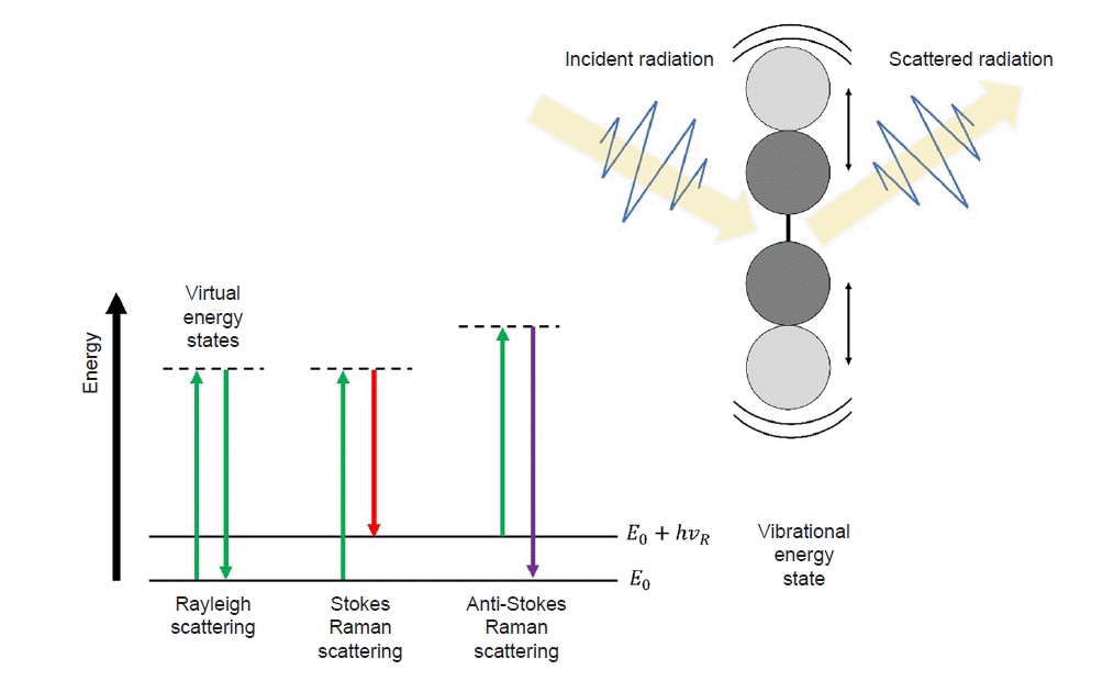
To explain the Raman scattering phenomenon, it is necessary to first describe Rayleigh scattering. Various interactions, such as absorption, reflection, and scattering, occur when light interacts with a material. Scattering refers to the deviation of light from its original path and its propagation in different directions. Rayleigh scattering occurs when the energies of the incident light and light emitted in different directions are equal. This type of scattering is also known as elastic scattering, owing to its characteristic nature. However, there are cases in which the scattered light possesses more or less energy than the original energy. For example, a portion of the incident energy may be utilized for the vibrational motion of atoms or the rotational motion of molecules, whereas the remaining energy is scattered as light. In this scenario, only energy lower than the incident energy is emitted, resulting in the emission of light with relatively longer wavelengths compared to Rayleigh scattering. This process is referred to as Stokes Raman scattering. Conversely, when the material is already in a high-energy state upon receiving light, more energy is emitted than incident energy. Consequently, the wavelength of the scattered light shortens; this phenomenon is known as anti-Stokes Raman scattering (Fig. 1) [38].
2. Raman spectra
The incident photons interact with molecules in the tissue. Rayleigh occurs when the energy of the scattered photon is equal to that of the incident photon. The rare occurrence of a difference in energy between a scattered photon and an incident photon is called inelastic scattering and is known as the Raman effect. Only one photon in 108 undergoes the Raman effect [30,39].
A laser is used to provide high-quality monochromatic light and induce Raman scattering. To collect light that reacts with the tissue, appropriate optics must be configured in the optical path. Recent developments in Raman detectors include the use of highly sensitive detectors and gratings. A schematic of the Raman spectroscopy system is shown in Fig. 2.
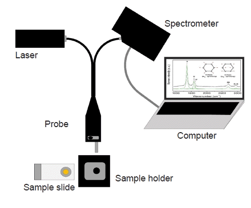 | Fig. 2.Monochromatic excitation light generated by a laser source passing through a narrow-band filter within a probe. After interacting with the sample, the laser light re-enters the probe. When the Raman operation is activated, the incident light is directed to an optical system and transmitted to a spectrometer. The spectrometer’s grating separates the collected light, which is then detected and analyzed through software. A sample holder can be used to minimize ambient light noise.
|
In general, Raman spectra have characteristic chemical fingerprints that depend on the wavelength of the incident laser, molecular composition, and bonding form. Raman spectroscopic techniques have been developed in several types. Spontaneous Raman spectroscopy, when used in combination with a fiber probe or microscope, is characterized by being label-free, noninvasive, and nondestructive. Resonance Raman spectroscopy, which matches the excitation wavelength to the electron resonance of molecules, increases the signal-to-noise ratio by 103 to 105. Surface-enhanced Raman spectroscopy applied to rough metal surfaces results in a 106-fold increase in the signal-to-noise ratio and has been applied to cell-based assays and immunoassays. Spatially offset Raman spectroscopy technology, which collects diffusely scattered photons, can acquire information even in relatively thick tissues and has been applied to cancer detection in breast tissue [30].
The Raman spectrum provides the fingerprint of a material; however, it is not possible to directly interpret the composition of the material from this chemical fingerprint. A database of reference spectra is required to use Raman spectroscopy in the analysis of materials. A large number of Raman spectra have been published for this purpose [40-42]. Also, simulations and deep learning continue to be studied [39,43]. Unlike single materials, human tissues are a complex assembly of various molecular structures. Raman spectra of protein structures in human tissues are continuously being studied [44,45].
Go to :
Raman spectroscopy of breast cancer surgery
1. Frozen section analysis in breast cancer surgery
Partial mastectomy combined with postoperative radiation therapy has become the gold standard treatment for patients with early-stage breast cancer, offering equivalent survival and improved quality of life compared to patients undergoing total mastectomy [46,47]. Complete resection of tumors is essential for partial mastectomy to reduce the recurrence rate after surgical treatment [48,49]. During partial mastectomy, the surgeon may request rapid pathological information of marginal status and determine whether additional resection is required. SLNB is also important for the surgical treatment of early breast cancer. The surgeon checks the progress of the disease through SLNB during surgery and makes a dicision of the surgical scope of the axillary region. As the rate of early breast cancer increases, the need for a method that can quickly confirm pathological results during surgery has increased. Currently, frozen section analysis is the most frequently performed method for confirming the pathological results of intraoperative biopsies. When a surgeon sends a tissue that needs to be inspected during surgery to a pathologist, the tissue is analyzed through frozen sectioning and notified of the result, which reduces the frequency of reoperations that occur after surgery [50,51]. However, This method has limitation in sensitivity. Studies analyzing marginal frozen sections obtained from partial mastectomy have reported sensitivities of 77%–81% [52,53]. In SLNB, the sensitivity of macro-metastasis and micro-metastasis was different. The sensitivity for diagnosis of macro-metastasis was over 90%, but the sensitivity for diagnosis of micro-metastasis was reported to be 30%–40% [8,54-56]. If the results of frozen section analysis confirmed during surgery and the final biopsy results confirmed after surgery are different, the patient may experience the inconvenience of having to repeat the operation, which leads to an increase in complications, hospitalization days, and medical expenses [57-60].
2. Raman spectroscopy of surgical margins
In 2006, a study that implemented Raman spectroscopy of breast tissue in an in vivo environment was reported for the first time. Haka et al. [61] obtained and analyzed 31 Raman spectra from nine patients who underwent partial mastectomy. In that study, cancer tissues were accurately distinguished from normal and benign tissues using Raman spectroscopy. These researchers later reported a negative predictive value of 99% using 129 tissue samples in a new prospective study [62]. However, to confirm the presence or absence of cancerous tissue on the surgical cut surface using Raman spectroscopy in actual clinical practice, an accurate location must be specified, and the single-point method using a probe causes sampling errors. To solve this problem, Zhang et al. [63] conducted a comparative study using Raman spectral mapping. A total of 53 sets of mapping data and 2,597 Raman spectra were analyzed and compared, and the data obtained using the mapping technology displayed excellent diagnostic performance. Raman microspectroscopy studies have also been reported. Raman microspectroscopy makes diagnosis without staining based on the morphological and biochemical contrast between normal and tumor tissue. Kong et al. [64] reported that using Raman microspectroscopy to detect invasive ductal carcinoma within breast tissue with 95.6% sensitivity and 96.2% specificity. Zhang et al. [65] reported characterization of biochemical properties and structural alterations of breast cancer tissues at various TNM stages and grades by Raman microspectroscopy. Early Raman microspectroscopy studies had limitations in that the scanning method used to construct Raman spectral images for tumor diagnosis was very slow [66]. However, with the development of various technologies, such as the use of selective sampling based on integrated autofluorescence imaging, the possibility of its clinical application as an intraoperative method has been demonstrated [64,67,68].
3. Raman spectroscopy of SLNB
Raman spectroscopy is noninvasive and can provide detailed chemical information about tissue, thereby making it a very suitable test to check the status of the sentinel lymph nodes in real time during surgery. In 2003, Smith et al. [69] first identified axillary lymph nodes in breast cancer using Raman spectroscopy. After that, Horsnell et al. [70] reported a sensitivity of 81% and specificity of 97% using the method of examining 10 points in the lymph node. Unfortunately, studies using Raman spectroscopy as a diagnostic tool for sentinel lymph node evaluation have not yet been conducted. Most studies were limited to small sample sizes and were laboratory-based. However, recently, studies using new technologies, such as a tissue mapping protocol obtained by analyzing the spectra of each cell [22] and studies using a nontoxic Raman nanoparticle tracer [21] have been reported, confirming the possibility that sentinel lymph node diagnosis through Raman spectroscopy can be used in clinical practice.
Go to :
Conclusions
Research on Raman spectroscopy has been conducted in various fields, ranging from basic to clinical applications. The high sensitivity of Raman spectroscopy was previously regarded as a disadvantage that made it difficult to apply in clinical practice; however, these limitations are now being overcome by the incorporation of various technologies and the development of spectrum analysis. Studies analyzing Raman spectra to identify cancerous tissue at the surgical margin and lymph node during breast cancer surgery are ongoing, and the positive results of the studies show the possibility of supplementing the frozen section method. In the surgical treatment of breast cancer, if it becomes possible to distinguish malignant tissue from normal tissue in vivo using Raman spectroscopy, unnecessary surgical biopsies during surgery will be reduced.
Go to :
 XML Download