

 PDF
PDF Citation
 Print
Print


Introduction
In the field of orthopedic surgery, distal radial fractures are common in elderly patients, especially women. A distal radius fracture is a posterior lateral dislocation fracture that occurs in the distal radius due to the external force of extension. The fracture lines may also involve the radiocarpals and the distal radioulnar joint [1]. Since bioresorbable implants were clinically introduced in 1984, various types have been developed using materials such as polyglycolides and polylactides [2]. Such bioresorbable devices have the advantage of not requiring a second operation for removal [3].
Recently, a new generation of materials such as bioresorbable screws or wires made of magnesium has been developed [4]. Compared with polylactide-co-glycolide implants, magnesium is a promising new biomaterial with reduced implant sizes and improved mechanical properties to support fracture healing in a load-sharing environment [5]. As such, bioresorbable implants may have several advantages in orthopedic surgical procedures and may have additional advantages in clinical performance.
Distal radius fractures account for approximately 25% of fractures in the pediatric population and up to 18% of all fractures in the elderly [6]. Therefore, in this study, group A, which was fixed only with a metal plate, and group B, which was additionally fixed using bioabsorbable screws or wires, were compared to patients with complex distal radius fractures to determine that there was no difference in the maintenance of correction and that it was acceptable to use.
Go to : 
Methods
Ethics statement: This retrospective study was conducted with the approval of the Institutional Bioethics Committee of Gwangju Christian Hospital (No. KVH-M-2022-12-02). Written informed consent was obtained from the patients for the publication of this article including all clinical images.
From January 2018 to March 2021, 232 patients with distal radial fractures visited our hospital, and 105 patients underwent surgical treatment. Of 105 patients, 57 had complex radius fractures. A complex distal radius fracture was considered a case of Fernandez classification grade V with intraarticular involvement. The 23 patients who visited the hospital between January 2018 and December 2019, were placed in group A and underwent open reduction using a metal plate (locking compression plate [LCP]; one-third anatomical locking plate [Arix; JEIL Medical Corp., Seoul, Korea]). The 22 patients who visited the hospital between January 2020 and March 2021, were placed in group B and underwent fixation using a metal plate, wire, and screw [Resomet; U&I Corp., Seoul, Korea]) (Fig. 1). For statistical comparison, group A was used first and group B was used later. All surgeries were performed by a single skilled surgeon (Figs. 2–6).
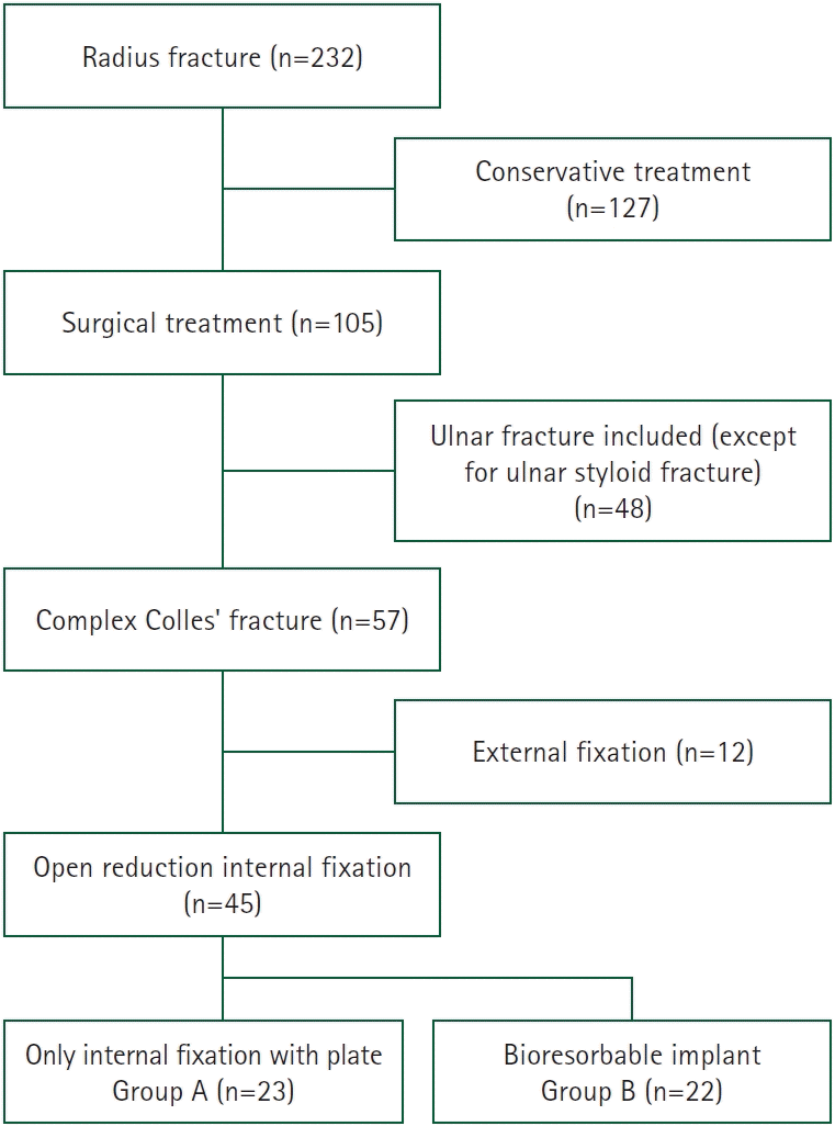 | Fig. 1.Flow chart of patients with radius fractures. In total, 232 patients with distal radius fractures were enrolled, and two groups appropriate for the goals of this study were selected as the study subjects: group A, internal fixation with a plate only (n=23) and group B, bioresorbable implant (n=22).
|
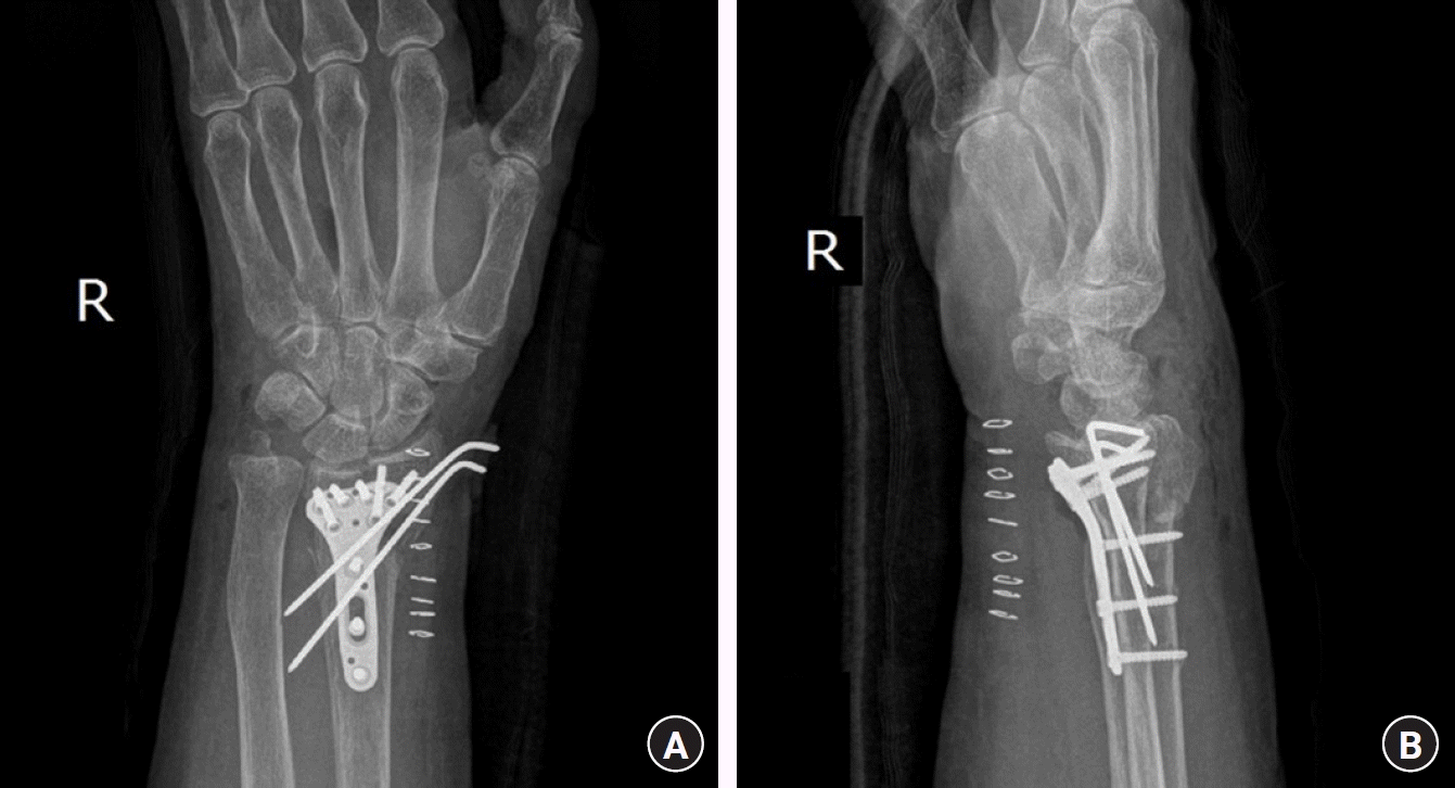
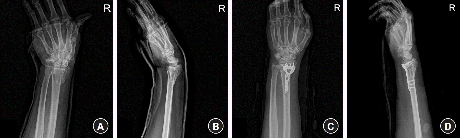 | Fig. 2.A 74-year-old female patient in group B (bioresorbable implant). (A, B) Preoperative simple radiology. (C, D) Postoperative radiology using internal fixation with a plate and absorbable implant for a distal radius fracture. (A, C) Anteroposterior view, (B, D) lateral view.
|
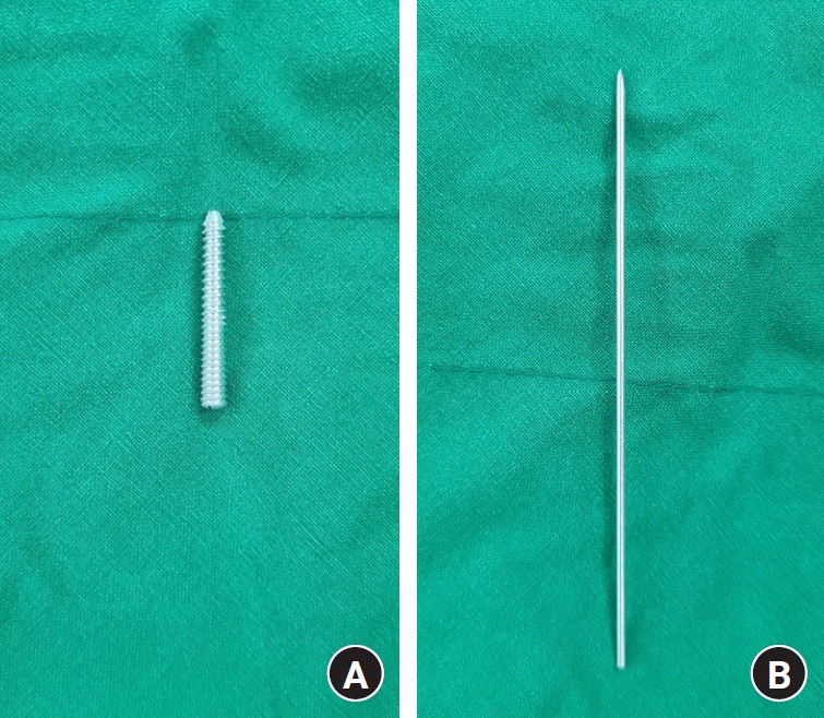 | Fig. 3.Bioresorbable implant used in this study (Resomet; U&I Corp., Seoul, Korea). (A) Bioresorbable screw, (B) bioresorbable wire.
|
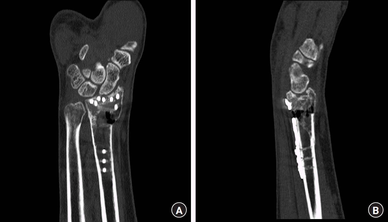
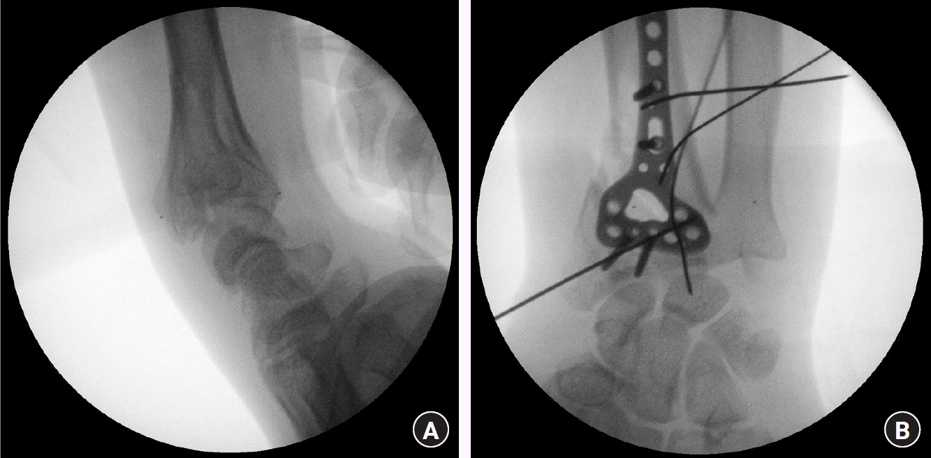 | Fig. 4.A 74-year-old female patient in group B (bioresorbable implant). Intraoperative radiology using internal fixation with plate and absorbable implant for distal radius fracture (A, lateral view; B, anteroposterior view).
|
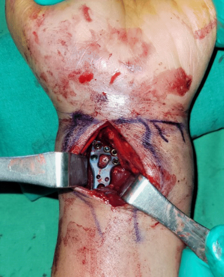
The surgical method used was a modified Henry’s approach. The radial artery was protected and accessed through the aponeurosis of the flexor carpi radialis tendon. The pronator quadratus was exposed and dissected radially, and the fracture site was exposed by pulling the pronator quadratus to the ulnar side, reduced with traction, and temporarily fixed using a Kirschner wire (K-wire). A metal plate was placed in the radius and the cortical screw was first inserted into the elongated hole. After the metal plate was positioned, a distal locking screw was inserted and fixed to the LCP at one-third of the anatomical locking plate. In group B, after fixation, as in group A, after checking the fracture line under the C-arm guide, a guidewire was inserted perpendicular to the fracture line. After removing the guidewire, a bioresorbable screw or wire was inserted into the site and cut to the desired size. After fixation, the pronator muscle was sutured to the brachioradialis tendon as closely as possible [7].
After confirming the age, sex, and follow-up period of both groups, the radial inclination, radial height, and volar tilt were evaluated using simple radiography before and after the operation to measure the degree of radius reduction.
We compared the radiographs immediately after surgery and at the latest outpatient follow-up to determine the degree of reduction maintenance. Postoperatively, the time to union was measured using simple radiography in groups A and B. In group B, the bioabsorption period of the bioresorbable screw or wire was confirmed using simple radiography.
For clinical evaluation, we used the Disabilities of the Arm, Shoulder, and Hand (DASH) score, measured at the last outpatient clinic visit. In addition, the DASH score can detect and differentiate small and large changes in disability over time after surgery in patients with upper-extremity musculoskeletal disorders [8]. The occurrence of complications such as infection, nonunion, and malunion was confirmed. In both groups, diabetes mellitus and smoking were identified as underlying diseases that could influence bone union and infection.
All measurements were performed by an orthopedic surgeon. For statistical tests, sex, right or left side, diabetes mellitus, smoking, osteoporosis, and cause of fracture were analyzed using the Fisher exact test. Age, DASH score, and time of union were analyzed using the Mann-Whitney U-test. An independent sample t-test was used for age, radial inclination, radial height, and volar tilt, and a paired t-test was used to compare the postoperative radiographs with the latest follow-up radiographs (IBM SPSS Statistics ver. 26.0; IBM Corp., Armonk, NY, USA). The statistical significance level was defined as the case where the p-value was less than 0.05.
Go to :
Results
The two groups showed a similar distribution in terms of sex, age, history, cause of fracture, and complications (infection, malunion, and nonunion) (Table 1). There were no differences between the two groups in radial inclination, radial height, and volar tilt, which evaluated the degree of reduction of radial fractures before and after surgery in both groups (Table 2). When comparing post-X-ray and the latest follow-up X-ray of each patient, reduction was maintained well in both groups, and there was no statistically significant change (p>0.05) (Table 3). The union time of radial fractures was found to be an average of 160 days (standard deviation [SD], ±47.71 days; range, 47–567 days) in group A and 181 days (SD, ±49.12 days; range, 47–576 days) in group B. There was no significant difference in the degree of maintenance of reduction in the radial inclination, radial height, and volar tilt evaluated by X-ray performed postoperative day 1, and at the latest outpatient follow-up (Table 3). Also, there were no statistically significant differences (p=0.54) (Table 4).
Table 1.
Preoperative demographics in the two groups
![]()
Table 2.
Preoperative, postoperative, and latest follow-up radiological values in the two groups
![]()
Table 3.
Differences between parameters on POD 1 X-rays and the latest follow-up X-rays
| Variable | p-valuea) | |
|---|---|---|
| Group A (n=23) | Group B (n=22) | |
| Radial inclination | 0.65 | 0.42 |
| Radial height | 0.45 | 0.33 |
| Volar tilt | 0.51 | 0.28 |
![]()
Table 4.
DASH scores and period until complete union of the radius in both groups and complete absorption time in group B
![]()
In the DASH score performed at the last outpatient follow-up for clinical evaluation, group A scored 14.8 (SD, ±6.21; range, 10–19) and group B scored 13.2 (SD, ±5.23; range, 10–17); and there was no significant difference between the two groups (Table 4). In group A and group B, infection, nonunion, and malunion were not observed after surgery.
The bioabsorption period of bioresorbable screws or wires was evaluated in group B, and the average was 407 days (SD, ±79.82 days; range, 110–640 days) (Table 4).
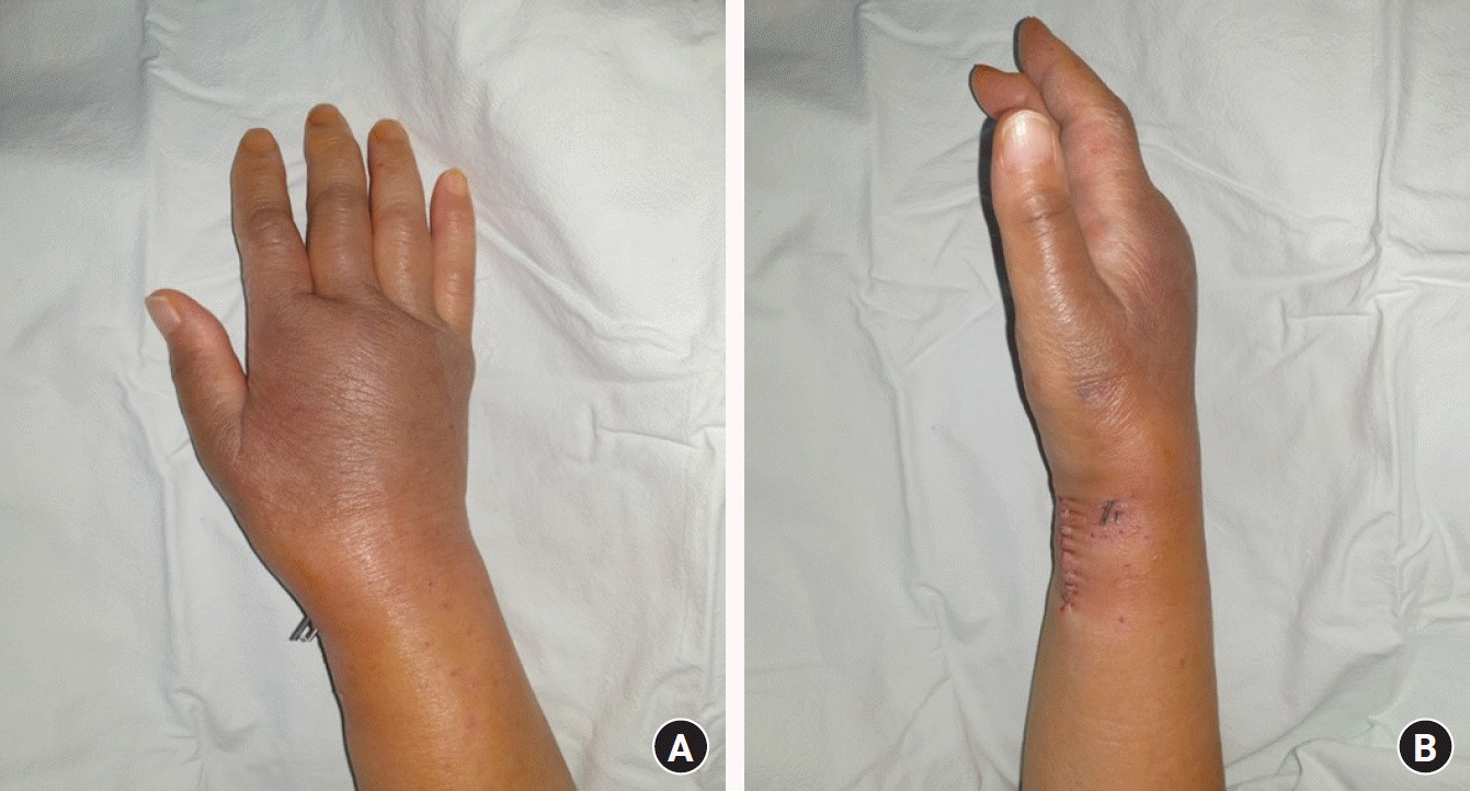
When clinically performed, it was possible to avoid the collision of the instrument during fixation due to the flexibility of the bioresorbable screw and wire and do more appropriate handling. In addition, as an alternative method to the external fixation and K-wire for complex distal radius surgical management, the discomfort of additional dressing and removal management could be alleviated (Figs. 7, 8). More metal device usage like a locked plate suggests increased potential for complications. Currently, hardware removal is secondary to tendon irritation or rupture [9]. Since the number of metallic devices is small when the usage of the bioresorbable screw and wire, it has the advantage of reducing the difficulty of hardware removal due to damage to the device including galvanic corrosion, cold-welding, and tendon irritation or rupture.
Go to :
Discussion
The incidence of distal radius fractures is increasing worldwide, and its prevalence in the United States has increased by approximately 17% over the past 40 years [6]. Treatment of distal radius fractures is treated using rush rods, percutaneous pin fixation, external fixation devices, open reduction, and internal fixation [10]. Surgical treatment rather than conservative treatment is recommended for radial shortening of >3 mm, dorsal tilt of >10°, and intraarticular displacement of >2 mm in distal radial fractures [11]. There are many cases of osteoporosis underlying this disease, and most of these patients have complex rather than simple fractures [12]. Malunion of distal radius fractures is uncommon but can occur quite often when rigid fixation is not achieved in these complex fractures [13]. It is debatable that more screws result in firmer fixation, however, previous biomechanical studies have shown that a stronger fixation is possible by inserting multiple screws into the distal portion [14,15].
In this study, the plate was firmly fixed using additional bioresorbable screws or wires after reduction using a metal plate to prevent malunion. During a follow-up period of approximately 2 years, no special complications were observed, and complete union was achieved in all cases.
Magnesium-based biomaterials have been used as orthopedic implants for more than 100 years because of their desirable mechanical and osteopromotive properties [16]. No allergic effects have been observed [17], even in a major study that reported the fixation of 13 chevron osteotomies using magnesium screws. No disadvantages were identified when comparing the results to a control group fixed with titanium alloy screws [18].
In general, in the case of a bioresorbable screw or wire, it is difficult to see well in a C-arm image because it is transparent. Therefore, we used a bioresorbable material with a guide. Because the bioresorbable material was not visible in the C-arm image, the operation time was longer in group B. However, the fracture line was clearly visible, and correct reduction surgery could be performed.
Previous studies have reported that bioresorbable implants, such as polydioxanone, may interfere with bone union because of foreign body reactions [19]. The bioresorbable implant used in this study was made of magnesium and was much weaker than the screws and K-wires made of metal; therefore, it should be handled carefully. Additionally, when bioresorbable implants are absorbed, CO2 gas is generated, which can be observed using radiography during follow-up. It must be carefully distinguished from gas gangrene, which indicates infection. Bioresorbable materials are naturally absorbed by the body and cause no complications. The bioabsorption period is approximately 1 year and 6 months on average. Although this varies between patients, it should be sufficient to ensure the union of the radial fracture [20].
This study had several limitations. First, the number of patients included was small. Among patients with radial fractures, the study was conducted with patient groups that excluded conservative treatment and external fixation devices. Therefore, further case studies are required. Second, because bioresorbable implants are transparent and radiolucent, they may be difficult to observe on radiographic examination. If no bioresorbable implants were observed on follow-up radiological examination, it was judged that all of them were absorbed; however, there was a possibility that they were actually left in the radius. Third, this is a retrospective study. Randomization was not performed in either group and there may have been a bias.
Go to :
Conclusion
There were no differences in bone union timing or complications when additional fixation using bioresorbable materials was performed after open reduction using a metal plate for complex fractures of the distal radius. Clinically, it is considered a useful method to compensate for the shortcomings of metal implants in complex distal radial fractures. Therefore, additional fixation using bioresorbable screws or wires is considered an appropriate treatment for preventing nonunion in complex fractures of the distal radius.
Go to :
 XML Download