Journal List > Korean Circ J > v.53(11) > 1516084741

Kim, Cho, Ko, Lee, Lee, Hong, Ha, and Shim: Early Aortic Valve Replacement in Symptomatic Normal-Flow, Low-Gradient Severe Aortic Stenosis: A Propensity Score–Matched Retrospective Cohort Study

Author's summary

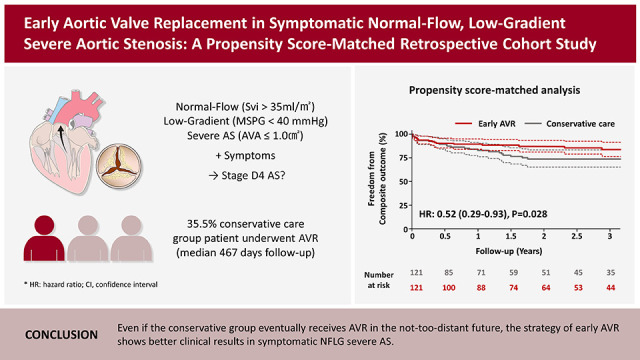
The benefits of aortic valve replacement (AVR) in symptomatic normal-flow, low-gradient (NFLG) severe aortic stenosis (AS) are controversial and current guidelines do not recommend AVR in these patients. Through this study, we found that the early AVR group had a significantly lower incidence of a composite of all-cause death and heart failure hospitalization. In conservative care group, 35.5% underwent delayed AVR during follow-up. These findings suggest that an early AVR strategy might offer a benefit for NFLG severe AS.

Abstract

Background and Objectives

Aortic valve replacement (AVR) is considered a class I indication for symptomatic severe aortic stenosis (AS). However, there is little evidence regarding the potential benefits of early AVR in symptomatic patients diagnosed with normal-flow, low-gradient (NFLG) severe AS.

Methods

Two-hundred eighty-one patients diagnosed with symptomatic NFLG severe AS (stroke volume index ≥35 mL/m2, mean transaortic pressure gradient <40 mmHg, peak transaortic velocity <4 m/s, and aortic valve area <1.0 cm2) between January 2010 and December 2020 were included in this retrospective study. After performing 1:1 propensity score matching, 121 patients aged 75.1±9.8 years (including 63 women) who underwent early AVR within 3 months after index echocardiography, were compared with 121 patients who received conservative care. The primary outcome was a composite of all-cause death and heart failure (HF) hospitalization.

Results

During a median follow-up of 21.9 months, 48 primary outcomes (18 in the early AVR group and 30 in the conservative care group) occurred. The early AVR group demonstrated a significantly lower incidence of primary outcomes (hazard ratio [HR], 0.52; 95% confidence interval [CI], 0.29–0.93; p=0.028); specifically, there was no significant difference in all-cause death (HR, 0.51; 95% CI, 0.23–1.16; p=0.110), although the early AVR group showed a significantly lower incidence of hospitalization for HF (HR, 0.43; 95% CI, 0.19–0.95, p=0.037). Subgroup analyses supported the main findings.

Conclusions

An early AVR strategy may be beneficial in reducing the risk of a composite outcome of death or hospitalization for HF in symptomatic patients with NFLG severe AS. Future randomized studies are required to validate and confirm our findings.

Graphical Abstract

kcj-53-744-abf001.jpg

INTRODUCTION

Severe aortic stenosis (AS) is associated with a high mortality rate if left untreated after symptoms begin. Aortic valve replacement (AVR) is strongly recommended for patients with symptomatic severe AS.1)2) Severe AS is defined as an aortic valve area (AVA) <1.0 cm2 and a mean transaortic pressure gradient (PG) ≥40 mmHg (or peak transaortic jet velocity [Vmax] ≥4 m/s), which are derived from echocardiographic measurement. However, there is often discrepancy between echocardiographic criteria for grading of AS severity, and one-third of patients with severe AS receive discordant gradings.3)4) Current guidelines and expert consensus classify low-gradient AS into several categories according to left ventricular (LV) ejection fraction (EF) and stroke volume (SV) index. Among low-gradient AS patients, those with normal-flow, low-gradient (NFLG) severe AS are often recognized as moderate AS.1)2)5) Nevertheless, recent studies have shown that the number of symptomatic NFLG AS patients is increasing, and have a poor prognosis.6)7) Prolonged LV ejection time, increased systolic blood pressure, and/or reduced systemic arterial compliance may explain the onset of NFLG severe AS.8) NFLG severe AS is a real and frequent entity that should receive special attention. However, the current guidelines offer no specific recommendations for this population because previous studies have reported conflicting results on the efficacy of AVR in patients with NFLG severe AS; consequently, AVR has been underutilized in such individuals.7)9)10)11)12) We aimed to assess the impact of early AVR on clinical outcomes compared to conservative care in symptomatic NFLG severe AS patients. We also aimed to determine which subgroup receives the greatest benefit from early AVR.

METHODS

Ethical statement

The study was approved by the Institutional Review Board of Severance Hospital (4-2022-1242), and the need for informed consent was waived due to the retrospective study design.

Study population

This was a retrospective cohort study performed at a single tertiary hospital in South Korea. Between January 2010 and December 2020, a total of 795 consecutive NFLG severe AS cases (AVA <1.0 cm2, mean transaortic PG <40 mmHg, peak transaortic velocity <4 m/s, and SV index ≥35 mL/m2) was reviewed. Patients without cardinal symptoms (dyspnea, angina, or syncope) (n=155) and those with a history of cardiac surgery (n=311), significant disease of other valve issues (e.g., mitral stenosis with mean PG >5.0 mmHg or any significant regurgitation grade ≥3) (n=32), active malignancies (n=13), or active infective endocarditis (n=3) were excluded. After exclusion, 281 symptomatic NFLG severe AS patients were analyzed. The study population was classified into early AVR and conservative care groups; patients who underwent AVR within 3 months after index echocardiography were classified into the early AVR group. All patient cases were discussed by a multidisciplinary heart team, which decided whether to perform AVR and what type (surgical or transcatheter) of procedure to perform. The index date was defined as the day of index echocardiography. A total of 121 patients was included in the early AVR group, and 1:1 propensity score matching was performed. The matching variables were age, sex, body surface area, body mass index, history of hypertension, diabetes mellitus treated with insulin, chronic kidney disease (stage ≥3), dialysis status, peripheral artery disease, coronary artery disease, old cerebrovascular accident, atrial fibrillation, bicuspid aortic valve, chronic lung disease, chronic liver disease, presence of a pacemaker, and other components of the European System for Cardiac Operative Risk Evaluation (EuroSCORE II) like estimated pulmonary artery systolic pressure or New York Heart Association (NYHA) class.13) Data on baseline characteristics and comorbidities were obtained via medical records. Concomitant multimodal imaging was defined as cardiac computed tomography or transesophageal echocardiography performed within one month after index echocardiography. Several subgroup analyses were performed according to comorbidities and echocardiographic parameters.

Echocardiography

All echocardiographic studies were performed using commercially available equipment, and standard measurements and AS assessments were performed according to current guidelines.14)15) AVA was calculated using the continuity equation formula. The left ventricular outflow tract (LVOT) was assessed in the apical 5-chamber view, and its diameter was measured mid-systole at a point 0.5–1 cm below the aortic annulus on the parasternal long-axis view. The velocity-time integral of the LVOT (VTILVOT) was assessed by pulse-wave Doppler. Assuming a circular geometry of LVOT, SV was calculated using the following formula: LVOT diameter2×0.785×VTILVOT. The highest peak velocity across the aortic valve, the mean transaortic PG, and the VTI of the aortic valve were measured in multiple acoustic windows (apical, suprasternal, or right parasternal) using continuous-wave Doppler. Concomitant valvular regurgitation was classified according to current guidelines.16) All measurements represent an average of 3 cardiac cycles for patients with sinus rhythm or an average of 5 cardiac cycles for patients with atrial fibrillation.

Clinical outcomes

The primary outcome was the composite of all-cause death and unplanned hospitalization for heart failure (HF). The secondary outcomes were individual components of the primary outcome. HF hospitalization was defined as an unplanned urgent admission for management of HF (including intravenous diuretics) lasting ≥24 hours. Patients were censored if primary outcome events occurred. Patients receiving conservative care underwent AVR during follow-up according to the heart team’s discussion and patient preference. Patients who were admitted for planned AVR with symptom and severity progression to classic severe AS were not considered as HF hospitalizations. We also performed 2 additional analyses, comparing (1) the early AVR group with the conservative care group with delayed AVR and (2) the early AVR group with the conservative care group without delayed AVR.

Statistical analyses

Continuous variables are expressed as mean±standard deviation or median (interquartile range) and were compared using Student’s t-test or the Wilcoxon rank-sum test according to distribution. Categorical variables were expressed as number with (percentage) and compared using the χ2 test. The 1:1 propensity score was estimated using logistic regression with all variables in the model and then matched using the greedy nearest-neighbor matching method, in which patients were matched on the logit of the propensity score using a caliper width of 0.2 of the standard deviation. Standardized mean differences were estimated to assess pre- and post-match balance, and <0.2 was considered small.17) Survival rates from clinical outcomes between 2 groups were estimated using the Kaplan-Meier method and compared with the log-rank test. The incidence rates of clinical outcomes were presented as %/year, and 95% confidence intervals (CIs) were calculated. Univariable and multivariable Cox proportional hazards models were used to calculate the hazard ratios (HRs) and 95% CIs for clinical outcomes. In the multivariable model, we included variables that achieved p<0.20 in univariate analyses. Several subgroup analyses were performed according to age, sex, comorbidities, and echocardiographic parameters and represented using forest plots. A density plot was used to show the balance for matched variables in the populations from before and after propensity score–matching. To assess the potential effect of unmeasured confounders, we calculated the E-value, which is defined as the minimum strength of association on the risk ratio scale. A large E-value implies that considerable unmeasured confounding would be needed to explain away an effect estimate, while a small E-value indicates that little unmeasured confounding would be necessary to explain an effect estimate.18)19) All tests were 2-sided, and p<0.05 was considered statistically significant. All statistical analyses were performed using R version 4.1.0 (The R Foundation for Statistical Computing, Vienna, Austria; www.R-project.org).

RESULTS

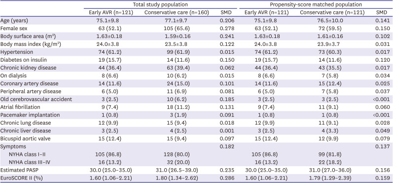
Baseline characteristics

Table 1 shows the baseline characteristics of the early AVR group and conservative care group before and after propensity score matching. All covariates were appropriately matched (Supplementary Figure 1). The mean age of the matched cohort was 75.8±9.9 years, and 135 participants (55.8%) were female. Dyspnea was the major cardinal symptom, and most patients had NYHA class II dyspnea. The median EuroSCORE was 1.57 (1.03–2.13) in the early AVR group and 1.75 (1.25–2.34) in the conservative care group. In the early AVR group, the interval between index echocardiography and AVR was 8 (5–25) days; 67 patients (55.4%) underwent surgical AVR (SAVR) and 54 (44.6%) underwent transcatheter AVR (TAVR). Concomitant coronary revascularization was performed in 11 patients (9.1%) in the early AVR group.
Table 1

Baseline characteristics of overall and propensity-score matched population

kcj-53-744-i001
Total study population Propensity-score matched population
Early AVR (n=121) Conservative care (n=160) SMD Early AVR (n=121) Conservative care (n=121) SMD
Age (years) 75.1±9.8 77.1±9.7 0.206 75.1±9.8 76.5±10.0 0.141
Female sex 63 (52.1) 105 (65.6) 0.278 63 (52.1) 72 (59.5) 0.150
Body surface area (m2) 1.63±0.18 1.59±0.16 0.241 1.63±0.18 1.61±0.16 0.102
Body mass index (kg/m2) 24.0±3.8 23.5±3.8 0.122 24.0±3.8 23.9±3.7 0.031
Hypertension 74 (61.2) 99 (61.9) 0.015 74 (61.2) 73 (60.3) 0.017
Diabetes on insulin 19 (15.7) 14 (11.6) 0.150 19 (15.7) 14 (11.6) 0.120
Chronic kidney disease 44 (36.4) 63 (39.4) 0.062 44 (36.4) 43 (35.5) 0.017
On dialysis 8 (6.6) 10 (6.2) 0.015 8 (6.6) 7 (5.8) 0.034
Coronary artery disease 14 (11.6) 24 (15.0) 0.101 14 (11.6) 15 (12.4) 0.025
Peripheral artery disease 6 (5.0) 11 (6.9) 0.081 6 (5.0) 7 (5.8) 0.037
Old cerebrovascular accident 3 (2.5) 10 (6.2) 0.185 3 (2.5) 3 (2.5) <0.001
Atrial fibrillation 9 (7.4) 18 (11.2) 0.131 9 (7.4) 11 (9.1) 0.060
Pacemaker implantation 1 (0.8) 3 (1.9) 0.091 1 (0.8) 1 (0.8) <0.001
Chronic lung disease 12 (9.9) 15 (9.4) 0.018 12 (9.9) 11 (9.1) 0.028
Chronic liver disease 3 (2.5) 4 (2.5) 0.001 3 (2.5) 4 (3.3) 0.049
Bicuspid aortic valve 15 (12.4) 15 (9.4) 0.097 15 (12.4) 12 (9.9) 0.079
Symptoms 0.182 0.137
NYHA class I–II 105 (86.8) 128 (80.0) 105 (86.8) 99 (81.8)
NYHA class III–IV 16 (13.2) 32 (20.0) 16 (13.2) 22 (18.2)
Estimated PASP 30.0 (25.0–35.0) 31.0 (26.5–39.0) 0.235 30.0 (25.0–35.0) 31.0 (27.0–36.0) 0.156
EuroSCORE II (%) 1.60 (1.06–2.21) 1.80 (1.34–2.62) 0.286 1.60 (1.06–2.21) 1.79 (1.29–2.39) 0.159
Values are presented as number (%), mean±standard deviation or median (range).
AVR = aortic valve replacement; EuroSCORE = European System for Cardiac Operative Risk Evaluation; NYHA = New York Heart Association, PASP = pulmonary artery systolic pressure; SMD = standardized mean difference.
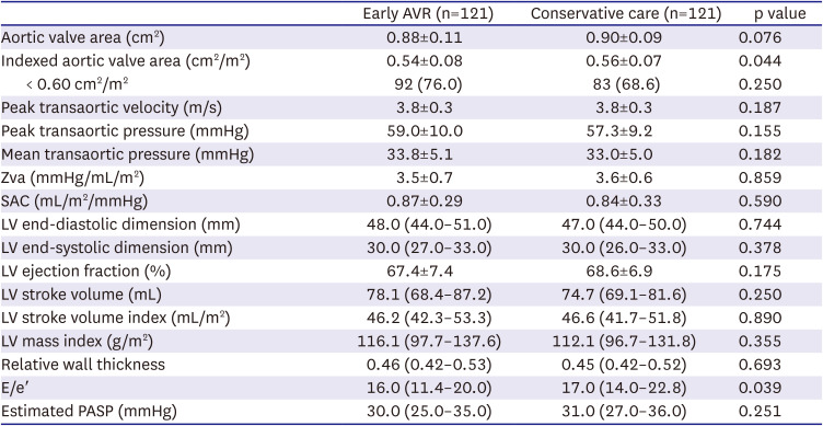
Table 2 shows the echocardiographic characteristics of the 2 groups. Most echocardiographic parameters including AVA, transaortic velocity, transaortic PG, SV index, and LVEF, were similar between the groups. However, indexed AVA (0.54±0.08 vs. 0.56±0.07 cm2/m2, p=0.044) and E/e′ (16.0 [11.4–20.0] vs. 17.0 [14.0–22.8], p=0.039) were lower in the early AVR group than in the conservative care group. Patients in the early AVR group underwent multimodal imaging for in-depth structural or functional evaluation prior to intervention more frequently than patients in the conservative care group (75.2% vs. 21.5%, p<0.001).
Table 2

Echocardiographic parameters

kcj-53-744-i002
Early AVR (n=121) Conservative care (n=121) p value
Aortic valve area (cm2) 0.88±0.11 0.90±0.09 0.076
Indexed aortic valve area (cm2/m2) 0.54±0.08 0.56±0.07 0.044
< 0.60 cm2/m2 92 (76.0) 83 (68.6) 0.250
Peak transaortic velocity (m/s) 3.8±0.3 3.8±0.3 0.187
Peak transaortic pressure (mmHg) 59.0±10.0 57.3±9.2 0.155
Mean transaortic pressure (mmHg) 33.8±5.1 33.0±5.0 0.182
Zva (mmHg/mL/m2) 3.5±0.7 3.6±0.6 0.859
SAC (mL/m2/mmHg) 0.87±0.29 0.84±0.33 0.590
LV end-diastolic dimension (mm) 48.0 (44.0–51.0) 47.0 (44.0–50.0) 0.744
LV end-systolic dimension (mm) 30.0 (27.0–33.0) 30.0 (26.0–33.0) 0.378
LV ejection fraction (%) 67.4±7.4 68.6±6.9 0.175
LV stroke volume (mL) 78.1 (68.4–87.2) 74.7 (69.1–81.6) 0.250
LV stroke volume index (mL/m2) 46.2 (42.3–53.3) 46.6 (41.7–51.8) 0.890
LV mass index (g/m2) 116.1 (97.7–137.6) 112.1 (96.7–131.8) 0.355
Relative wall thickness 0.46 (0.42–0.53) 0.45 (0.42–0.52) 0.693
E/e′ 16.0 (11.4–20.0) 17.0 (14.0–22.8) 0.039
Estimated PASP (mmHg) 30.0 (25.0–35.0) 31.0 (27.0–36.0) 0.251
Values are presented as number (%), mean±standard deviation or median (range).
AVR = aortic valve replacement; LV = left ventricle; PASP = pulmonary artery systolic pressure; SAC = systemic arterial compliance; Zva = valvuloarterial impedance.

Clinical outcomes

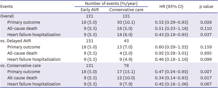
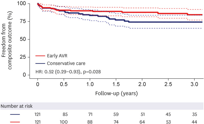
The median follow-up duration was 21.3 (6.5–47.5) months. Table 3 shows the comparisons of clinical events in the early AVR group and the conservative care group. A total of 48 primary outcomes (18 in the early AVR group vs. 30 in the conservative care group) occurred during follow-up. Three patients (2.5%) in the early AVR group died within 30 days after surgery. The numbers of primary outcome events within 3 months after index echocardiography were similar in the 2 groups (7 in the early AVR group vs. 7 in the conservative care group). The early AVR group had a significantly lower incidence of primary outcomes than the conservative care group (5.0 vs. 10.1%/year; HR, 0.52; 95% CI, 0.29–0.93; p=0.028; Figure 1). The E-value for the primary outcome was 2.52. Considering individual outcomes, the early AVR group had a lower incidence of all-cause death than the conservative-care group, but this result did not reach statistical significance (2.5 vs. 5.0%/year; HR, 0.51; 95% CI, 0.23–1.16; p=0.110; Figure 2A). Patients in the early AVR group had a significantly lower incidence of HF hospitalizations than conservative care group patients (2.5 vs. 6.7%/year; HR, 0.43; 95% CI, 0.19–0.95; p=0.037; Figure 2B).
Table 3

Clinical outcomes

kcj-53-744-i003
Events Number of events (%/year) HR (95% CI) p value
Early AVR Conservative care
Overall 121 121
Primary outcome 18 (5.0) 30 (10.1) 0.52 (0.29–0.93) 0.028
All-cause death 9 (2.5) 16 (5.0) 0.51 (0.23–1.16) 0.110
Heart failure hospitalization 9 (2.5) 18 (6.0) 0.43 (0.19–0.95) 0.037
vs. Delayed AVR 121 43
Primary outcome 18 (5.0) 13 (7.0) 0.60 (0.29–1.22) 0.159
All-cause death 9 (2.5) 4 (2.0) 0.92 (0.28–3.01) 0.895
Heart failure hospitalization 9 (2.5) 9 (4.9) 0.46 (0.18–1.16) 0.099
vs. Conservative care 121 78
Primary outcome 18 (5.0) 17 (15.1) 0.47 (0.24–0.92) 0.027
All-cause death 9 (2.5) 12 (10.0) 0.34 (0.14–0.83) 0.017
Heart failure hospitalization 9 (2.5) 9 (7.9) 0.42 (0.16–1.06) 0.067
AVR = aortic valve replacement; CI = confidence interval; HR = hazard ratio.
Figure 1

Survival from composite outcomes of all-cause death or heart failure hospitalization.

AVR = aortic valve replacement; HR = hazard ratio.
kcj-53-744-g001
Figure 2

Survival from individual outcomes of all-cause death (A) and heart failure hospitalization (B).

AVR = aortic valve replacement; HR = hazard ratio.
kcj-53-744-g002
During follow-up, 43 patients (35.5%) in the conservative care group ultimately underwent AVR, and the time interval between indexed echocardiography and AVR was 467 (217–859) days. Progression to high-gradient severe AS (73.3%) was the most common reason for AVR in the conservative care group. Other patients (26.7%) underwent delayed AVR for symptom progression without hemodynamic progression (Supplementary Table 1).
When comparing the early AVR subgroup and delayed AVR subgroup, the former group had a tendency for fewer HF hospitalization events (2.5 vs. 4.9%/year; HR, 0.46; 95% CI, 0.18–1.16; p=0.099). Also, when comparing the early AVR subgroup and conservative care subgroup (excluding patients who underwent delayed AVR), the former group had a significantly lower incidence of the primary outcome (5.0 vs. 15.1%/year; HR, 0.47; 95% CI, 0.24–0.92; p=0.027) and all-cause death (2.5 vs. 7.9%/year; HR, 0.34; 95% CI, 0.14–0.83; p=0.017).
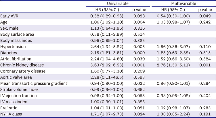
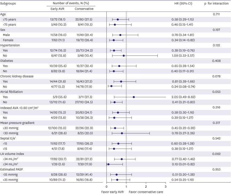
According to multivariable Cox regression modeling, early AVR was independently associated with a lower risk of the primary outcome (adjusted HR, 0.54; 95% CI, 0.30–1.00; p=0.049), while chronic kidney disease was independently associated with a higher risk of the primary outcome (adjusted HR, 2.76; 95% CI, 1.50–5.11; p=0.001; Table 4). Subgroup analysis offered similar results to the main findings (No p value for any interaction was significant). However, the atrial fibrillation subgroup showed a trend toward a greater risk of early AVR (Figure 3). In the multivariable analysis, there was no significant difference between early TAVR and SAVR (adjusted HR, 2.66; 95% CI, 0.63–11.1; p=0.181).
Table 4

Predictors of primary outcomes in Cox regression analysis

kcj-53-744-i004
Univariable Multivariable
HR (95% CI) p value HR (95% CI) p value
Early AVR 0.52 (0.29–0.93) 0.028 0.54 (0.30–1.00) 0.049
Age 1.06 (1.02–1.10) 0.004 1.03 (0.98–1.07) 0.242
Sex, male 1.13 (0.64–1.96) 0.816
Body surface area 0.58 (0.11–2.99) 0.514
Body mass index 0.96 (0.89–1.04) 0.325
Hypertension 2.64 (1.34–5.22) 0.005 1.86 (0.86–3.97) 0.110
Diabetes 2.15 (1.21–3.81) 0.009 1.23 (0.63–2.32) 0.515
Atrial fibrillation 2.24 (1.04–4.80) 0.039 1.52 (0.66–3.50) 0.324
Chronic kidney disease 3.63 (2.02–6.53) <0.001 2.76 (1.50–5.11) 0.001
Coronary artery disease 1.60 (0.77–3.30) 0.209
Aortic valve area 2.28 (0.11–46.5) 0.593
Mean transaortic pressure gradient 0.94 (0.90–1.00) 0.032 0.96 (0.90–1.01) 0.284
Stroke volume index 0.99 (0.96–1.03) 0.662
LV ejection fraction 0.96 (0.94–1.00) 0.053 0.98 (0.95–1.02) 0.404
LV mass index 1.00 (0.99–1.01) 0.825
E/e′ ratio 1.04 (1.01–1.08) 0.021 1.02 (0.98–1.07) 0.285
NYHA class 1.71 (1.07–2.73) 0.024 1.38 (0.85–2.24) 0.191
AVR = aortic valve replacement; CI = confidence interval; HR = hazard ratio; LV = left ventricular; NYHA = New York Heart Association.
Figure 3

Forest plot of subgroup analysis.

AVR = aortic valve replacement; CI = confidence interval; HR = hazard ratio; LV = left ventricular; PASP = pulmonary artery systolic pressure.
kcj-53-744-g003

DISCUSSION

The main findings of this study are as follows: (1) early AVR was associated with a significantly lower rate of the composite outcome of all-cause death and HF hospitalization; (2) about 33% of patients in the conservative care group eventually underwent AVR during a median 21.3-month follow-up; (3) patients in the conservative care group patients who did not undergo delayed AVR had significantly higher incidence rates of the primary outcome and all-cause death. Even the patients receiving delayed AVR in the conservative group had a higher tendency for HF hospitalization compared to patients in the early AVR group; and (4) subgroup analysis consistently showed the benefits of early AVR in patients with symptomatic NFLG severe AS. These findings provide evidence supporting the potential benefit of early AVR in patients with symptomatic NFLG severe AS.
Unlike the situation with classical high-gradient severe AS, evidence of surgical and TAVR in low-gradient severe AS is limited.1)2) According to current guidelines, NFLG severe AS patients are usually recognized to have only moderate AS, and AVR is not indicated.2) However, recent studies have reported that NFLG severe AS patients have specific characteristics, such as female predominance, small body size, and reduced arterial compliance combined with high blood pressure, which are related to a poor prognosis.7)20)21) Compared to previous studies, our study enrolled patients with similar characteristics, although the mortality rate was low.9)10)11) This might be related to the presence of fewer comorbidities and few ethnic differences in our study population.22)23)
Several previous studies have reported conflicting results on the benefits of early AVR (Supplementary Table 2). Saeed et al.10) documented mortality benefits of AVR in small numbers of a matched population (80 matched pairs) and a higher proportion of NYHA III–IV patients. A study by Zusman et al.11) enrolled a much older population (median age, 83 years) and matched only conservatively treated patients and TAVR patients, yielding a small sample of 94 matched pairs. The meta-analysis by Dayan et al.24) also reported the benefits of AVR in NFLG severe AS. Our study documented a benefit of AVR in the composite outcome but not in all-cause death. These differences have several possible explanations. First, our study population might have received more meticulous follow-up care. In the present study, 43 of 121 patients (35.5%) in the conservative care group underwent delayed AVR, whereas only 2.7% of conservative group patients underwent AVR in the study by Zusman et al.11) Second, as mentioned above, the mortality rate of our study was less than those of previous studies. Thus, our study sample might be too small to show statistical significance in reducing mortality. Additionally, the mortality benefit from AVR might be smaller for NFLG severe AS patients than for those with conventional high-gradient AS because NFLG severe AS might actually be an “intermediate” stage of the condition between moderate and severe AS.25)
Kang et al.9) showed similar mortality rates from AVR to watchful waiting in similar study subjects as ours, but they enrolled a markedly small population of TAVR patients (n = 5) and allowed differences in echocardiographic parameters between the groups. Eleid et al.26) showed that NFLG severe AS patients had favorable survival with standard medical treatment with no benefit or damage from AVR, but the NFLG group had significantly fewer comorbidities.
In addition, neither of these previous studies examined HF hospitalization events. Our study showed significant benefits of early AVR in HF hospitalization. Notably, the conservative care group had a higher rate of HF hospitalization than the early AVR group despite delayed AVR being performed during follow-up. In this study, the conservative group patients without delayed AVR showed higher mortality rates. Although timely performed AVR appeared to reduce adverse outcomes, there was a tendency for more HF hospitalization events. These findings suggest that early AVR performed before advanced cardiac remodeling and comorbidity progression might be helpful in NFLG severe AS patients. Considering composite outcomes of death or HF hospitalization as major primary outcomes in recent cardiology trials, our study has strength in showing the benefit of early AVR in composite outcomes with a well-matched, larger study population.27)28)
We expected to find specific subgroups in which the effect of early AVR was magnified, but we did not. Our subgroup analysis showed consistent benefits of early AVR, and these findings suggest that early AVR might be performed for most symptomatic NFLG severe AS patients. Only the atrial fibrillation subgroup showed a trend that did not favor early AVR, but we included very few patients in this subgroup, so this result should not be over-interpreted. In addition, the beat-to-beat variability and low-flow status of atrial fibrillation might induce bias in AS assessment.29) Finally, we were not able to examine the roles of multimodal imaging because of the small number of subjects who underwent multimodal imaging. Future prospective studies with combined multimodal imaging and larger populations are needed to confirm our findings.
This study has several limitations. First, this was a retrospective study from a single tertiary center in South Korea; thus, it carries inherent limitations and ethnical biases. Second, the selection bias needs to be acknowledged. It is likely that early AVR group members had more advanced aortic valve disease. Third, the severity of AS was only assessed by Doppler echocardiography; multimodal imaging was not performed in many patients, especially in the conservative care group. However, the method of choice for assessing AS severity is Doppler echocardiography, and the role of other imaging modalities in this situation is inconclusive. Fourth, although we considered and matched all available clinical variables from previous studies, there might be unmeasured confounders that affected clinical events. Fifth, although AVA and E/e' were different between the 2 groups, we could not include echocardiographic parameters in the calculation of the propensity score due to the small number of patients. Instead, we performed a multivariable analysis including such echocardiographic variables and the results were consistent. These limitations should be taken into account when interpreting the results. Instead, we performed univariable and multivariable analysis including such echocardiographic variables, and the results showed that early AVR also showed meaningful results. However, despite these limitations, we believe that the results observed in this study are meaningful in addressing unmet clinical needs.
In patients with symptomatic NFLG severe AS, early AVR may improve the composite outcome of all-cause death and HF hospitalization compared to conservative care. Future large-scale prospective studies are needed to confirm these findings.

ACKNOWLEDGMENTS

The authors would like to thank Medical Illustration & Design, part of the Medical Research Support Services of Yonsei University College of Medicine, for all artistic support related to this work.

Notes

Funding: This research was supported by the Korean Cardiac Research Foundation (202202-02).

Conflict of Interest: The authors have no financial conflicts of interest.

Data Sharing Statement: The data generated in this study is available from the corresponding author(s) upon reasonable request.

Author Contributions:

  • Conceptualization: Kim K, Cho I, Shim CY.

  • Data curation: Kim K, Cho I, Hong GR, Ha JW, Shim CY.

  • Formal analysis: Kim K, Ko KY, Shim CY.

  • Funding acquisition: Kim K.

  • Investigation: Ko KY.

  • Methodology: Kim K.

  • Project administration: Shim CY.

  • Resources: Ko KY, Lee SH, Lee S.

  • Supervision: Cho I, Hong GR, Ha JW, Shim CY.

  • Validation: Kim K, Lee SH, Lee S, Hong GR, Ha JW, Shim CY.

  • Visualization: Shim CY.

  • Writing - original draft: Kim K, Shim CY.

  • Writing - review & editing: Kim K, Shim CY.

References

1. Otto CM, Nishimura RA, Bonow RO, et al. 2020 ACC/AHA guideline for the management of patients with valvular heart disease: executive summary: a report of the American College of Cardiology/American Heart Association joint committee on clinical practice guidelines. Circulation. 2021; 143:e35–e71. PMID: 33332149.
2. Vahanian A, Beyersdorf F, Praz F, et al. 2021 ESC/EACTS guidelines for the management of valvular heart disease. Eur Heart J. 2022; 43:561–632. PMID: 34453165.
3. Minners J, Allgeier M, Gohlke-Baerwolf C, Kienzle RP, Neumann FJ, Jander N. Inconsistencies of echocardiographic criteria for the grading of aortic valve stenosis. Eur Heart J. 2008; 29:1043–1048. PMID: 18156619.
crossref
4. Berthelot-Richer M, Pibarot P, Capoulade R, et al. Discordant grading of aortic stenosis severity: echocardiographic predictors of survival benefit associated with aortic valve replacement. JACC Cardiovasc Imaging. 2016; 9:797–805. PMID: 27209111.
5. Clavel MA, Burwash IG, Pibarot P. Cardiac imaging for assessing low-gradient severe aortic stenosis. JACC Cardiovasc Imaging. 2017; 10:185–202. PMID: 28183438.
crossref
6. Strange G, Stewart S, Celermajer D, et al. Poor long-term survival in patients with moderate aortic stenosis. J Am Coll Cardiol. 2019; 74:1851–1863. PMID: 31491546.
crossref
7. Li SX, Patel NK, Flannery LD, et al. Trends in utilization of aortic valve replacement for severe aortic stenosis. J Am Coll Cardiol. 2022; 79:864–877. PMID: 35241220.
crossref
8. Clavel MA, Guzzetti E, Annabi MS, Salaun E, Ong G, Pibarot P. Normal-flow low-gradient severe aortic stenosis: myth or reality? Struct Heart. 2018; 2:180–187.
crossref
9. Kang DH, Jang JY, Park SJ, et al. Watchful observation versus early aortic valve replacement for symptomatic patients with normal flow, low-gradient severe aortic stenosis. Heart. 2015; 101:1375–1381. PMID: 26105038.
crossref
10. Saeed S, Vamvakidou A, Seifert R, Khattar R, Li W, Senior R. The impact of aortic valve replacement on survival in patients with normal flow low gradient severe aortic stenosis: a propensity-matched comparison. Eur Heart J Cardiovasc Imaging. 2019; 20:1094–1101. PMID: 31327014.
crossref
11. Zusman O, Pressman GS, Banai S, Finkelstein A, Topilsky Y. Intervention versus observation in symptomatic patients with normal flow low gradient severe aortic stenosis. JACC Cardiovasc Imaging. 2018; 11:1225–1232. PMID: 29055632.
crossref
12. Taniguchi T, Morimoto T, Shiomi H, et al. High- versus low-gradient severe aortic stenosis: demographics, clinical outcomes, and effects of the initial aortic valve replacement strategy on long-term prognosis. Circ Cardiovasc Interv. 2017; 10:e004796. PMID: 28500139.
13. Nashef SA, Roques F, Sharples LD, et al. EuroSCORE II. Eur J Cardiothorac Surg. 2012; 41:734–744. PMID: 22378855.
crossref
14. Lang RM, Badano LP, Mor-Avi V, et al. Recommendations for cardiac chamber quantification by echocardiography in adults: an update from the American Society of Echocardiography and the European Association of Cardiovascular Imaging. J Am Soc Echocardiogr. 2015; 28:1–39.e14. PMID: 25559473.
crossref
15. Baumgartner H, Hung J, Bermejo J, et al. Recommendations on the echocardiographic assessment of aortic valve stenosis: a focused update from the European Association of Cardiovascular Imaging and the American Society of Echocardiography. J Am Soc Echocardiogr. 2017; 30:372–392. PMID: 28385280.
crossref
16. Zoghbi WA, Adams D, Bonow RO, et al. Recommendations for noninvasive evaluation of native valvular regurgitation: a report from the American Society of Echocardiography developed in collaboration with the Society for Cardiovascular Magnetic Resonance. J Am Soc Echocardiogr. 2017; 30:303–371. PMID: 28314623.
crossref
17. Harris H, Horst SJ. A brief guide to decisions at each step of the propensity score matching process. Pract Assess Res Eval. 2016; 21:4.
18. Zhao QY, Luo JC, Su Y, Zhang YJ, Tu GW, Luo Z. Propensity score matching with R: conventional methods and new features. Ann Transl Med. 2021; 9:812. PMID: 34268425.
crossref
19. VanderWeele TJ, Ding P. Sensitivity analysis in observational research: introducing the E-value. Ann Intern Med. 2017; 167:268–274. PMID: 28693043.
crossref
20. Mantovani F, Fanti D, Tafciu E, et al. When aortic stenosis is not alone: epidemiology, pathophysiology, diagnosis and management in mixed and combined valvular disease. Front Cardiovasc Med. 2021; 8:744497. PMID: 34722676.
crossref
21. Tribouilloy C, Rusinaru D, Maréchaux S, et al. Low-gradient, low-flow severe aortic stenosis with preserved left ventricular ejection fraction: characteristics, outcome, and implications for surgery. J Am Coll Cardiol. 2015; 65:55–66. PMID: 25572511.
crossref
22. Wilson JB, Jackson LR 2nd, Ugowe FE, et al. Racial and ethnic differences in treatment and outcomes of severe aortic stenosis: a review. JACC Cardiovasc Interv. 2020; 13:149–156. PMID: 31973792.
crossref
23. Ryu DR, Park SJ, Han H, et al. Progression rate of aortic valve stenosis in Korean patients. J Cardiovasc Ultrasound. 2010; 18:127–133. PMID: 21253361.
crossref
24. Dayan V, Vignolo G, Magne J, Clavel MA, Mohty D, Pibarot P. Outcome and impact of aortic valve replacement in patients with preserved LVEF and low-gradient aortic stenosis. J Am Coll Cardiol. 2015; 66:2594–2603. PMID: 26670058.
crossref
25. Chadha G, Bohbot Y, Lachambre P, et al. Progression of normal flow low gradient “severe” aortic stenosis with preserved left ventricular ejection fraction. Am J Cardiol. 2020; 128:151–158. PMID: 32650909.
crossref
26. Eleid MF, Sorajja P, Michelena HI, Malouf JF, Scott CG, Pellikka PA. Flow-gradient patterns in severe aortic stenosis with preserved ejection fraction: clinical characteristics and predictors of survival. Circulation. 2013; 128:1781–1789. PMID: 24048203.
crossref
27. Banovic M, Putnik S, Penicka M, et al. Aortic valve replacement versus conservative treatment in asymptomatic severe aortic stenosis: the AVATAR trial. Circulation. 2022; 145:648–658. PMID: 34779220.
28. Solomon SD, McMurray JJ, Claggett B, et al. Dapagliflozin in heart failure with mildly reduced or preserved ejection fraction. N Engl J Med. 2022; 387:1089–1098. PMID: 36027570.
29. Alsidawi S, Khan S, Pislaru SV, et al. High prevalence of severe aortic stenosis in low-flow state associated with atrial fibrillation. Circ Cardiovasc Imaging. 2021; 14:e012453. PMID: 34250815.
crossref

SUPPLEMENTARY MATERIALS

Supplementary Table 1

Details of delayed AVR in conservative care group
kcj-53-744-s001.xls

Supplementary Table 2

Details on previous and current studies on normal-flow low-gradient severe aortic stenosis
kcj-53-744-s002.xls

Supplementary Figure 1

Density plots showing the distribution balance for variables before and after propensity score matching.
kcj-53-744-s003.ppt
TOOLS
Similar articles